Patient-ventilator asynchrony (PVA)—also termed patient-ventilator dyssynchrony (PVD)—is defined as a failure of mechanical breath timing, flow delivery, or pressure assistance to match the patient's intrinsic neural respiratory drive and muscular effort. In the pediatric intensive care unit (PICU), PVA is remarkably prevalent, affecting up to $30-50\%$ of mechanically ventilated children and infants during assisted breathing modes.
Unlike adult patients with mature respiratory mechanics and large lung volumes, infants and young children possess distinctive neurodevelopmental and mechanical vulnerabilities that amplify both the frequency of asynchronies and their deleterious physiological sequelae. High baseline respiratory frequencies, short neural inspiratory times ($T_{i,\text{neural}} \approx 0.3 - 0.6\text{ s}$), miniature tidal volumes ($20 - 100\text{ mL}$), highly compliant chest walls, and frequent uncuffed/micro-cuff peritubular airway leaks create an exceptionally challenging environment for conventional pneumatic triggering and cycling algorithms.
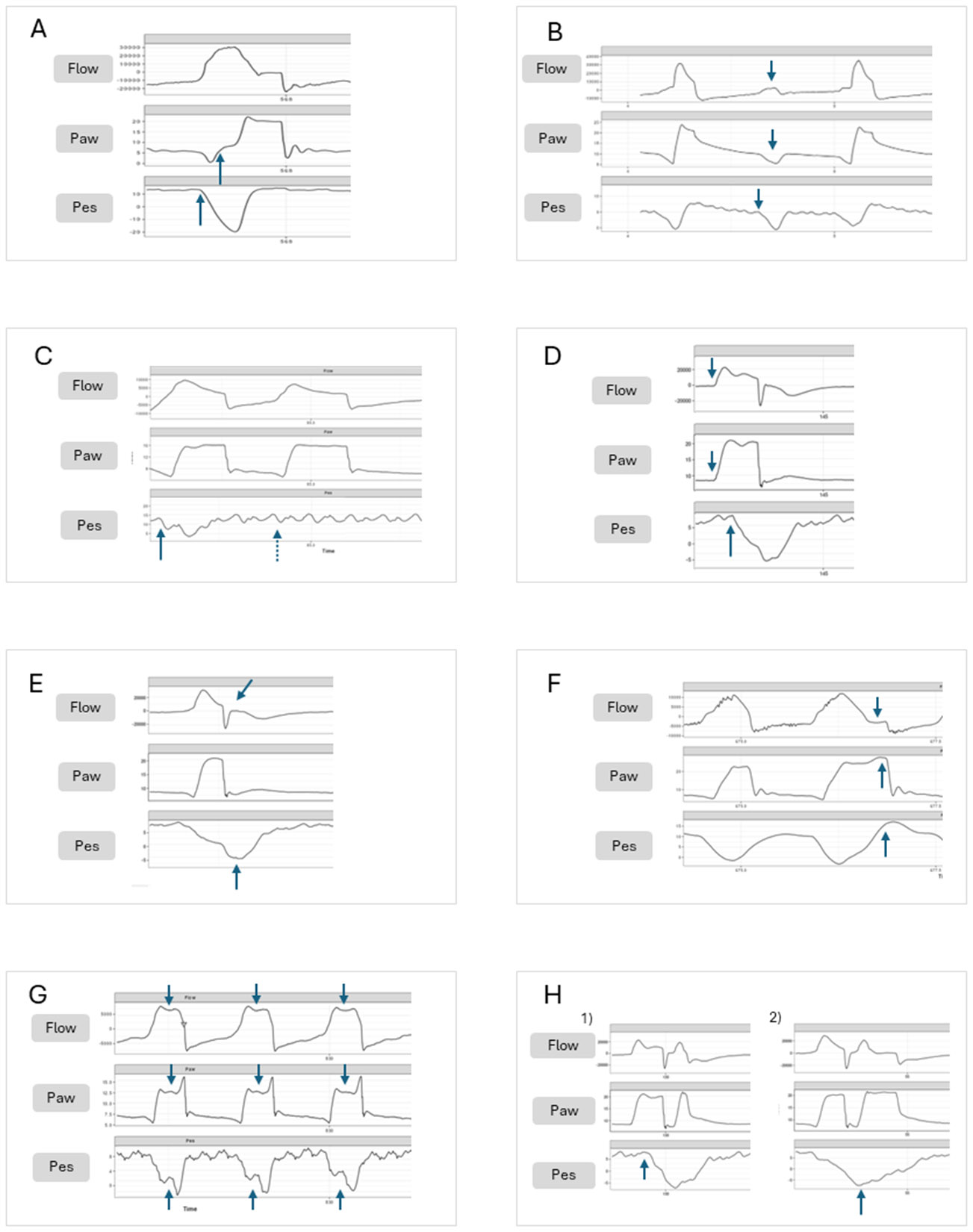 Figure 1: Comprehensive scalar tracings demonstrating major patient-ventilator asynchrony (PVA) subtypes in pediatric mechanical ventilation, recorded with simultaneous airway flow (top row), airway pressure ($P_{\text{aw}}$, middle row), and esophageal pressure ($P_{\text{es}}$, bottom row). (A) Delayed Triggering: Time lag between diaphragmatic effort onset ($P_{\text{es}}$ drop) and machine pressurization. (B) Ineffective Triggering: Patient inspiratory effort fails to cross the pneumatic threshold; flow and $P_{\text{aw}}$ deflect without machine breath delivery. (C) Auto-Triggering: Machine breath delivered in the absence of true neural effort (flat $P_{\text{es}}$). (D) Reverse Triggering: Reflex diaphragmatic contraction entrained after machine-delivered breath initiation. (E) Premature Cycling/Termination: Neural inspiration persists beyond set $T_i$, producing a post-cycling flow dip and bounce. (F) Delayed Cycling/Termination: Machine $T_i$ exceeds neural $T_i$, causing active expiratory braking against the inspiratory phase ($P_{\text{aw}}$ spike and $P_{\text{es}}$ rise). (G) Flow Undershoot / Starvation: Patient flow demand exceeds delivery, scooping $P_{\text{aw}}$ and creating a W-shaped $P_{\text{es}}$ deflection. (H) Double Cycling / Breath Stacking: Two consecutive mechanical cycles triggered by either reverse triggering or prolonged neural inspiratory effort. Source: Yoon B, Blokpoel R, Khemani RG, Emeriaud G, Kneyber MCJ, et al. Expert Rev Respir Med. 2025;19(5):371-384. DOI:
10.1080/17476348.2025.2487165
. PMCID: PMC12109136.
Unique Pediatric Vulnerabilities to Dyssynchrony
The interaction between a critically ill child and a mechanical ventilator is governed by the equation of motion. However, pediatric anatomical and developmental physiology introduces substantial non-linear perturbations into this system:
flowchart TD
    subgraph Anatomical & Developmental Factors
        A1["High Baseline RR & Short Ti,neural
(Ti ≈ 0.3 - 0.6 s)"] --> P1["Extreme Sensitivity to Trigger & Cycling Delays
(50-100 ms delay consumes 20-30% of Ti)"]
        A2["Uncuffed / Micro-cuff ETT Leaks
(Up to 20-40% peritubular loss)"] --> P2["Pneumatic Trigger Distortion
(Auto-triggering vs Missing True Effort)"]
        A3["Low Tidal Volumes (20-100 mL)
& High Circuit Compliance"] --> P3["Cardiogenic & Water Oscillation Artifact
(Pneumatic sensor misinterpretation)"]
        A4["Highly Compliant Ribcage
(High Ccw relative to adult)"] --> P4["Chest Wall Distortion & Inward Sucking
(Dissipation of Pmus without airway flow)"]
    end

    P1 & P2 & P3 & P4 --> ASYNC["Severe Patient-Ventilator Asynchrony (PVA)
(Trigger, Flow, Cycling, & Breath Stacking Errors)"]
1. High Baseline Respiratory Rate & Truncated Neural Inspiratory Time
An infant breathing at $40 - 60\text{ breaths/min}$ exhibits a neural inspiratory time ($T_{i,\text{neural}}$) of merely $0.30 - 0.50\text{ seconds}$ (compared to $0.80 - 1.20\text{ seconds}$ in adults). A pneumatic trigger delay of $80 - 120\text{ milliseconds}$—readily tolerated by an adult—represents up to $25 - 35\%$ of the infant's entire inspiratory cycle. By the time the ventilator senses the trigger and accelerates inspiratory flow, the infant's neural drive may have already terminated, precipitating immediate cycling mismatch and profound patient agitation.
2. Uncuffed and Micro-Cuff Endotracheal Tube Leaks
In neonates and infants, uncuffed or subglottic-sealing micro-cuff endotracheal tubes (ETTs) frequently allow peritubular air escape during positive-pressure inspiration.
- In flow-triggered systems, the baseline bias flow ($1.0 - 6.0\text{ L/min}$) passing through the circuit is depleted by the peritubular leak. The return flow sensor at the expiratory valve detects a flow deficit ($\dot{V}_{\text{insp}} > \dot{V}_{\text{exp}}$), misinterpreting the leak as patient inspiratory effort and generating rapid-fire auto-triggering runaway.
- When leak compensation algorithms attempt to offset this baseline leak, small spontaneous patient efforts may be filtered out as "leak variance", leading to ineffective triggering.
3. Miniature Tidal Volumes and Sensor Artifacts
Infant tidal volumes range from $20\text{ mL}$ to $100\text{ mL}$ (or $< 10\text{ mL}$ in neonates). Under these diminutive flow regimes:
- Cardiogenic Oscillations: The physical impact of cardiac systole against adjacent lung lobes creates cyclic pressure swings ($\Delta P \approx 0.5 - 1.5\text{ cmH}_2\text{O}$) and flow oscillations ($\Delta \dot{V} \approx 0.2 - 0.8\text{ L/min}$). In a sensitive trigger setting, each heartbeat auto-triggers a mechanical breath.
- Circuit Condensation: Water droplets pooled in heated inspiratory or expiratory limbs generate turbulent pressure-flow fluttering that frequently auto-triggers the ventilator at the machine's maximum respiratory rate.
4. Highly Compliant Chest Wall
The infant ribcage consists primarily of pliable cartilaginous ribs. During vigorous diaphragmatic contraction (elevated muscular pressure, $P_{\text{mus}}$), the highly compliant chest wall sucks inward (subcostal and intercostal retractions) rather than expanding outwards. A large fraction of $P_{\text{mus}}$ is dissipated into deforming the ribcage rather than generating a negative airway opening pressure ($\Delta P_{\text{ao}}$) or inspiratory airflow ($\dot{V}$). Consequently, the ventilator fails to detect the breath, culminating in ineffective triggering.
The mechanical equation of motion during spontaneous patient-triggered breathing is:
$$P_{\text{aw}}(t) + P_{\text{mus}}(t) = E_{\text{rs}} \cdot V(t) + R_{\text{aw}} \cdot \dot{V}(t) + \text{PEEP}_{\text{tot}}$$Prior to trigger opening ($t_0 \to t_{\text{trigger}}$), airway pressure is supported only by the baseline circuit PEEP, and gas flow is zero ($\dot{V} = 0$). The total work of breathing performed exclusively by the child's diaphragm just to activate the ventilator valve is expressed as:
$$WOB_{\text{trigger}} = \int_{t_0}^{t_{\text{trigger}}} P_{\text{mus}}(t) \cdot \dot{V}(t) \, dt + \Delta P_{\text{es}} \cdot V_{\text{trig}}$$When intrinsic PEEP ($\text{PEEP}_i$) is present, the child must generate an additional threshold pressure:
$$P_{\text{mus,required}} \ge \text{PEEP}_i + \text{Trigger Sensitivity Threshold}$$In infants with bronchiolitis or severe asthma, $\text{PEEP}_i$ can exceed $8 - 12\text{ cmH}_2\text{O}$, forcing the child to perform enormous isometric work before the ventilator delivers a single milliliter of gas.
Comprehensive Asynchrony Taxonomy in Pediatrics
Patient-ventilator asynchronies are systematically classified based on the phase of the mechanical breath cycle in which the disharmony occurs: Triggering, Flow Delivery, Cycling (Termination), and Phase Coupling (Breath Stacking).
flowchart TD
    PVA["Patient-Ventilator Asynchrony (PVA)"]

    PVA --> T["1. Trigger Phase Asynchronies"]
    PVA --> F["2. Flow Phase Asynchronies"]
    PVA --> C["3. Cycling Phase Asynchronies"]
    PVA --> S["4. Phase-Coupling / Stacking"]

    T --> T1["Ineffective Triggering
(PEEPi, Weak Drive, High Threshold)"]
    T --> T2["Auto-Triggering
(Condensation, Leaks, Cardiogenic)"]
    T --> T3["Trigger Delay
(Pneumatic transmission lag)"]

    F --> F1["Flow Starvation
(Dished-out Paw in VCV / Low Rise)"]
    F --> F2["Excessive Flow / Overshoot
(Pressure Spike in PCV / Fast Rise)"]

    C --> C1["Premature Cycling
(Tiset < Tineural or high %Esens)"]
    C --> C2["Delayed Cycling
(Tiset > Tineural or low %Esens in COPD/Asthma)"]

    S --> S1["Double Triggering
(High neural drive vs short set Ti)"]
    S --> S2["Reverse Triggering
(Mechanical stretch entraining reflex Pmus)"]
1. Trigger Phase Asynchronies
| Asynchrony Type | Pathophysiological Mechanism | Scalar Waveform Signature ($P_{\text{aw}}$, Flow, $P_{\text{es}}$ / $E_{\text{di}}$) | Bedside Corrective Action |
|---|---|---|---|
| Ineffective Triggering (Wasted Effort) | Patient generates inspiratory effort ($P_{\text{mus}} > 0$), but $P_{\text{aw}}$ or flow deflection fails to reach the set trigger threshold. Primary causes: high dynamic intrinsic PEEP ($\text{PEEP}_i$), diaphragmatic fatigue, oversedation, or blunt trigger sensitivity. | Negative deflection on $P_{\text{es}}$ or upward spike on $E_{\text{di}}$ during expiration accompanied by a transient upward deflection in expiratory flow toward baseline, without a machine breath delivery. | 1. Apply extrinsic PEEP to match $\approx 75-85\%$ of dynamic $\text{PEEP}_i$ (counteracting the threshold load). 2. Increase trigger sensitivity. 3. Decrease excessive pressure support / tidal volume (which promotes dynamic hyperinflation). |
| Auto-Triggering (False Triggering) | Ventilator delivers a mechanical breath triggered by non-patient mechanical artifacts rather than true neuro-muscular effort. Primary causes: large circuit leaks, uncuffed ETT gas escape, water condensation fluttering in tubing, or cardiogenic pressure-flow oscillations. | Mechanical breath initiated with zero preceding negative deflection on $P_{\text{es}}$ and zero $E_{\text{di}}$ signal. Unexplained tachypnea at machine's maximum set backup rate. | 1. Drain circuit condensation from water traps. 2. Inflate ETT micro-cuff or activate intelligent leak compensation. 3. Decrease trigger sensitivity (e.g., raise flow trigger from $0.5$ to $1.0-1.5\text{ L/min}$). 4. Switch to NAVA ($E_{\text{di}}$ triggering). |
| Trigger Delay | Prolonged time interval between the onset of neural inspiration (drop in $P_{\text{es}}$ or surge in $E_{\text{di}}$) and the initial rise in airway pressure ($P_{\text{aw}}$). Trigger delay $> 100\text{ ms}$ severely impairs pediatric breathing. | Visible temporal gap between $E_{\text{di}} / P_{\text{es}}$ deflection onset and machine $P_{\text{aw}}$ upstroke. $P_{\text{aw}}$ displays a deep negative dip prior to pressurization. | 1. Transition from pressure triggering to flow triggering. 2. Increase flow trigger sensitivity. 3. Minimize circuit compliance and eliminate ETT resistance bottlenecks. |
Pressure triggering requires the infant to depressurize the closed circuit volume, demanding significant isometric work ($\Delta P \approx 0.5 - 2.0\text{ cmH}_2\text{O}$). In contrast, Flow Triggering maintains a continuous bias flow ($\dot{V}_{\text{bias}} \approx 2 - 6\text{ L/min}$) through the circuit. As soon as the infant inhales even $0.2 - 0.5\text{ L/min}$, the expiratory sensor detects the instantaneous drop in returning flow, opening the inspiratory valve with $< 40-60\text{ ms}$ delay and reducing trigger work of breathing by $40-60\%$. Flow triggering is the undisputed standard of care in pediatric mechanical ventilation.
2. Flow Phase Asynchronies
flowchart LR
    subgraph Flow Starvation / Under-Assist
        FS1["High Patient Drive & Peak Flow Demand"] --> FS2["Fixed Constant Flow in VCV or Slow Rise Time in PCV"]
        FS2 --> FS3["Pmus pulls Paw downward
(Dished-out Paw scalar & W-shaped Pes)"]
        FS3 --> FS4["Patient Agitation & Respiratory Muscle Exhaustion"]
    end

    subgraph Excessive Flow / Overshoot
        EF1["Overly Aggressive Rise Time in PCV
or Excessive PSV level"] --> EF2["Flow exceeds airway impedance capability"]
        EF2 --> EF3["Sharp Pressure Spike at Breath Onset
(Ringing / Pressure Overshoot)"]
        EF3 --> EF4["Early Flow Termination & Premature Cycling"]
    end
| Asynchrony Type | Pathophysiological Mechanism | Scalar Waveform Signature | Bedside Corrective Action |
|---|---|---|---|
| Flow Starvation (Flow Undershoot) | Delivered inspiratory gas flow fails to satisfy the patient's instantaneous muscular flow demand ($\dot{V}_{\text{patient}} > \dot{V}_{\text{ventilator}}$). Highly prevalent in Volume Control Continuous Mandatory Ventilation (VC-CMV) with fixed square flow patterns. | Airway pressure ($P_{\text{aw}}$) scalar displays a sunken, scooped-out, or concave profile during inspiration. Esophageal pressure ($P_{\text{es}}$) exhibits an exaggerated negative W-shaped plunge. | 1. Switch from VC-CMV to Pressure-Controlled (PC) modes or Pressure Support (PSV) where flow is decelerating and variable. 2. In PC modes, shorten the pressure rise time / slope ($0.05 - 0.10\text{ s}$) to allow immediate peak flow delivery. 3. In VC modes, increase set peak inspiratory flow rate. |
| Excessive Flow (Pressure Overshoot) | Inspiratory flow accelerates too rapidly into high-resistance pediatric airways, creating turbulence and transient pressure spikes exceeding the set target pressure. | Sharp, transient spike ("pressure ringing") at the opening limb of the $P_{\text{aw}}$ waveform exceeding the set inspiratory plateau by $> 2 - 4\text{ cmH}_2\text{O}$, followed by rapid oscillation. | 1. Lengthen the inspiratory rise time / slope ($0.10 - 0.20\text{ s}$). 2. Reduce inspiratory pressure level if tidal volumes are excessive. |
3. Cycling (Termination) Phase Asynchronies
Cycling defines the transition from mechanical inspiration to expiration. In pressure support ventilation (PSV), cycling is governed by the Expiratory Trigger Sensitivity ($\%E_{\text{sens}}$)—the percentage of peak inspiratory flow at which the ventilator terminates the breath.
flowchart TD
    A["Patient Neural Effort (T_i,neural) vs Ventilator Set Mechanical (T_i,set)"] --> B{"Compare Timings"}

    B -->|"T_i,set < T_i,neural
or %Esens too high (e.g. 50%)"| C["Premature Cycling (Early Termination)"]
    C --> C_Sign["Waveform: Sudden drop in flow with upward bounce
Pes / Edi continues downward into expiration
Risk: Triggers immediate second breath (Double Trigger)"]

    B -->|"T_i,set > T_i,neural
or %Esens too low (e.g. 10%)"| D["Delayed Cycling (Late Termination)"]
    D --> D_Sign["Waveform: Expiratory braking spikes Paw at end-inspiration
Pes rises sharply while flow is still positive
Risk: Dynamic hyperinflation, PEEPi, patient fighting vent"]
Premature Cycling ($T_{i,\text{set}} < T_{i,\text{neural}}$)
- Pathophysiology: The ventilator terminates the mechanical breath while the child's diaphragm is still actively contracting.
- Waveform Signature: The flow scalar drops abruptly toward zero, then displays a positive inspiratory "bounce" or inflection. Esophageal pressure ($P_{\text{es}}$) remains deeply negative into mechanical expiration.
- Clinical Hazard: The ongoing neural effort immediately re-triggers the ventilator, producing Double Triggering.
- Corrective Action: Lengthen set $T_i$ in time-cycled modes; decrease $\%E_{\text{sens}}$ threshold (e.g., from $50\%$ to $25\%$) in flow-cycled PSV.
Delayed Cycling ($T_{i,\text{set}} > T_{i,\text{neural}}$)
- Pathophysiology: The ventilator continues to deliver pressurized gas after the child's neural inspiration has ceased and active expiration has commenced. Common in pediatric obstructive lung disease (bronchiolitis, asthma) where elevated airway resistance ($R_{\text{aw}}$) prolongs the expiratory time constant ($\tau = R_{\text{aw}} \times C_{\text{rs}}$), slowing flow decay.
- Waveform Signature: A sharp terminal upward spike on the $P_{\text{aw}}$ scalar ("expiratory kick") as the child actively exhales against the closed inspiratory valve; $P_{\text{es}}$ rises above baseline during the end of machine inspiration.
- Corrective Action: Shorten set $T_i$; increase $\%E_{\text{sens}}$ threshold (e.g., from $15\%$ to $40-50\%$) so the breath terminates at a higher flow cutoff.
4. Phase-Coupling & Complex Asynchronies
flowchart TD
    subgraph Double Triggering (Patient-Initiated)
        DT1["High Central Respiratory Drive
(Sepsis, ARDS, Severe Acidosis)"] --> DT2["T_i,neural significantly exceeds T_i,set"]
        DT2 --> DT3["Breath 1 Cycles Off while Diaphragm is still Contracting"]
        DT3 --> DT4["Persistent Negative Pes instantly triggers Breath 2
(Zero Expiratory Pause)"]
        DT4 --> DT5["BREATH STACKING:
Cumulative VT = 12-16 mL/kg
Alveolar Rupture & Barotrauma"]
    end

    subgraph Reverse Triggering (Ventilator-Entrained)
        RT1["Deeply Sedated Child on Controlled Mode (PC/VC)"] --> RT2["Time-Triggered Mandatory Machine Breath Delivered"]
        RT2 --> RT3["Diaphragmatic Stretch Reflex / Mechanoreceptor Activation
(Hering-Breuer Reflex Entrainment)"]
        RT3 --> RT4["Reflex Diaphragmatic Contraction (Pmus) triggered mid/late-breath"]
        RT4 --> RT5["Mid-breath Pes drop or Double Cycle
Eccentric Muscle Contraction & Biotrauma"]
    end
Double triggering delivers two consecutive mechanical tidal volumes within a single neural breath cycle without allowing time for expiratory alveolar emptying ($T_e \approx 0\text{ s}$). In a child ventilated at $6\text{ mL/kg}$, a double-triggered breath stacks tidal volume to $12 - 15\text{ mL/kg}$, escalating transpulmonary pressure ($P_L$) and generating acute localized volutrauma, alveolar rupture (pneumothorax/pneumomediastinum), and shearing strain.
Asynchrony Index & Clinical Outcomes in Pediatrics
The frequency and severity of patient-ventilator dyssynchrony is quantified clinically using the Asynchrony Index (AI):
- Major Asynchronies: Ineffective triggering, auto-triggering, double triggering, and reverse triggering.
- Minor Asynchronies: Flow starvation, premature cycling, and delayed cycling.
flowchart LR
    AI["Asynchrony Index (AI) >= 10%"] --> D1["Diaphragmatic Myotrauma
(Eccentric Load Injury)"]
    AI --> D2["Excessive Sedation & Paralytic Escalation
(Opioid/Benzodiazepine Delirium)"]
    AI --> D3["Dynamic Hyperinflation & Hemodynamic Compromise
(Impaired Venous Return)"]

    D1 & D2 & D3 --> OUT["ADVERSE CLINICAL OUTCOMES:
- Prolonged Duration of Mechanical Ventilation
- Extubation Failure & Reintubation
- Prolonged PICU & Hospital Length of Stay"]
Clinical Evidence in Pediatric Cohorts
- The $\text{AI} \ge 10\%$ Threshold: Landmark pediatric studies demonstrate that critically ill children exhibiting an $\text{AI} \ge 10\%$ experience significantly longer durations of mechanical ventilation, increased need for sedative dose escalation, higher rates of ICU delirium, and higher rates of failed extubation compared to synchronous patients ($\text{AI} < 10\%$).
- Diaphragmatic Myotrauma & Eccentric Injury: In premature cycling and reverse triggering, the diaphragm is forced to lengthen while actively contracting (eccentric contraction), generating direct sarcomeric micro-tears, severe inflammatory cytokine expression within myofibers, and subsequent ventilator-induced diaphragmatic dysfunction (VIDD).
Advanced Triggering Solutions: NAVA & Neural Synchronization
Conventional pneumatic triggering relies on airway pressure or flow sensors situated either in the ventilator chassis or at the proximal airway (Y-piece). Pneumatic signal transmission across compliant breathing circuits, humidifiers, and high-resistance narrow ETTs imposes unavoidable physiological delays ($50 - 150\text{ ms}$).
flowchart TD
    subgraph Pneumatic Triggering Pathway (Delay: 80-150 ms)
        N1["Brainstem Respiratory Center"] --> N2["Phrenic Nerve Impulse"]
        N2 --> N3["Diaphragm Depolarization (Edi)"]
        N3 --> N4["Diaphragm Contraction (Pmus)"]
        N4 --> N5["Pleural Pressure Drop (Pes)"]
        N5 --> N6["Alveolar Expansion"]
        N6 --> N7["Airway Pressure / Flow Drop at ETT"]
        N7 --> N8["Gas Movement along Circuit Tubing"]
        N8 --> N9["Ventilator Pneumatic Sensor Detects Deflection"]
        N9 --> N10["Machine Pressurization Delivers Breath"]
    end

    subgraph NAVA Neural Triggering Pathway (Delay: < 20-30 ms)
        N1 --> N2
        N2 --> N3
        N3 -->|Direct Micro-Voltage Sensing| NAVA1["Edi Micro-array Catheter (16-electrode array in Esophagus)"]
        NAVA1 --> NAVA2["Ventilator Microprocessor Triggers & Proportionalizes Assist"]
        NAVA2 --> N10
    end
Neurally Adjusted Ventilatory Assist (NAVA)
NAVA utilizes a specialized nasogastric/orogastric feeding tube embedded with a distal array of miniaturized bipolar electrodes positioned at the level of the crural diaphragm to continuously monitor the Electrical Activity of the Diaphragm ($E_{\text{di}}$).
$$\text{Airway Pressure Delivered: } P_{\text{aw}}(t) = \text{PEEP} + (\text{NAVA Level} \times E_{\text{di}}(t))$$| Feature / Dimension | Conventional Flow/Pressure Triggering | Neurally Adjusted Ventilatory Assist (NAVA) |
|---|---|---|
| Trigger Source | Pneumatic pressure drop ($\Delta P_{\text{aw}}$) or flow displacement ($\Delta \dot{V}$) at airway opening | Direct micro-voltage diaphragmatic action potential ($E_{\text{di}}$, measured in $\mu\text{V}$) |
| Trigger Delay | $80 - 150\text{ milliseconds}$ | $< 20 - 30\text{ milliseconds}$ (instantaneous electromechanical coupling) |
| Cycling Mechanism | Fixed time ($T_i$) or arbitrary flow percentage threshold ($\%E_{\text{sens}}$) | Neural Cycling: Terminates exactly when $E_{\text{di}}$ drops to $70\%$ of its peak value ($E_{\text{di,peak}}$) |
| Vulnerability to ETT Leaks | High (Uncuffed tube leaks cause massive auto-triggering or missed triggers) | Completely Immune: Uncuffed ETT leaks do not affect diaphragmatic action potentials |
| Auto-PEEP Overcoming | Patient must generate isometric $P_{\text{mus}} \ge \text{PEEP}_i$ to crack open the trigger | Zero Threshold Load: Breath triggers at the instant phrenic firing occurs, before gas even moves |
| Cardiogenic / Water Artifacts | Frequent false triggering from cardiac pulse and circuit condensation | Zero false triggering (cardiac electrical signals are filtered via spatial array processing) |
| Proportionality of Assist | Fixed target pressure or volume regardless of patient breath-to-breath effort | Dynamic breath-by-breath proportionality matched to instantaneous patient neuro-respiratory demand |
In neonates and infants managed on non-invasive ventilation (NIV via nasal prongs or mask), interface leaks routinely exceed $50-80\%$, rendering pneumatic flow triggering virtually useless. NIV-NAVA bypasses interface leaks entirely, delivering perfectly synchronized non-invasive pressure support synchronized with phrenic nerve firing, markedly reducing intubation rates in severe bronchiolitis and neonatal respiratory distress syndrome.
Bedside Diagnostic & Optimization Algorithm
flowchart TD
    Start["Child Fighting Ventilator / Unexplained Tachypnea / Agitation"] --> Step1["Examine Flow-Time & Pressure-Time Waveforms at Bedside"]

    Step1 --> CheckT{"Examine Expiratory Flow Base
& Breath Initiation"}

    CheckT -->|"Expiratory Flow spikes upward with no breath"| Ineff["INEFFECTIVE TRIGGERING"]
    Ineff --> Act1["1. Assess for dynamic PEEPi (increase extrinsic PEEP)
2. Increase flow trigger sensitivity (e.g. 0.5 L/min)
3. Decrease PSV level to reduce air trapping"]

    CheckT -->|"Breaths trigger with NO patient effort"| Auto["AUTO-TRIGGERING"]
    Auto --> Act2["1. Drain water condensation from circuit tubing
2. Check for uncuffed ETT leak / inflate micro-cuff
3. Decrease trigger sensitivity (e.g. raise to 1.0-1.5 L/min)"]

    CheckT -->|"Paw scooped out / concave during inspiration"| FlowStarv["FLOW STARVATION"]
    FlowStarv --> Act3["1. Switch to Pressure Control / PSV
2. Shorten Rise Time (0.05-0.10 s)
3. Increase set peak flow in VC mode"]

    CheckT -->|"Two consecutive breaths without expiratory pause"| DoubleT["DOUBLE TRIGGERING"]
    DoubleT --> Act4["1. Lengthen set Ti in PC/VC modes
2. Lower %Esens (e.g. to 20%) in PSV
3. Check for Reverse Triggering if deeply sedated"]

    CheckT -->|"Terminal inspiratory Paw spike / Kick"| DelCycle["DELAYED CYCLING"]
    DelCycle --> Act5["1. Shorten set Ti in PC mode
2. Raise %Esens (e.g. to 40-50%) in obstructive disease"]
Initial Settings & Clinical Titration Protocol
The following standardized parameter matrix guides initial synchrony-optimized ventilator configuration across pediatric age groups:
| Parameter | Neonate ($< 3\text{ kg}$) | Infant ($3 - 10\text{ kg}$) | Child ($10 - 30\text{ kg}$) | Adolescent ($> 30\text{ kg}$) | Clinical Titration Goal |
|---|---|---|---|---|---|
| Trigger Mode | Flow Triggering (or NAVA) | Flow Triggering (or NAVA) | Flow Triggering | Flow Triggering | Avoid pressure triggering across all pediatric age cohorts. |
| Flow Trigger Threshold | $0.2 - 0.5\text{ L/min}$ | $0.5 - 1.0\text{ L/min}$ | $1.0 - 1.5\text{ L/min}$ | $1.5 - 2.0\text{ L/min}$ | Set as sensitive as possible without inducing auto-triggering. |
| Inspiratory Rise Time | $0.05 - 0.08\text{ s}$ | $0.08 - 0.12\text{ s}$ | $0.10 - 0.15\text{ s}$ | $0.15 - 0.20\text{ s}$ | Rapid pressurization to satisfy initial flow demand without pressure ringing. |
| Mechanical $T_{i,\text{set}}$ (Mandatory) | $0.30 - 0.45\text{ s}$ | $0.45 - 0.65\text{ s}$ | $0.60 - 0.85\text{ s}$ | $0.80 - 1.10\text{ s}$ | Match mechanical $T_i$ closely to age-appropriate $T_{i,\text{neural}}$. |
| Expiratory Trigger ($\%E_{\text{sens}}$) | $25 - 30\%$ | $25 - 35\%$ | $25 - 30\%$ | $25 - 30\%$ | Titrate to $40 - 50\%$ in obstructive disease; lower to $15 - 20\%$ in restrictive disease. |
| NAVA Level ($E_{\text{di}}$) | $0.5 - 1.5\text{ cmH}_2\text{O}/\mu\text{V}$ | $0.5 - 1.2\text{ cmH}_2\text{O}/\mu\text{V}$ | $0.3 - 1.0\text{ cmH}_2\text{O}/\mu\text{V}$ | $0.2 - 0.8\text{ cmH}_2\text{O}/\mu\text{V}$ | Target $E_{\text{di,peak}} \approx 5 - 15\mu\text{V}$ to ensure diaphragmatic unloading without over-assist. |
Bedside Pearls & Dangerous Pitfalls
When a ventilated child fights the ventilator, the reflexive clinical response is often to administer additional sedatives, opioids, or neuromuscular blockers. This is frequently counterproductive. Sedation blunt central respiratory drive without fixing underlying mechanical mismatch, transforming double-triggering or flow starvation into severe ineffective triggering, worsening $\text{AI}$, prolonging ICU length of stay, and inducing drug tolerance. Always optimize ventilator synchrony (rise time, $T_i$, trigger sensitivity, and $\text{PEEP}$) before escalating pharmacological sedation.
In infants with severe RSV bronchiolitis, small airway mucus plugging and dynamic air trapping create high intrinsic PEEP ($\text{PEEP}_i \approx 8-12\text{ cmH}_2\text{O}$). If extrinsic PEEP is set at only $4\text{ cmH}_2\text{O}$, the infant must generate $8\text{ cmH}_2\text{O}$ of muscular effort just to equalize circuit pressure before triggering can begin, resulting in $> 50\%$ ineffective trigger rate. Increasing extrinsic PEEP to $8-10\text{ cmH}_2\text{O}$ neutralizes this threshold load, immediately abolishing wasted efforts and resting the infant's exhausted respiratory muscles.
- Disconnect and Bag (DOPE Algorithm): If acute hypoxia or bradycardia occurs, immediately disconnect the child from the ventilator and manually ventilate with $100\%\text{ O}_2$ via a bag-valve-mask with a PEEP valve to rule out Displacement, Obstruction, Pneumothorax, or Equipment failure.
- Check for Water in Circuit Tubing: Elevate and drain all heated wire circuit limbs away from the patient into collection traps.
- Assess ETT Cuff Pressure / Leak: Verify cuff pressure ($20 - 25\text{ cmH}_2\text{O}$); re-seat displaced or shallow endotracheal tubes.
- Evaluate Expiratory Flow Scalar: Confirm whether expiratory flow returns completely to zero before the next breath initiates; if flow is truncated, lengthen $T_e$ and treat dynamic hyperinflation.
- Adjust Rise Time and Cycling Sensitivity: Convert fixed-flow modes to pressure-targeted modes, accelerate rise time, and tailor $\%E_{\text{sens}}$ to patient mechanics.
Key Takeaways & Summary
- Pediatric Physiology Amplifies Dyssynchrony: Short neural inspiratory times ($T_{i,\text{neural}} \approx 0.3-0.6\text{ s}$), rapid respiratory rates, compliant chest walls, and uncuffed ETT leaks render infants extraordinarily vulnerable to trigger delays, auto-triggering, and cycling mismatches.
- Flow Triggering is Mandatory: Flow triggering ($0.2-1.5\text{ L/min}$) significantly reduces inspiratory trigger delay and diaphragmatic work compared to pressure triggering, serving as the universal standard of care in pediatrics.
- Ineffective Triggering is Driven by Auto-PEEP: Dynamic hyperinflation creates an unmeasured threshold load; applying extrinsic PEEP to balance $\approx 80\%$ of $\text{PEEP}_i$ restores trigger responsiveness.
- Beware Double-Triggering Volutrauma: Persistent neural effort beyond machine $T_i$ triggers two consecutive cycles, stacking tidal volumes up to $12-16\text{ mL/kg}$ and risking acute alveolar rupture.
- Treat the Ventilator, Not Just the Sedation Level: Escalating sedation to suppress patient-ventilator conflict exacerbates muscle weakness, delirium, and duration of ventilation; bedside waveform optimization of rise time, $T_i$, and $\%E_{\text{sens}}$ must precede pharmacotherapy.
- NAVA Provides Direct Electromechanical Coupling: Utilizing diaphragmatic electrical activity ($E_{\text{di}}$) eliminates pneumatic trigger delays, renders triggering completely leak-immune, and provides dynamic, physiologically proportional ventilatory assistance.