Pediatric long-term home mechanical ventilation (HMV) represents an advanced continuum of specialized multidisciplinary care designed to support infants, children, and adolescents with chronic respiratory insufficiency. The overarching paradigm of pediatric HMV has shifted from mere life preservation to optimizing somatic growth, neurodevelopmental potential, school attendance, and family-centered quality of life while mitigating the catastrophic risks of accidental decannulation, critical airway obstruction, and interface-related craniofacial morbidity.
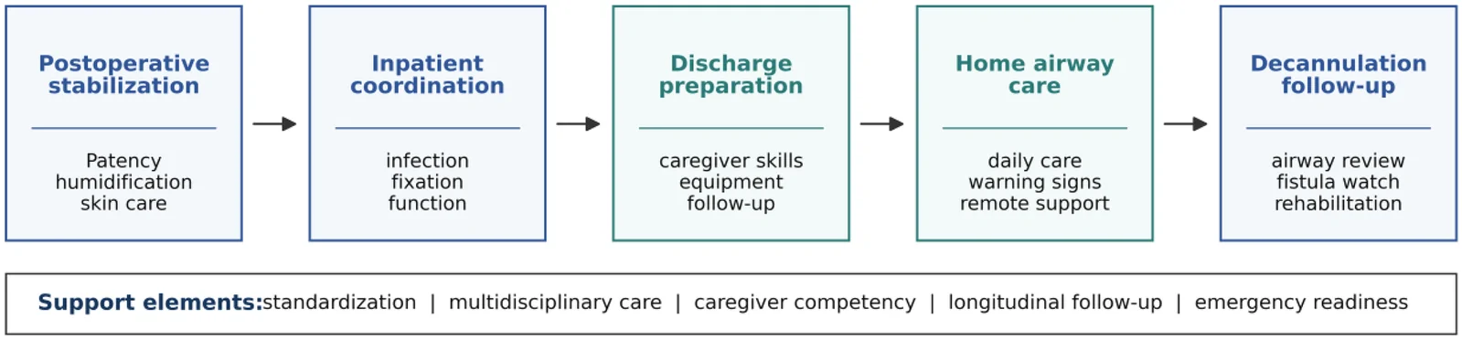 Figure 1: Comprehensive implementation pathway and longitudinal care trajectory for pediatric tracheostomy and long-term home mechanical ventilation. Demonstrates the staged multidisciplinary continuum from postoperative stabilization in the pediatric intensive care unit (PICU) to caregiver competency certification, simulated independent home care, emergency go-bag verification, community resource mobilization, and long-term decannulation evaluation (Frontiers in Pediatrics, 2026).
graph TD
    A["Pediatric Chronic Respiratory Insufficiency"] --> B{"Etiological Phenotype & Bulbar Competence"}
    B -->|"Neuromuscular (SMA, DMD, CMD)<br>Chest Wall Deformities (Severe Scoliosis)<br>Stable Upper Airway"| C["Trial of Non-Invasive Ventilation (NIV / BiPAP / AVAPS)"]
    B -->|"Severe BPD with Tracheobronchomalacia<br>Impaired Bulbar Function / Aspiration<br>CCHS (Infant/24h Support)<br>High Spinal Cord Injury (C1-C3)"| D["Invasive Mechanical Ventilation via Tracheostomy"]

    C --> E{"Nocturnal Hypoventilation Controlled?<br>Normal Daytime PtcCO2 & Growth?"}
    E -->|"Yes"| F["Home NIV Program:<br>Interface Rotation (Prevent Midface Hypoplasia)<br>Daytime Mouthpiece Ventilation (if older)<br>MI-E / CoughAssist Airway Clearance"]
    E -->|"No / Failure to Thrive / Aspiration"| D

    D --> G["Pre-Discharge Tracheostomy & Ventilator Program:<br>Dual-Caregiver Independent Competency & CPR<br>Bedside Emergency Go-Bag Standardized<br>24-48 Hour Hospital Simulation Stay"]
    G --> H["Successful Community / Home Discharge & Longitudinal Follow-Up"]
Clinical Indications and Pathophysiological Substrates
Chronic respiratory failure in children arises from five primary physiological substrates: neuromuscular failure, severe parenchymal and structural lung disease of prematurity, fixed or dynamic upper/lower airway collapse, severe chest wall distortion, and central respiratory drive dysfunction.
graph LR
    subgraph "Neuromuscular & Chest Wall"
        NMD["SMA, DMD, Congenital Myopathies"] --> N1["Pump Failure: $\downarrow V_T$, Diaphragmatic Fatigue"]
        Scol["Severe Early-Onset Scoliosis"] --> N2["Restricted Thoracic Compliance ($C_{cw}$)"]
    end

    subgraph "Parenchymal & Airway Dynamics"
        BPD["Severe BPD / Chronic Lung Disease"] --> P1["Heterogeneous $R_{aw}$, Alveolar Simplification"]
        TBM["Tracheobronchomalacia"] --> P2["Dynamic Expiratory Airway Collapse"]
    end

    subgraph "Central Controller"
        CCHS["CCHS (PHOX2B Mutation)"] --> C1["Absent Central Chemoreceptor Sensitivity to $CO_2$"]
    end

    N1 & N2 & P1 & P2 & C1 --> CRF["Nocturnal & Diurnal Alveolar Hypoventilation:<br>$\dot{V}_A = (V_T - V_D) \times RR$<br>Hypercapnia, Hypoxemia, Cor Pulmonale"]
Neuromuscular Disorders
Pediatric neuromuscular disorders (NMDs)—including Spinal Muscular Atrophy (SMA Type 1 and 2), Duchenne Muscular Dystrophy (DMD), and Congenital Muscular Dystrophies (CMD)—are characterized by progressive respiratory muscle pump failure. The diaphragm and accessory intercostal muscles fail to generate sufficient negative intrathoracic pressure to overcome baseline respiratory impedance.
During spontaneous respiration, muscular pressure ($P_{mus}$) drives airflow and volume expansion:
$$P_{mus}(t) + P_{vent}(t) = E_{rs} \cdot V_T(t) + R_{aw} \cdot \dot{V}(t) + \text{PEEP}_i$$Where $E_{rs} = 1/C_{rs}$ is total respiratory system elastance, $R_{aw}$ is airway resistance, $\dot{V}$ is inspiratory flow, and $\text{PEEP}_i$ is intrinsic PEEP. When $P_{mus}$ diminishes secondary to anterior horn cell degeneration or dystrophin deficiency, $V_T$ collapses, resulting in severe microatelectasis, loss of lung compliance ($C_L$), and rapid dead-space breathing:
$$\dot{V}_A = (V_T - V_D) \times RR$$As $V_T \to V_D$, effective alveolar ventilation ($\dot{V}_A$) plummets toward zero, precipitating profound nocturnal hypercapnia during rapid eye movement (REM) sleep when physiological intercostal atonia leaves respiration entirely dependent on an already compromised diaphragm.
Severe Bronchopulmonary Dysplasia and Airway Malacia
Infants with severe bronchopulmonary dysplasia (sBPD) and chronic lung disease of prematurity (CLD) exhibit alveolar simplification, dysmorphic pulmonary vascular beds, and marked ventilation-perfusion ($\dot{V}/\dot{Q}$) mismatch. Over 50% of infants with sBPD requiring tracheostomy possess concurrent central airway collapse, including severe laryngomalacia, tracheomalacia, and bronchomalacia (TBM).
In these infants, positive end-expiratory pressure (PEEP) acts as an intraluminal pneumatic stent. Insufficient PEEP leads to complete dynamic tracheal collapse during expiration, generating flow limitation, severe gas trapping, and acute hypoxemic "dying spells."
Central Hypoventilation and Chest Wall Deformities
- Congenital Central Hypoventilation Syndrome (CCHS): Caused by polyalanine repeat expansion mutations (PARMs) or non-PARMs in the PHOX2B gene, CCHS leads to dysgenesis of the autonomic nervous system and retrotrapezoid nucleus. Affected children lack ventilatory responsiveness to hypercapnia and hypoxemia during non-REM and REM sleep (and in severe genotypes, during wakefulness). Long-term home mechanical ventilation (via tracheostomy in infancy or non-invasive interfaces/diaphragmatic pacing in older children) is mandatory to prevent severe neurocognitive deficits, hypoxic pulmonary hypertension, and cor pulmonale.
- Chest Wall Disorders & Severe Scoliosis: Thoracic deformities (e.g., early-onset neuromuscular scoliosis, Jeune asphyxiating thoracic dystrophy, spondylothoracic dysplasia) markedly reduce chest wall compliance ($C_{cw}$), severely restrict thoracic cage expansion, and mechanically disadvantage diaphragmatic excursion, leading to restrictive chronic respiratory failure.
Modalities of Pediatric Home Mechanical Ventilation
The choice between Non-Invasive Positive Pressure Ventilation (NIV) and Invasive Mechanical Ventilation via Tracheostomy is dictated by age, diagnosis, bulbar stability, secretion burden, and the daily hours of ventilatory dependence.
graph TD
    subgraph "Home NIV Architecture"
        NIV1["Interfaces: Nasal Mask, Nasal Pillows, Full-Face"] --> NIV2["Circuit: Single-Limb with Intentional Leak (Exhalation Port)"]
        NIV2 --> NIV3["Algorithms: BiPAP S/T, Target-Volume / AVAPS, Dynamic Leak Compensation"]
    end

    subgraph "Invasive Tracheostomy Architecture"
        TR1["Dedicated Portable Turbine Ventilator (Astral, Trilogy)"] --> TR2["Circuit Modality: Active Exhalation Valve, Dual-Limb, or Passive Leak"]
        TR2 --> TR3["Emergency Safety: External Battery Backups + Dual Internal Batteries (>= 8h Total)"]
    end
Non-Invasive Positive Pressure Ventilation
Pediatric home NIV is the first-line therapeutic modality in children with intact bulbar function, manageable secretions, and neuromuscular or chest wall disorders who primarily manifest sleep-disordered breathing or nocturnal hypoventilation.
Pediatric Interfaces and Craniofacial Preservation
The pediatric facial skeleton undergoes rapid development. The cartilaginous nasal septum, maxilla, and premaxillary sutures are exquisitely vulnerable to ischemic necrosis and abnormal compressive remodelling.
graph LR
    A["Tight Nasal Mask Straps"] --> B["Continuous Posterior Force on Nasofrontal & Maxillary Sutures"]
    B --> C["Hypoperfusion & Chondrocyte Growth Arrest"]
    C --> D["Midface Hypoplasia, Flat Maxillary Profile & Class III Malocclusion"]

    E["Interface Rotation Strategy"] --> F["Alternate Nasal Cushion $\leftrightarrow$ Nasal Pillows $\leftrightarrow$ Full-Face"]
    F --> G["Shift Contact Pressure Vectors Across Frontal, Malar & Alar Tissues"]
Chronic nocturnal application of nasal and oronasal masks exerts continuous dorsal pressure on the anterior nasal spine and maxillary dental arch. In toddlers and young children, this can induce severe midface hypoplasia, maxillary retrusion, and retrognathia (Class III malocclusion). Clinical Mitigation Strategies:
- Interface Rotation: Alternate nightly between custom-molded nasal masks, nasal pillows/prongs, and total face masks to redistribute pressure points away from the nasion and philtrum.
- Orthodontic Surveillance: Implement routine bi-annual craniofacial and pediatric dental evaluations.
- Tension Management: Adjust headgear tension using the finger-slide rule (two fingers easily pass beneath straps) while relying on ventilator software leak algorithms rather than strap overtightening to control leaks.
Ventilator Algorithms and Modes: BiPAP, AVAPS, and Daytime Mouthpiece Ventilation
- Bi-Level Positive Airway Pressure (BiPAP S/T): Delivers a set Inspiratory Positive Airway Pressure (IPAP) and Expiratory Positive Airway Pressure (EPAP) with a backup respiratory rate. IPAP augments $V_T$ to clear $CO_2$, while EPAP maintains functional residual capacity (FRC) and counteracts upper airway obstruction.
- Average Volume Assured Pressure Support (AVAPS) / iVAPS: Closed-loop hybrid algorithms that automatically titrate IPAP breath-by-breath between a predefined minimum ($IPAP_{min}$) and maximum ($IPAP_{max}$) to achieve a target tidal volume ($V_{Tt}$) or alveolar minute ventilation ($MV_A$), compensating for changes in sleep architecture, chest wall impedance, and mask leaks.
- Daytime Mouthpiece Ventilation (MPV): In older pediatric patients with progressive neuromuscular disease (e.g., DMD, SMA Type 2) who experience daytime hypercapnia and dyspnea, an angled mouthpiece is mounted on the wheelchair. The child triggers open-circuit, volume-preset assist breaths on demand via glossopharyngeal action, maintaining daytime speech and independence while avoiding daytime tracheostomy.
Invasive Home Mechanical Ventilation via Tracheostomy
Invasive home mechanical ventilation is required when children require $\ge 16\text{--}24\text{ hours/day}$ of ventilatory support, fail non-invasive interfaces due to severe facial deformities, exhibit severe bulbar dysfunction with unmanageable aspiration, or have severe airway collapse (e.g., severe sBPD with dynamic tracheobronchomalacia).
graph TD
    A["Dedicated Home Turbine Ventilator"] --> B["Active Exhalation Valve Circuit"]
    A --> C["Dual-Limb Circuit"]
    A --> D["Passive Leak Circuit (Single-Limb with Whispertube / Vent)"]

    B --> B1["True Expiratory Flow Measurement<br>No Rebreathing Risk<br>Ideal for Small Infants & High Airway Resistance"]
    C --> C1["Complete Separation of Inspiratory & Expiratory Limbs<br>Precise Low $V_T$ Delivery ($< 50\text{ mL}$)<br>Bulky Tubing in Mobile Wheelchairs"]
    D --> D1["Continuous Bias Flow Flushes $CO_2$<br>Risk of $CO_2$ Rebreathing if EPAP < 4-5 cmH2O<br>Lightweight Single Tubing"]
Comparison of Home Ventilator Circuit Configurations
| Circuit Modality | Circuit Architecture | Advantages | Disadvantages / Safety Hazards | Optimal Clinical Cohort |
|---|---|---|---|---|
| Active Exhalation Valve | Single-limb with proximal pneumatic/electronic drive line controlling a balloon exhalation valve at the airway. | - True proximal pressure sensing. - Prevents exhaled gas rebreathing. - Accurate low $V_T$ monitoring. | - Risk of valve assembly misconnection. - Condensation occlusion in exhalation drive line. | Infants and young children with low tidal volumes ($V_T < 100\text{ mL}$) and high airway resistance. |
| Dual-Limb | Separate inspiratory and expiratory limbs connecting to an internal expiratory flow sensor. | - Absolute separation of gases. - Gold standard spirometry and flow triggering. - Zero rebreathing. | - Increased circuit weight and bulk. - High dead space if Y-piece is oversized. - Complex wheelchair routing. | Hospitalized pediatric step-down patients; severe BPD requiring precise PEEP and FiO2 titration. |
| Passive Leak (Vented) | Single limb with calibrated exhalation port / whisper swivel near the tracheostomy. | - Lightweight and flexible. - Single tubing reduces drag on tracheostomy. | - Rebreathing hazard if EPAP falls below $4\text{ cmH}_2\text{O}$. - Cannot deliver $100\%\text{ FiO}_2$. - Exhaled volume is estimated by leak algorithms. | Older children and adolescents with stable neuromuscular disorders and uncuffed/cuffed tracheostomy tubes. |
Power and Battery Redundancy Standards
Every child discharged on invasive home mechanical ventilation must possess an uninterrupted power system (UPS) with multiple layers of redundancy:
- Internal Lithium-Ion Battery: Integrated into the ventilator chassis (typically $3\text{--}4\text{ hours}$ operational time).
- Hot-Swappable External Battery Pack: Detachable secondary battery providing an additional $4\text{--}8\text{ hours}$ of autonomy (total $\ge 8\text{--}12\text{ hours}$ off-grid operational runtime).
- Backup Ventilator: A secondary, identical home ventilator programmed with matching settings is mandatory for all children dependent on ventilation $\ge 12\text{--}16\text{ hours/day}$ or who are clinically unable to sustain spontaneous respiration for $\ge 2\text{ hours}$.
- Municipal Priority Grid Registry: Formal registration with local electrical utilities, emergency medical services (EMS), and municipal fire departments as a life-support priority address.
Comprehensive Tracheostomy Care and Emergency Safety Protocols
Pediatric tracheostomies carry a baseline catastrophic obstruction and accidental decannulation mortality risk. Meticulous protocolization of bedside equipment, emergency algorithms, and caregiver drills is vital.
graph TD
    subgraph "Bedside Emergency Go-Bag (Always with Patient)"
        GB1["Same Size Tracheostomy Tube (with obturator)"]
        GB2["One-Half Size Smaller Tracheostomy Tube (with obturator)"]
        GB3["Suction Machine + Correct-Caliber Suction Catheters"]
        GB4["Water-Soluble Lubricant (Surgilube)"]
        GB5["Manual Resuscitator Bag + Appropriately Sized Mask"]
        GB6["Spare Tracheostomy Ties + Normal Saline Bullets + Scissors"]
    end
The Bedside Emergency "Go-Bag"
The emergency "Go-Bag" (or tracheostomy travel pack) must accompany the pediatric patient at all times—including during stroller rides, school transit, and bathroom transfers.
- Exact-Size Tracheostomy Tube: Complete with obturator and ties pre-threaded.
- One-Half Size Smaller Tracheostomy Tube: (e.g., Size 3.5 mm ID if child wears Size 4.0 mm ID) to guarantee stomal access in the event of parastomal edema or stomal constriction.
- Obturators: Matched specifically to both tracheostomy tubes.
- Water-Soluble Lubricating Jelly: For immediate tube lubrication prior to insertion.
- Suction Equipment: Portable battery-powered suction unit capable of generating $-80\text{ to }-100\text{ mmHg}$ in infants ($-100\text{ to }-120\text{ mmHg}$ in older children), plus spare suction canisters and calibrated suction catheters.
- Manual Resuscitation Equipment: Pediatric self-inflating bag with oxygen reservoir and a tight-fitting mask (to allow stomal or oral bag-mask rescue ventilation).
- Heat and Moisture Exchanger (HME) / Speaking valve / Saline bullets.
Emergency Troubleshooting Algorithm: The DOPE Framework
Acute desaturation, respiratory distress, bradycardia, or ventilator high-pressure alarms in a tracheostomy-dependent child must trigger the instantaneous execution of the DOPE algorithm.
graph TD
    A["Acute Respiratory Distress / Alarm in Tracheostomized Child"] --> B["Disconnect from Ventilator & Attach Manual Resuscitation Bag with 100% O2"]
    B --> C{"Is Bag Ventilation Easy and Chest Rising?"}

    C -->|"Yes"| D["Suspect Ventilator / Equipment Failure (E):<br>Check circuit, exhalation valve, power, settings.<br>Switch to backup ventilator."]
    C -->|"No / High Resistance"| E["Assess DOPE: Dislodgement, Obstruction, Pneumothorax"]

    E --> F["Attempt Suctioning through Tracheostomy Tube"]
    F --> G{"Does Suction Catheter Pass Smoothly Beyond Tip?"}

    G -->|"Yes, but child remains in distress"| H["Auscultate Chest:<br>Unilateral absent breath sounds, tracheal deviation, subcutaneous emphysema?<br>Suspect Pneumothorax (P) $\to$ Urgent Needle Decompression."]

    G -->|"No: Catheter Blocked"| I["Mucus Plug / Obstruction (O) or False Passage (D):<br>Instill 0.5-1 mL sterile saline & repeat suction."]
    I --> J{"Obstruction Cleared?"}

    J -->|"Yes"| K["Re-evaluate clinical stability and auscultate lungs."]
    J -->|"No"| L["IMMEDIATE EMERGENCY TUBE EXCHANGE:<br>Cut ties, remove tracheostomy tube.<br>Insert new lubricated SAME-SIZE tube with obturator.<br>Remove obturator immediately, ventilate."]

    L --> M{"Successful Insertion & Bag Ventilation?"}
    M -->|"Yes"| N["Secure ties, confirm bilateral chest rise & ETCO2."]
    M -->|"No / False Passage / Stoma Constriction"| O["Insert SMALLER SIZE (0.5 mm smaller) tube.<br>If still unable to pass, perform Bag-Mask Ventilation over Mouth/Nose while occluding stoma."]
- False Passage (Pre-Tracheal Dislodgement): Occurs when the tracheostomy tube inadvertently slips anterior or lateral to the true tracheal lumen into the pre-tracheal subcutaneous tissue. Attempting to ventilate through a false passage generates massive subcutaneous emphysema, pneumomediastinum, and complete asphyxiation.
- Clinical Hallmark: Resistance to passing a suction catheter beyond the tube tip, absence of expired $CO_2$ ($P_{et}CO_2 = 0$), inability to deliver breaths with a manual resuscitator bag, and progressive cervical crepitus.
- Immediate Action: Remove the malpositioned tube immediately. Do not attempt blind forceful re-advancement. Re-cannulate the stoma under direct visualization using a lubricated obturator, or pass a suction catheter/bougie into the true tracheal lumen as a Seldinger guide before sliding the tube over it.
Speaking Valves in Pediatric Tracheostomy
The Passy-Muir Valve (PMV) is a closed-position, bias-closed, one-way speaking valve that opens during inspiration and closes instantaneously at the onset of expiration, redirecting exhaled gas column upward through the vocal cords and oral/nasal cavities. This restores phonation, subglottic pressure, laryngeal sensation, and swallowing safety.
graph LR
    A["Inspiration"] -->|"Valve Opens"| B["Air Enters Tracheostomy Tube $\to$ Lungs"]
    C["Onset of Expiration"] -->|"Valve Snaps Shut"| D["One-Way Flow Blocked at Trachea"]
    D -->|"Gas Diverted Upward Around Tube"| E["Trans-Laryngeal Airflow $\to$ Phonation & Secretion Clearance"]
graph TD
    A["Evaluate for Passy-Muir Valve (PMV)"] --> B{"Is Tracheostomy Tube Cuffed?"}
    B -->|"Yes"| C["MANDATORY ACTION: FULLY DEFLATE CUFF FIRST"]
    B -->|"No (Uncuffed Tube)"| D["Assess Upper Airway Patency & Expiratory Space"]
    C --> D

    D --> E["Place PMV on Tracheostomy Hub"]
    E --> F["Monitor Manometer / Expiratory Airway Pressure & Breathing Work"]
    F --> G{"Signs of Expiratory Air Trapping?<br>Manometer Baseline > 5 cmH2O?<br>Retractions, Stridor, Agitation?"}

    G -->|"Yes"| H["IMMEDIATELY REMOVE PMV!<br>Upper airway too small or tube too large.<br>Risk of complete asphyxiation/air trapping."]
    G -->|"No"| I["Successful In-Line or Spontaneous PMV Application:<br>Promotes speech, swallow, and decannulation readiness."]
Fatal Danger Alert: NEVER place a speaking valve on a cuffed tracheostomy tube without 100% complete cuff deflation. If the cuff remains inflated, inspiratory gas enters through the one-way valve, but exhaled gas cannot escape around the cuff or out of the valve, producing catastrophic acute lung overdistension, tension pneumothorax, and immediate cardiac arrest within breaths.
Pediatric Speaking Valve Candidacy Criteria
| Criteria Category | Prerequisite Clinical Standard | Pathophysiological Rationale |
|---|---|---|
| Hemodynamic & Clinical Stability | Medically stable, afebrile, absence of acute bronchospasm or active pulmonary hemorrhage. | Prevents acute decompensation during upper airway resistance challenge. |
| Cuff Status | Uncuffed tube OR fully deflated cuffed tube with pilot balloon evacuated. | Guarantees an unobstructed annular space around the tube for cephalad airflow. |
| Airway Space / Tube Sizing | Tracheostomy tube outer diameter (OD) must occupy $\le 60\text{--}70\%$ of the internal tracheal lumen. | Ensures adequate peritubular cross-sectional area to minimize expiratory resistance. |
| Expiratory Manometry | Measured end-expiratory tracheal pressure $< 5\text{ cmH}_2\text{O}$ during valve trial. | Pressures $> 5\text{ cmH}_2\text{O}$ indicate peritubular airway stenosis, subglottic stenosis, or vocal cord paralysis. |
| Secretion Management | Thin, manageable secretions; child can swallow or tolerate redirection of oral secretions. | Thick, copious secretions foul the silicone diaphragm and increase choking risk. |
In-Home Airway Clearance and Physiological Monitoring
Impaired mucociliary clearance and ineffective cough mechanics are universal in children with neuromuscular disease and long-term tracheostomies, predisposing them to recurrent lobar atelectasis and pneumonia.
graph TD
    subgraph "Airway Clearance Armamentarium"
        MIE["Mechanical Insufflation-Exsufflation (CoughAssist)"] --> M1["Rapid Switch: $+20\text{ to }+40\text{ cmH}_2\text{O} \to -20\text{ to }-40\text{ cmH}_2\text{O}$<br>Simulates Peak Cough Flow ($> 160\text{--}270\text{ L/min}$)"]
        HFCWO["High-Frequency Chest Wall Oscillation (The Vest)"] --> H1["Oscillations (10-15 Hz) Shear Mucus from Airway Walls"]
        IPV["Intrapulmonary Percussive Ventilation (IPV)"] --> I1["High-Frequency Phasitron Pulses + Aerosolized Saline"]
    end

    subgraph "In-Home Physiological Monitoring"
        SpO2["Continuous Nocturnal Pulse Oximetry ($SpO_2$)"] --> Mon1["Detects Desaturations, Mucus Plugging, Hypoxemia"]
        PtcCO2["Transcutaneous Capnography ($PtcCO_2$)"] --> Mon2["Gold Standard for Titrating Nocturnal Ventilation & Alveolar Hypoventilation"]
    end
Mechanical Insufflation-Exsufflation and Airway Clearance Devices
- Mechanical Insufflation-Exsufflation (MI-E / CoughAssist): The gold standard for neuromuscular airway clearance. Delivers a positive pressure breath ($+20\text{ to }+40\text{ cmH}_2\text{O}$) to achieve maximum insufflation capacity, followed by a rapid, active transition to negative pressure ($-20\text{ to }-40\text{ cmH}_2\text{O}$) for $1.5\text{--}2.5\text{ seconds}$. This generates high peak expiratory cough flows ($\text{PECF} > 160\text{--}270\text{ L/min}$), shearing secretions from peripheral bronchi into the proximal trachea for easy suctioning or expectoration.
- High-Frequency Chest Wall Oscillation (HFCWO / The Vest): Inflatable vest connected to an air-pulse generator delivering rapid compressions at $10\text{--}15\text{ Hz}$. Reduces mucus viscoelasticity and creates transient expiratory airflow spikes, propelling secretions centrally in children with cystic fibrosis, primary ciliary dyskinesia, or cerebral palsy.
- Intrapulmonary Percussive Ventilation (IPV): Delivers dense, high-frequency gas mini-bursts ($100\text{--}300\text{ cycles/min}$) superimposed on spontaneous breathing while delivering heated saline aerosols, recruiting atelectatic lung units and mobilizing secretions in tracheostomized infants.
In-Home Monitoring: Nocturnal Pulse Oximetry and Transcutaneous Capnography
- Continuous Pulse Oximetry ($SpO_2$): Mandatory during sleep and all ventilator hours. Alarm limits are tailored ($SpO_2 < 92\%$ or $< 90\%$ in cyanotic heart disease).
- Transcutaneous $CO_2$ Monitoring ($PtcCO_2$): In children with non-invasive interfaces or uncuffed tracheostomies with large variable leaks, end-tidal capnography ($P_{et}CO_2$) significantly underestimates true arterial $PaCO_2$. Transcutaneous capnography heats the stratum corneum to $41\text{--}43^\circ\text{C}$, melting skin lipids and allowing gaseous $CO_2$ diffusion to an electrochemical sensor. $PtcCO_2$ is the gold standard for in-home or sleep lab nocturnal ventilation titration.
Caregiver Competency Certification and Discharge Readiness
Discharging a ventilator-dependent child requires a rigorous, multi-tiered caregiver training and institutional simulation program.
graph TD
    Stage1["Tier 1: Basic Tracheostomy Care & Suctioning Skills"] --> Stage2["Tier 2: Ventilator Circuit Assembly, Humidification & Power Systems"]
    Stage2 --> Stage3["Tier 3: Emergency Decannulation & DOPE Simulation on High-Fidelity Mannequins"]
    Stage3 --> Stage4["Tier 4: Certified Pediatric Basic Life Support (PBLS) for Both Primary Caregivers"]
    Stage4 --> Stage5["Tier 5: 24- to 48-Hour Independent Hospital Inpatient Simulation Stay"]
    Stage5 --> Stage6["Safe Discharge to Community Home Environment with Home Nursing Support"]
Caregiver Competency Matrix
| Domain | Required Core Skill | Objective Demonstration Standard |
|---|---|---|
| Routine Tracheostomy Care | Stoma cleaning, dressing change, and tie replacement. | Two-person technique performed without stoma trauma or accidental extubation. |
| Emergency Recannulation | Accidental decannulation drill. | Successful insertion of same-size tube and 0.5-size smaller tube within $< 20\text{ seconds}$ on a mannequin. |
| Airway Clearance | In-home suctioning and CoughAssist operation. | Proper sterile/clean catheter technique, correct vacuum limits, and MI-E cycle execution. |
| Ventilator Mechanics | Circuit change, filter replacement, heated humidifier maintenance. | Complete circuit assembly and leak self-test passed independently. |
| Power Management | Battery swapping and emergency power failure response. | Correct connection of external batteries, inverter, and manual bag ventilation initiation. |
| Resuscitation | Cardiopulmonary resuscitation (CPR) with artificial airway. | Current certified PBLS status; demonstration of stomal vs oral bag-mask rescue ventilation. |
Clinical Pearls and High-Yield Board Review
- Call for Help & Stay Calm: Never leave the infant unattended.
- Insert Lubricated Same-Size Tube with Obturator: Advance gently along the tract curved downward. Immediately pull the obturator out once seated.
- If Resistance Encountered, Drop 0.5 Size Immediately: Do not force a tight tube into an edematous stoma. Insert the smaller emergency tube.
- If Stoma Inaccessible: Place a pediatric resuscitation mask over the mouth/nose, occlude the neck stoma with a gloved finger, and deliver positive-pressure rescue breaths while preparing emergency surgical or fiberoptic airway access.
In infants and toddlers weighing $< 10\text{ kg}$, the addition of extraneous inline adapters, closed suction catheters, and oversized HMEs between the Y-piece and tracheostomy tube adds substantial mechanical dead space ($V_{D,app}$). Because infant baseline $V_T$ is small ($6\text{--}8\text{ mL/kg} = 30\text{--}40\text{ mL}$ in a $5\text{ kg}$ infant), adding just $10\text{ mL}$ of dead space can increase dead space fraction ($V_D/V_T$) from $30\%$ to $> 60\%$, inducing severe refractory hypercapnia. Always use low-dead-space pediatric-specific connectors.
Summary of Practice Recommendations
- Patient Selection: Match ventilatory interface to bulbar status, secretional load, and etiology. Preserve non-invasive interfaces in NMD whenever possible; proceed to tracheostomy for severe airway malacia, intractable aspiration, or 24-hour infant dependence.
- Craniofacial Protection: Prevent midface hypoplasia in pediatric NIV via nightly interface rotation and objective strap tension limits.
- Safety Redundancy: Mandate dual certified caregivers, standardized bedside go-bags, and $\ge 8\text{--}12\text{ hours}$ of battery autonomy for every invasive home ventilator patient.
- Speaking Valve Safety: Never apply a Passy-Muir valve without complete cuff deflation and prior manometric confirmation of adequate peritubular expiratory space.
- DOPE Execution: Standardize the emergency DOPE algorithm across hospital, school, and home environments to rapidly reverse life-threatening tracheostomy obstruction and false passages.