Prone positioning represents one of the most rigorously validated, physiology-driven non-pharmacological interventions for severe Pediatric Acute Respiratory Distress Syndrome (PARDS). By rotating the critically ill, mechanically ventilated child from the supine to the ventral decubitus posture, clinicians exploit gravitational, anatomical, and chest wall biomechanics to reverse refractory hypoxemia, reduce alveolar strain heterogeneity, optimize lung protective ventilation, and mitigate ventilator-induced lung injury (VILI).
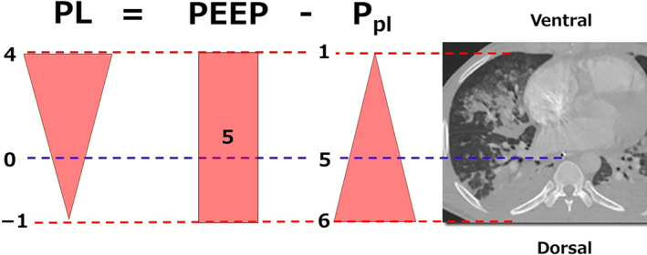 Figure 1: Gravitational Pleural Pressure Gradient and Regional Transpulmonary Pressure Homogenization in ARDS. In the supine posture, dependent dorsal lung regions experience elevated superimposed hydrostatic tissue pressure and ventral-to-dorsal pleural pressure ($P_{pl}$) gradients, resulting in negative end-expiratory transpulmonary pressure ($P_L = \text{PEEP} - P_{pl} < 0\text{ cmH}_2\text{O}$) and severe dorsal compressive atelectasis (right CT panel). Setting adequate positive end-expiratory pressure (PEEP) alongside prone positioning homogenizes regional transpulmonary distending pressures, re-expanding collapsed dorsal lung mass and stabilizing alveolar architecture. Adapted from Hoshino & Yoshida, Acute Medicine & Surgery, 2024; DOI: 10.1002/ams2.918 (PMC10761614).
Physiological Mechanisms of Prone Positioning in Children
The pediatric respiratory system possesses distinct anatomical and biomechanical features—including a highly compliant chest wall, a circular thoracic cross-section, smaller airway calibers with elevated peripheral resistance, and lower baseline functional residual capacity (FRC)—that amplify both the pathological collapse seen in supine ARDS and the therapeutic response to prone positioning.
graph TD
    subgraph "Supine ARDS Pathophysiology"
        A["Ventral-to-Dorsal Pleural Pressure Gradient"] --> B["Elevated Dorsal Superimposed Hydrostatic Pressure"]
        C["Ventral Cardiac & Mediastinal Compression"] --> B
        B --> D["Negative Dorsal Transpulmonary Pressure (P_L < 0)"]
        D --> E["Severe Dorsal Dependent Atelectasis"]
        E --> F["Persistent Dorsal Perfusion (Q) >> Ventilation (V)"]
        F --> G["Massive Intrapulmonary Shunt (Qs/Qt > 30%) & Hypoxemia"]
        E --> H["Ventral Overdistension & Volutrauma/Biotrauma"]
    end

    subgraph "Prone Therapeutic Mechanisms"
        I["Inversion of Thoracic Gravitational Vector"] --> J["Pleural Pressure (P_pl) Homogenization"]
        K["Ventral Decubitus Restricts Ventral Ribcage Expansion"] --> L["Redistribution of Tidal Volume to Dorsal Lung Mass"]
        J --> M["Positive Dorsal Transpulmonary Pressure (P_L > 0)"]
        M --> N["Dorsal Alveolar Recruitment without Hyperinflation"]
        N & F --> O["V/Q Matching Optimization & Shunt Reduction"]
        O --> P["PaO2/FiO2 Surge & Driving Pressure (ΔP) Reduction"]
        Q["Gravity-Assisted Postural Drainage"] --> R["Mobilization of Tracheobronchial Secretions"]
    end
Gravitational Pleural Pressure Gradient Redistribution
In the supine patient with PARDS, fluid-engorged, consolidated edematous lung tissue exerts a vertical hydrostatic force on dependent dorsal parenchyma. The vertical gradient of pleural pressure ($\Delta P_{pl} / \Delta h$) increases steeply from ventral to dorsal regions (typically $0.5\text{ to }1.0\text{ cmH}_2\text{O/cm}$ height in edematous lungs). Furthermore, the heavy cardiac mass rests directly atop the dependent lingula, left lower lobe, and medial segments of the right lower lobe, transmitting an additional localized compressive pressure of $3\text{ to }5\text{ cmH}_2\text{O}$.
The regional transpulmonary distending pressure ($P_L$) is defined as:
$$P_L = P_{ao} - P_{pl}$$Where:
- $P_{ao}$ is the airway opening pressure (equal to alveolar pressure $P_{alv}$ under zero-flow, static conditions).
- $P_{pl}$ is the local pleural surface pressure.
In the supine posture at end-expiration:
- Ventral Lung Units: $P_{pl,\text{ventral}} \approx -2\text{ to }0\text{ cmH}_2\text{O} \implies P_{L,\text{ventral}} = \text{PEEP} - (-2) = \text{PEEP} + 2\text{ cmH}_2\text{O}$ (patent, high risk of overdistension).
- Dorsal Dependent Units: $P_{pl,\text{dorsal}} \approx +8\text{ to }+12\text{ cmH}_2\text{O} \implies P_{L,\text{dorsal}} = \text{PEEP} - (+10) = \text{PEEP} - 10\text{ cmH}_2\text{O}$. If applied $\text{PEEP} \le 10\text{ cmH}_2\text{O}$, $P_{L,\text{dorsal}} \le 0\text{ cmH}_2\text{O}$, precipitating complete cyclical collapse and severe atelectrauma.
When turned prone, the vertical pleural pressure gradient flattens dramatically. Because the adult and pediatric dorsal lung parenchyma encompasses approximately $60\%\text{ to }70\%$ of total lung mass and cross-sectional surface area, the dorsal lung conforms to the flatter dorsal ribcage without the compressive weight of the heart and mediastinum (which now rests anteriorly against the sternum). Consequently:
$$\Delta P_{L,\text{dorsal}} \approx \Delta P_{L,\text{ventral}}$$Regional transpulmonary pressure becomes uniform along the ventrodorsal axis, reversing dorsal atelectasis and establishing a stable end-expiratory alveolar architecture.
Alveolar cyclic strain ($\epsilon$) is directly proportional to regional tidal volume distribution relative to baseline resting functional residual capacity ($\text{FRC}_{reg}$):
$$\epsilon_{reg} = \frac{V_{T,reg}}{\text{FRC}_{reg}} \propto \frac{\Delta P_L}{E_{L,reg}}$$In the supine position, extreme heterogeneity between collapsed dorsal units ($\text{FRC}_{reg} \to 0$) and over-expanded ventral units creates high localized shear stresses at the open-closed interface. Prone positioning eliminates this interface by homogenizing $P_L$, recruiting non-aerated dorsal mass, expanding functional lung size ("baby lung" enlargement), and decreasing dynamic global driving pressure ($\Delta P = P_{plat} - \text{PEEP}$).
Ventilation-Perfusion Matching & Intrapulmonary Shunt
Pulmonary blood flow distribution in mammals is determined primarily by non-gravitational anatomical vascular branching architecture rather than gravity alone. Perfusion capacity is structurally concentrated in dorsal and caudal lung segments due to larger cross-sectional vascular bed dimensions and lower regional vascular resistance.
In supine ARDS:
- Perfusion ($Q$) remains predominantly dorsal ($>65\%$).
- Ventilation ($V$) is diverted exclusively to open ventral alveoli ($>80\%$).
- Result: Severe ventilation-perfusion mismatch, characterized by massive physiological intrapulmonary shunt ($Q_s/Q_t \ge 30\text{–}50\%$) through unventilated dorsal zones, and high dead-space fraction ($V_D/V_T$) in overdistended ventral zones.
In prone ARDS:
- Perfusion ($Q$) remains anchored predominantly in dorsal lung regions.
- Ventilation ($V$) shifts to newly recruited dorsal parenchyma.
- Result: Superb spatial co-localization of ventilation and perfusion ($V/Q \approx 1.0$), resulting in an immediate and sustained drop in intrapulmonary shunt ($Q_s/Q_t$), enhanced arterial oxygenation ($\text{PaO}_2/\text{FiO}_2$), and more efficient carbon dioxide clearance ($\text{VCO}_2$).
graph LR
    subgraph "Supine Mismatch"
        S_V["Ventral Ventilation (High)\n Ventral Perfusion (Low)\n-> High V/Q & Dead Space"]
        S_D["Dorsal Ventilation (Zero/Atelectasis)\n Dorsal Perfusion (High)\n-> Low V/Q & Massive Shunt (Qs/Qt)"]
    end

    subgraph "Prone Alignment"
        P_V["Ventral Ventilation (Moderate)\n Ventral Perfusion (Low-Moderate)\n-> Balanced V/Q"]
        P_D["Dorsal Ventilation (Recruited/High)\n Dorsal Perfusion (High)\n-> Optimal V/Q (≈ 1.0) & Shunt Reversal"]
    end
Chest Wall Mechanics & Regional Compliance
The chest wall compliance ($C_{cw}$) of infants and young children is exceptionally high due to cartilaginous ribs and weak intercostal musculature.
- In the supine position, positive pressure ventilation easily pushes the anterior chest wall upward, preferentially inflating non-dependent ventral regions without generating sufficient transpulmonary pressure to open stiff, fluid-laden dorsal lung units.
- In the prone position, the sternum and anterior ribcage are mechanically constrained by the bed surface and positioning pads. This localized reduction in ventral chest wall compliance redirects delivered tidal volume into the more compliant, unconstrained dorsal lung units.
- Crucially, this dorsal redistribution occurs without increasing driving pressure ($\Delta P$) or peak inspiratory pressure, minimizing global mechanical power delivery ($\text{MP} = 0.098 \cdot \text{RR} \cdot V_T \cdot [P_{peak} - \frac{1}{2}\Delta P]$).
Secretion Drainage by Gravity
In supine position, standard pediatric endotracheal tubes and mainstem bronchi orient posteriorly downward, promoting secretional pooling in dependent segments, secondary micro-atelectasis, and microbial colonization. The prone position reverses this orientation, placing the posterior subsegmental bronchi in an upward-draining inclination toward the central tracheobronchial tree. Gravity-assisted clearance promotes mobilization of tenacious mucopurulent plugs, relieving localized airway obstruction and improving gas exchange kinetics.
PALICC-2 Guidelines & Landmark Evidence
The Pediatric Acute Lung Injury Consensus Conference (PALICC-2) 2023 updated recommendations provide evidence-based guidance for the clinical application of prone positioning in pediatric critical care.
graph TD
    A["Pediatric Acute Respiratory Distress Syndrome (PARDS) Diagnosed"] --> B{"Calculate Severity Index\n(Within 4-12h of Optimization)"}

    B -->|"Severe PARDS\n(OI >= 16 or OSI >= 12.3)"| C["STRONG PALICC-2 RECOMMENDATION:\nInitiate Invasive Prone Positioning Protocol"]
    B -->|"Moderate PARDS\n(OI 8-15.9 or OSI 7.5-12.2)"| D["CONSIDERATION / CONDITIONAL RECOMMENDATION:\nTrial Prone Positioning for Refractory Hypoxemia"]
    B -->|"Mild PARDS\n(OI 4-7.9 or OSI 5-7.4)"| E["Maintain Supine Lung-Protective Ventilation\nMonitor for Progression"]

    C & D --> F["Execute Pre-Proning PICU Safety Checklist"]
    F --> G["Multi-Disciplinary Envelope/Burrito Turn"]
    G --> H["Maintain Continuous Prone Position >= 16-24 Hours/Day"]

    H --> I{"Assess Response at 4 Hours"}
    I -->|"Responder:\nPaO2/FiO2 Increase >= 20% OR\nOI Decrease >= 20%"| J["Continue Daily Prone Cycles (16-24h/day)\nWean FiO2 < 0.60, PEEP <= 10"]
    I -->|"Non-Responder:\n< 20% Improvement & High Driving P"| K["Evaluate Mechanics, PEEP Titration, EIT\nConsider HFOV, iNO, or VV-ECMO"]
PALICC-2 Recommendation Matrix
| PARDS Category | Invasive Oxygenation Metric | Non-Invasive Metric | PALICC-2 Recommendation | Target Duration |
|---|---|---|---|---|
| Severe PARDS | $\text{OI} \ge 16$ | $\text{OSI} \ge 12.3$ | Strong Recommendation (Evidence-supported mortality/VILI reduction) | $\ge 16\text{ to }24\text{ consecutive hours/session}$ |
| Moderate PARDS | $8 \le \text{OI} < 16$ | $7.5 \le \text{OSI} < 12.3$ | Consideration / Weak Recommendation (Indicated if hypoxemia escalates or $\Delta P > 15\text{ cmH}_2\text{O}$) | $\ge 16\text{ consecutive hours/session}$ |
| Mild PARDS | $4 \le \text{OI} < 8$ | $5.0 \le \text{OSI} < 7.5$ | Not routinely indicated; maintain standard lung-protective ventilation | N/A |
Landmark Clinical Trials: PROSEVA and PROSpect
- PROSEVA Trial (Guerin et al., NEJM 2013): In adult severe ARDS ($\text{PaO}_2/\text{FiO}_2 < 150$), early application of prolonged prone positioning ($\ge 16\text{ h/day}$) demonstrated a dramatic 28-day mortality reduction ($16.0\%\text{ vs }32.8\%$, $p < 0.001$, hazard ratio $0.39$).
- Pediatric Prone Trials (Curley et al., JAMA 2005): Early pediatric multicenter RCT demonstrated significant short-term oxygenation improvement, though unstratified for severe PARDS.
- PROSpect Trial (Prone and Oscillation Pediatric Clinical Trial, Curley et al., 2023–2024): The largest international randomized trial in severe PARDS evaluating invasive mechanical ventilation with prone positioning versus conventional supine care, reinforcing the safety, feasibility, and role of prolonged prone positioning ($\ge 16\text{–}24\text{ hours/day}$) in reducing ventilator days and enhancing liberation.
Clinical Response Criteria
A patient is classified as a Prone Responder if within 4 hours of turning prone:
- Oxygenation Ratio: $\text{PaO}_2/\text{FiO}_2$ ratio increases by $\ge 20\%$.
- Oxygenation Index: $\text{OI}$ decreases by $\ge 20\%$.
- Mechanical Response: Static respiratory compliance ($C_{stat}$) increases by $\ge 15\%$ or driving pressure ($\Delta P$) decreases by $\ge 2\text{ cmH}_2\text{O}$ at constant tidal volume ($V_T = 4\text{–}6\text{ mL/kg}$).
Even in patients who do not exhibit immediate surge in $\text{PaO}_2/\text{FiO}_2$ ("oxygenation non-responders"), prone positioning preserves lung architecture by homogenizing mechanical strain and preventing ventral alveolar overdistension. Maintain the full 16-hour session unless life-threatening hemodynamic instability or severe desaturation occurs.
Step-by-Step PICU Prone Positioning Protocol
Safe execution of pediatric proning requires a dedicated, multidisciplinary team executing standardized checklists.
sequenceDiagram
    autonumber
    participant MD as PICU Lead Intensivist (Head/Airway)
    participant RT as Respiratory Therapist (Lines/Vent)
    participant N1 as Primary Nurse 1 (Right Torso)
    participant N2 as Primary Nurse 2 (Left Torso)

    Note over MD,N2: Phase 1: Pre-Proning Safety Checklist
    MD->>RT: Confirm ETT depth on CXR & retape endotracheal tube
    MD->>N1: Hold enteral nutrition (1h prior), aspirate gastric tube
    MD->>N2: Apply silicone/hydrocolloid dressings to pressure points
    RT->>MD: Suction ETT and oral pharynx thoroughly; pre-oxygenate (FiO2 1.0)

    Note over MD,N2: Phase 2: The Envelope / Burrito Wrap
    N1->>N2: Place new base sheet on patient; place top sheet over chest
    N1->>N2: Roll sheets tightly together at patient sides (Envelope wrap)

    Note over MD,N2: Phase 3: Synchronized Lateral Turn & Prone Placement
    MD->>MD: Securely grip ETT, circuit, and inline suction with both hands
    MD->>N1: Command: "Slide to opposite side on 3: 1, 2, 3"
    MD->>N2: Command: "Rotate to lateral decubitus on 3: 1, 2, 3"
    MD->>N2: Command: "Turn fully prone on 3: 1, 2, 3"

    Note over MD,N2: Phase 4: Post-Turn Optimization & Swimmer Positioning
    MD->>RT: Auscultate bilateral breath sounds; confirm EtCO2 & Vt return
    N1->>N2: Position in Swimmer's Posture; elevate head of bed 15-30 deg
    N1->>MD: Re-zero arterial line transducers to right atrium level
Phase 1: Pre-Proning Safety Checklist
- Airway Security:
- Verify ETT tip position at mid-trachea on recent chest radiograph ($T2\text{–}T3$ vertebral body level).
- Clean, re-tape, or re-clamp ETT with commercial pediatric tube fixation device.
- Perform thorough endotracheal and oral cavity suctioning.
- Pre-oxygenate with $\text{FiO}_2 = 1.0$ for 5 minutes.
- Gastrointestinal & Nutrition:
- Pause enteral nutrition 1 hour prior to turn.
- Aspirate nasogastric / orogastric tube to decompress stomach, then connect to free dependent drainage.
- Vascular Access & Monitoring:
- Identify, loop, and secure all central venous catheters, arterial lines, peripheral IVs, and chest tubes.
- Ensure adequate line slack with zero tension during full $180^\circ$ rotation.
- Relocate ECG leads from anterior chest wall to bilateral shoulders/back.
- Ocular & Facial Protection:
- Instill preservative-free lubricating ophthalmic ointment into both eyes.
- Tape eyelids completely closed in horizontal alignment to prevent corneal abrasions and exposure keratopathy.
- Skin & Pressure Point Preparation:
- Apply prophylactic multi-layer silicone foam / hydrocolloid dressings to:
- Forehead and malar prominences.
- Chin.
- Anterior clavicles and bilateral acromion processes.
- Anterior superior iliac spines (ASIS).
- Bilateral patellae and pretibial crests.
- Dorsal surface of feet/toes.
- Apply prophylactic multi-layer silicone foam / hydrocolloid dressings to:
- Sedation & Neuromuscular Blockade:
- Ensure deep analgesia and sedation (State Behavioral Scale SBS -2 to -3, or RASS -4 to -5).
- In patients with high respiratory drive or marginal airway stability, administer a bolus of neuromuscular blocking agent (e.g., cisatracurium $0.15\text{–}0.2\text{ mg/kg}$ or rocuronium $0.6\text{–}1.0\text{ mg/kg}$) immediately prior to turn.
Phase 2 & 3: The Envelope / Burrito Turning Technique
The team consists of minimum 4–5 personnel:
- Airway Lead (Intensivist or Senior RT): Positioned at the head of the bed; solely responsible for stabilizing the ETT, inline suction catheter, and ventilator circuit. Commands all movements.
- Nurse 1 (Bedside Left): Controls torso, left arm, pelvic turn, and left-sided lines.
- Nurse 2 (Bedside Right): Controls torso, right arm, pelvic turn, and right-sided lines.
- Staff 3 (Bed Foot): Manages lower extremities, urinary catheter, and fecal containment.
- Staff 4 (Auxiliary RT / Nurse): Manages ventilator tubing support arm and monitors arterial line trace.
Turning Procedure Steps:
- Prepare Sheet Wrap: Place a clean slide sheet underneath the patient and a clean top sheet over the patient's anterior torso (sandwiching chest/pelvic foam positioning pads).
- Tight Roll ("Envelope"): Roll the edges of the top and bottom sheets together tightly along both sides of the child, creating a secure "burrito" wrap.
- Step 1 (Horizontal Slide): On the airway lead's count ("1, 2, 3, slide"), shift the child horizontally toward the side of the bed opposite the planned turn direction.
- Step 2 (Lateral Decubitus): On the count, rotate the wrapped child $90^\circ$ into the lateral decubitus position, maintaining spine alignment.
- Step 3 (Complete Prone Rotation): On the count, complete the $90^\circ$ rotation to place the child fully prone onto the bed. Remove the top wrapping sheet.
Phase 4: Positioning Optimization ("Swimmer's Position")
graph LR
    subgraph "Swimmer's Position Configuration"
        Head["Head: Rotated to Right\n(Ear/Cheek on Gel Donut Cushion)"]
        ArmR["Right Arm (Direction of Face):\nAbducted 80 deg, Elbow Flexed 90 deg\n('UP Position')"]
        ArmL["Left Arm (Opposite Face):\nExtended alongside torso, Palm Up\n('DOWN Position')"]
        Chest["Chest & Pelvis:\nFoam Bolsters under Clavicle & Iliac Crests\n-> Abdomen Completely Free"]
        Legs["Lower Extremities:\nPillow under Lower Shins\n-> Knees & Toes Suspended Free"]
    end
Pressure Point & Position Rotation Schedule:
- Frequency: Every 2 to 4 hours.
- Rotation: Rotate the head to the opposite side, simultaneously alternating arm positions (the arm on the face side moves "UP", contralateral arm moves "DOWN").
- Abdominal Suspension: Verify that the abdomen hangs completely free between the chest roll (supporting clavicles/sternum) and pelvic roll (supporting anterior iliac crests). Abdominal compression elevates intra-abdominal pressure (IAP), transmits force across the diaphragm, increases intrathoracic pressure, and impedes inferior vena cava venous return.
- Head Elevation: Place bed in Reverse Trendelenburg position ($15^\circ\text{ to }30^\circ$) to reduce facial and airway edema.
| Body Region | Prophylactic Measure | Repositioning / Offloading Interval |
|---|---|---|
| Forehead / Cheek / Ear | Hydrocolloid dressing + closed-cell gel donut ring | Reposition and reverse head rotation every 2–4 hours |
| Eyes / Cornea | Lubricating eye ointment + tape eyelids shut horizontally | Inspect pupils and conjunctiva every 4 hours |
| Clavicles / Anterior Ribs | High-density foam bolster beneath upper chest | Verify bolster alignment every 4 hours |
| Abdomen | Void space created between chest and pelvic bolsters | Continuous clearance (ensure flat hand passes under abdomen) |
| Anterior Iliac Spines | Foam bolster across pelvis; silicone dressing | Verify ASIS offloaded from hard mattress |
| Patellae & Pre-tibial Crests | Silicone foam dressings on patellae | Reposition shin pillow every 2–4 hours |
| Toes & Feet | Pillow beneath lower shins keeping toes suspended | Prevent plantar hyper-extension and bed friction |
Complications & Emergency Resuscitation
While prone positioning is highly effective, adverse events can occur during turns or prolonged ventral decubitus posture.
graph TD
    A["Emergency in Prone Position"] --> B{"Identify Primary Event"}

    B -->|"Unplanned Extubation"| C["IMMEDIATE ACTION:\n1. Call for Immediate Help & Code Button\n2. Open Airway, Manual Bag-Valve-Mask at FiO2 1.0\n3. Flip Emergent Supine onto Backboard\n4. Direct Laryngoscopy / Video Re-Intubation"]

    B -->|"Cardiac Arrest / PEA / Asystole"| D{"Patient Size & Provider Feasibility"}
    D -->|"Infant / Small Child\n(Immediate Effective Prone CPR)"| E["Initiate PRONE CPR:\nCompress Mid-Thoracic Spine (T4-T7)\nCounter-support Sternum with Hand or Pad\nPrepare Emergent Supine Flip"]
    D -->|"Older Child / Ineffective Prone CPR"| F["Execute IMMEDIATE EMERGENCY FLIP to Supine\nPlace CPR Backboard\nStandard PALS Resuscitation"]

    B -->|"Acute Hemodynamic Collapse / Line Pull"| G["1. Clamp/Hold Bleeding Line Sites\n2. Rapid Fluid Bolus (10-20 mL/kg) & Vasopressor Push\n3. Check for Tension Pneumothorax (Needle Decompression)\n4. If Unresponsive -> Emergent Supine Turn"]
Complication Identification & Management Protocol
- Unplanned Extubation (Accidental Decannulation):
- Incidence: $<1.5\%$ in standardized PICU protocols.
- Emergency Protocol:
- Immediately place bag-valve-mask (BVM) with PEEP valve over mouth/nose and attempt two-person mask ventilation.
- Call code team.
- Turn patient supine immediately using the emergency flip maneuver.
- Perform immediate re-intubation using video laryngoscopy with stylet loaded.
- Accidental Vascular Line Dislodgement:
- Apply immediate direct pressure with sterile gauze to central arterial/venous exit site.
- Establish rapid peripheral access or intraosseous (IO) needle insertion if central lines are lost during shock.
- Severe Facial Edema & Macroglossia:
- Universal in prolonged proning ($\ge 16\text{ h}$); benign and typically resolves within 24–48 hours of return to supine.
- Maintained via $20^\circ\text{–}30^\circ$ Reverse Trendelenburg tilt.
- Avoid routine endotracheal extubation immediately following a prone cycle until facial and airway edema resolve (perform quantitative endotracheal tube cuff leak test prior to extubation).
- Ocular Hypertension & Ischemic Optic Neuropathy:
- Direct globe compression can precipitate central retinal artery occlusion.
- Strictly verify that ocular globes are free of any contact with pillows, donuts, or bed surfaces.
The single most vulnerable interval during proning is the 10-second transition through lateral decubitus. The Airway Lead MUST hold the ETT adapter firmly against the patient's maxilla with both hands, moving synchronously with the child's head. Ventilator tubing should be disconnected or supported on a swivel arm to eliminate circuit torque.
Cardiac Arrest in the Prone Position (Prone CPR vs. Emergent Flip)
When sudden cardiac arrest occurs in a prone pediatric patient:
- Prone CPR Protocol (First 60–90 Seconds):
- Call code button and activate resuscitation team.
- If the patient is an infant or small child, initiate immediate posterior chest compressions by placing the heel of one hand over the mid-thoracic spine ($T4\text{–}T7$ level, interscapular region) with the other hand providing counter-pressure beneath the sternum.
- In older children, deliver two-handed compressions directly over the mid-thoracic spine.
- Attach defibrillator pads in anterior-posterior or bi-axillary orientation.
- Verify EtCO2 waveform on ventilator monitor to assess compression quality.
- Emergent Supine Flip Protocol:
- If compressions in prone do not generate adequate EtCO2 ($>10\text{–}15\text{ mmHg}$) or arterial line pulsatility ($\text{DBP} > 25\text{–}30\text{ mmHg}$), or if advanced airway/vascular access is required:
- Perform rapid emergency flip to supine position with CPR backboard pre-positioned beneath the back.
- Resume standard PALS high-quality chest compressions and epinephrine administration ($0.01\text{ mg/kg}$ IV/IO).
- Call code team and press emergency alarm.
- Increase $\text{FiO}_2$ to $1.0$ and switch to manual bag ventilation if dyssynchrony or ventilator high-pressure alarm occurs.
- Deliver posterior mid-thoracic compressions ($T4\text{–}T7$) with sternal counter-support.
- Apply defibrillator pads (Anterior-Posterior or Bi-Axillary).
- Prepare bed for Emergent Supine Flip if no immediate ROSC or if vascular/airway access is compromised.
Daily Liberation & Discontinuation Protocol
graph TD
    A["Daily Evaluation after 16-24 Hours Prone"] --> B{"Assess Clinical Stability & Gas Exchange"}

    B -->|"Criteria Met:\n• PaO2/FiO2 >= 150 on Supine Post-Turn\n• PEEP <= 10 cmH2O & FiO2 <= 0.60\n• OI < 8 or OSI < 7.5\n• Hemodynamically Stable"| C["DISCONTINUE PRONE CYCLES\nMaintain Supine Lung-Protective Ventilation\nInitiate Weaning / SBT Protocol"]

    B -->|"Criteria NOT Met:\n• PaO2/FiO2 Drops on Supine Turn\n• PEEP > 10 or FiO2 > 0.60\n• OI >= 8"| D["Rest Supine for 4-8 Hours\nPerform Skin/Airway Care\nInitiate Next 16-24 Hour Prone Session"]
Prone positioning is maintained in daily cycles ($16\text{ to }24\text{ hours prone} / 4\text{ to }8\text{ hours supine}$) until definitive recovery criteria are satisfied:
- $\text{PaO}_2/\text{FiO}_2 \ge 150\text{ mmHg}$ maintained for $\ge 4\text{ hours}$ in the supine position following a prone session.
- $\text{PEEP} \le 10\text{ cmH}_2\text{O}$ and $\text{FiO}_2 \le 0.60$.
- $\text{OI} < 8$ or $\text{OSI} < 7.5$.
- Resolution of acute underlying pulmonary trigger and absence of refractory respiratory acidosis.
Practical Clinical Pearls & Bedside Rules
- 16-Hour Minimum Dose: Prone positioning sessions shorter than 12–16 hours fail to demonstrate survival benefit and increase mechanical turning risks without clinical payoff.
- Abdominal Free-Hanging Rule: Never allow the pediatric abdomen to touch the bed surface directly. Use chest and pelvic bolsters to keep the abdomen freely suspended, preventing diaphragmatic splinting and inferior vena cava compression.
- Transducer Re-Zeroing: Always re-zero arterial catheter and central venous pressure transducers to the mid-axillary line (phlebostatic axis) immediately after turning prone, as hydrostatic reference points shift.
- Early Initiation: Prone positioning achieves maximal therapeutic efficacy when initiated within the first 24 to 48 hours of severe PARDS onset before irreversible fibroproliferative remodeling develops.