Pediatric neuromuscular diseases (NMDs) and central hypoventilation syndromes encompass a heterogeneous spectrum of congenital and acquired conditions characterized by pump failure—the failure of the central respiratory controller, anterior horn cells, peripheral motor nerves, neuromuscular junctions, or respiratory skeletal muscles to generate sufficient alveolar ventilation. Unlike intrinsic parenchymal lung diseases (e.g., pediatric ARDS, severe bronchiolitis) where gas exchange is impaired by ventilation-perfusion mismatch and intrapulmonary shunt, respiratory failure in neuromuscular and central disorders is fundamentally a failure of minute ventilation ($V_E$) and airway clearance in the setting of initially normal lung parenchyma.
As disease progresses, secondary parenchymal changes (micro-atelectasis, thoracic cage stiffening, restrictive thoracic remodeling) compound the primary neuromuscular deficit, precipitating a dangerous cycle of chronic nocturnal hypercapnia, daytime respiratory failure, and lethal aspiration pneumonia.
Spectrum of Pediatric Neuromuscular & Central Disorders
flowchart TD
    classDef cortex fill:#4a154b,stroke:#e0a96d,stroke-width:2px,color:#fff;
    classDef ahc fill:#1e3d59,stroke:#17b978,stroke-width:2px,color:#fff;
    classDef nerve fill:#17223b,stroke:#6b778d,stroke-width:2px,color:#fff;
    classDef junc fill:#3d0c11,stroke:#d1512d,stroke-width:2px,color:#fff;
    classDef muscle fill:#2c5d63,stroke:#a8e6cf,stroke-width:2px,color:#fff;

    C1["Central Controller / Brainstem<br/>(CCHS / PHOX2B, Brainstem Glioma, Encephalitis)"]:::cortex
    C1 --> C2["Anterior Horn Cells<br/>(SMA Type 1, 2, 3 / SMN1 Deletion, Poliomyelitis)"]:::ahc
    C2 --> C3["Peripheral Motor Nerves<br/>(Guillain-Barré Syndrome, CIDP, Phrenic Nerve Injury)"]:::nerve
    C3 --> C4["Neuromuscular Junction<br/>(Infant Botulism, Congenital Myasthenic Syndromes, Myasthenia Gravis)"]:::junc
    C4 --> C5["Respiratory Skeletal Musculature<br/>(DMD, Congenital Myopathies, Nemaline, Pompe Disease)"]:::muscle
Anterior Horn Cell Disorders: Spinal Muscular Atrophy
Spinal Muscular Atrophy (SMA) is an autosomal recessive neurodegenerative disorder resulting from homozygous deletion or mutation of the Survival Motor Neuron 1 (SMN1) gene on chromosome 5q13. Deficiency of SMN protein leads to progressive degeneration of alpha motor neurons in the anterior horns of the spinal cord.
- SMA Type 1 (Werdnig-Hoffman Disease): Onset before 6 months of age. Characterized by profound generalized hypotonia, absent deep tendon reflexes, tongue fasciculations, and paradoxical breathing. The diaphragm (innervated by cervical phrenic motor neurons C3–C5) is relatively spared early in the disease, whereas intercostal and accessory respiratory muscles (innervated by thoracic cord segments) suffer devastating denervation. This severe muscle imbalance causes a characteristic bell-shaped chest deformity and pectus excavatum. Without proactive ventilatory support, historical median survival was $<2$ years.
- SMA Type 2 (Intermediate): Onset between 6 and 18 months; children achieve independent sitting but never walk. Progressive intercostal muscle weakness leads to restrictive lung disease, severe thoracic scoliosis, and nocturnal hypoventilation during early childhood.
- SMA Type 3 (Kugelberg-Welander): Onset after 18 months; patients achieve independent ambulation. Respiratory involvement is slower and typically manifests during late adolescence or adulthood as diaphragmatic fatigue during intercurrent respiratory tract infections.
Peripheral Neuropathies & Neuromuscular Junction Disorders
- Guillain-Barré Syndrome (GBS / AIDP): Acute post-infectious inflammatory polyradiculoneuropathy presenting with ascending flaccid paralysis. Rapid involvement of phrenic and intercostal nerves leads to acute neuromuscular respiratory failure in $20\text{--}30\%$ of pediatric cases, necessitating emergency endotracheal intubation or timely initiation of non-invasive ventilatory support.
- Infant Botulism: Caused by intestinal colonization with Clostridium botulinum and systemic absorption of botulinum neurotoxin (typically types A or B), which irreversibly cleaves SNARE proteins (SNAP-25 or synaptobrevin) at the presynaptic neuromuscular junction, blocking vesicular acetylcholine exocytosis. Manifests in infants $<12$ months with constipation, weak cry, loss of head control, descending flaccid paralysis, ptosis, and sudden hypoventilation.
- Congenital Myasthenic Syndromes (CMS): Heterogeneous group of genetic defects involving presynaptic (e.g., CHAT), synaptic basal lamina (e.g., COLQ), or postsynaptic (e.g., CHRNE, RAPSN) machinery, predisposing infants to episodic, life-threatening apneas provoked by mild viral infections.
- Juvenile Myasthenia Gravis (JMG): Autoantibody-mediated (anti-AChR, anti-MuSK) destruction and blockade of postsynaptic acetylcholine receptors, presenting with fluctuating fatigable weakness, bulbar incompetence, and myasthenic crises.
Primary Myopathies & Muscular Dystrophies
- Duchenne Muscular Dystrophy (DMD): X-linked recessive dystrophinopathy caused by out-of-frame mutations in the DMD gene. Absence of the sarcolemmal structural protein dystrophin results in contraction-induced membrane micro-tearing, calcium overload, progressive myofiber necrosis, and fibrofatty replacement. Diaphragmatic and expiratory abdominal muscle force decline predictably by $5\text{--}10\%$ annually once ambulation is lost, typically leading to sleep-disordered breathing in mid-adolescence and respiratory failure by the late teens or early twenties.
- Congenital Myopathies (Nemaline, Central Core, Myotubular / Centronuclear): Structural protein gene mutations leading to non-dystrophic myofiber weakness, often presenting with severe congenital hypotonia, early diaphragmatic dysfunction, rigid spine syndrome, and early-onset nocturnal hypercapnia.
Central Hypoventilation Syndromes
- Congenital Central Hypoventilation Syndrome (CCHS / Ondine's Curse): Rare autosomal dominant neurodevelopmental disorder of the autonomic nervous system caused by heterozygous mutations in the Paired-Like Homeobox 2B (PHOX2B) gene on chromosome 4p12. Over $90\%$ of cases are polyalanine repeat expansion mutations (PARMs; genotypes 20/24 to 20/33), while the remainder are non-polyalanine repeat mutations (NPARMs; frameshift, missense, nonsense). Patients exhibit absent or severely blunted central and peripheral chemosensitivity to hypercapnia ($P_a\text{CO}_2$) and hypoxemia ($P_a\text{O}_2$), failing to augment minute ventilation or experience dyspnea during sleep (or continuously in severe genotypes).
- Acquired Central Hypoventilation: Caused by structural, ischemic, or inflammatory destruction of the pontomedullary respiratory chemoreceptor centers (e.g., brainstem low-grade gliomas, Chiari II malformation, transverse myelitis, autoimmune encephalitis).
Pathophysiological Mechanics of Neuromuscular Respiratory Failure
flowchart TD
    classDef primary fill:#1f4068,stroke:#e43f5a,stroke-width:2px,color:#fff;
    classDef pathway fill:#162447,stroke:#1b1b2f,stroke-width:1.5px,color:#fff;
    classDef failure fill:#8b0000,stroke:#ff4b5c,stroke-width:2px,color:#fff;

    A["Primary Neuromuscular Weakness"]:::primary --> B["Inspiratory Muscle Weakness<br/>(Diaphragm, Parasternal, Scalenes)"]:::pathway
    A --> C["Expiratory Muscle Weakness<br/>(Rectus Abdominis, Obliques)"]:::pathway
    A --> D["Bulbar Muscle Incompetence<br/>(Glossopharyngeal, Vagus)"]:::pathway

    B --> E["Decreased Tidal Volume (V_T)<br/>Rapid Shallow Breathing Index (RSBI) ↑"]:::pathway
    E --> F["Widespread Micro-Atelectasis<br/>Lung Compliance (C_L) ↓"]:::pathway
    F --> G["Increased Work of Breathing (WOB)<br/>Elastic Load Dissipation"]:::pathway
    G --> H["Nocturnal REM Hypoventilation<br/>P_aCO2 ↑ & Hypercapnic Chemoreceptor Blunting"]:::failure

    C --> I["Depressed Peak Cough Flow (PCF < 160 L/min)<br/>Max Expiratory Pressure (MEP) ↓"]:::pathway
    I --> J["Mucociliary Stasis & Retained Secretions"]:::pathway
    J --> K["Lobar Atelectasis & Recurrent Pneumonia"]:::failure

    D --> L["Impaired Swallowing & Laryngeal Closure<br/>Salivary Aspiration & Upper Airway Collapse"]:::failure

    H --> M["Acute-on-Chronic Overt Hypercapnic Respiratory Failure"]:::failure
    K --> M
    L --> M
1. Inspiratory Muscle Failure, Restrictive Physics, and Chest Wall Mechanics
Under normal physiological conditions, the respiratory system operates along the linear segment of its pressure-volume relationship. The dynamic compliance of the total respiratory system ($C_{rs}$) is the harmonic sum of lung compliance ($C_L$) and chest wall compliance ($C_{cw}$):
$$\frac{1}{C_{rs}} = \frac{1}{C_L} + \frac{1}{C_{cw}}$$In children with neuromuscular disorders, chronic hypoventilation and shallow tidal breathing prevent periodic deep sighs, resulting in progressive loss of alveolar surfactant distribution and diffuse micro-atelectasis. Consequently, lung compliance ($C_L$) plummets. Concurrently, prolonged shallow excursions promote costovertebral joint ankylosis, tendon shortening, and restrictive thoracic remodeling (exacerbated by neuromuscular scoliosis), substantially reducing chest wall compliance ($C_{cw}$).
The elastic work of breathing ($W_{elastic}$) per breath is defined by:
$$W_{elastic} = \int_{0}^{V_T} \frac{V}{C_{rs}} \, dV = \frac{V_T^2}{2 C_{rs}}$$To minimize mechanical work and muscle fatigue, the child's central neural drive shifts towards a rapid shallow breathing pattern (high frequency $f$, low tidal volume $V_T$). However, because anatomical dead space ($V_D$) remains fixed, alveolar minute ventilation ($V_A$) drops precipitously:
$$V_A = f \cdot (V_T - V_D)$$$$\Delta P_a\text{CO}_2 \propto \frac{\dot{V}_{\text{CO}_2}}{V_A}$$This mechanical inefficiency leads to carbon dioxide retention, initially during Rapid Eye Movement (REM) sleep (where physiological skeletal muscle atonia removes accessory muscle compensation), progressing over months to persistent daytime hypercapnia.
In infantile spinal muscular atrophy, diaphragmatic excursion remains intact while thoracic intercostal muscles are paralyzed. During inspiration, caudal contraction of the diaphragm generates negative intrapleural pressure ($\Delta P_{pl} < 0$). In the absence of intercostal tone to stabilize the rib cage, the negative intrathoracic pressure sucks the compliant cartilaginous ribs inward during inspiration while the abdomen expands outward.
This classic paradoxical thoracoabdominal breathing results in an hourglass or "bell-shaped" chest deformity, secondary pectus excavatum, and marked reduction in functional residual capacity (FRC).
2. Expiratory Muscle Weakness and the Cough Cascade
An effective physiologic cough requires three distinct, sequential mechanical phases:
- Inspiratory Phase: Full inflation of the lungs to at least $85\text{--}90\%$ of Total Lung Capacity (TLC) to optimize the initial resting length and elastic recoil of expiratory muscles.
- Compressive Phase: Tight glottic closure for $0.20\text{ s}$ while rectus abdominis, transversus abdominis, and internal intercostal muscles contract vigorously, elevating transient intrathoracic and intra-abdominal pressures up to $+100\text{ to }+200\text{ cmH}_2\text{O}$.
- Expulsive Phase: Sudden, explosive glottic opening, generating high peak expiratory air velocities (transient Peak Cough Flow) that produce high kinetic shearing forces along the tracheobronchial mucosa to dislodge retained secretions.
Where $\tau_{shear}$ is the mucosal wall shear stress, $\rho_{gas}$ is gas density, and $v_{linear}$ is gas velocity through dynamically compressed central airways.
In neuromuscular weakness, inspiratory weakness limits initial lung volume recruitment, expiratory muscle weakness prevents intra-abdominal pressure generation, and bulbar weakness prevents glottic closure. As a result, Peak Cough Flow falls below the critical thresholds required for clearance.
| Peak Cough Flow (PCF) | Clinical Status & Functional Capacity | Indicated Airway Clearance Strategy |
|---|---|---|
| $> 270\text{ L/min}$ | Normal / Preserved cough clearance reserve | Routine clinical monitoring and annual spirometry |
| $160\text{--}270\text{ L/min}$ | Impaired cough reserve; vulnerable during viral URI | Air-stacking / Lung volume recruitment + assisted cough |
| $< 160\text{ L/min}$ | Severe cough incompetence; inability to clear secretions | Mandatory Mechanical Insufflation-Exsufflation (CoughAssist) |
Delivering unmonitored high-flow supplemental oxygen ($F_i\text{O}_2$) to a child with neuromuscular weakness or central hypoventilation without positive pressure ventilatory support is potentially fatal.
Hypoxemia in pure pump failure is entirely secondary to alveolar hypoventilation ($P_A\text{O}_2 = P_i\text{O}_2 - \frac{P_a\text{CO}_2}{R}$). Supplemental oxygen normalizes $S_p\text{O}_2$ and masks severe underlying alveolar hypoventilation, while eliminating the remaining hypoxemic peripheral chemoreceptor drive (carotid bodies). This precipitates catastrophic acute carbon dioxide narcosis, coma, and cardiorespiratory arrest. Always treat pump failure with non-invasive ventilation (BiPAP), not oxygen alone!
Comprehensive Diagnostic Matrix & Physiological Thresholds
| Diagnostic Parameter | Normal Pediatric Value | Vulnerability Threshold | Critical Action / Failure Threshold | Clinical Significance |
|---|---|---|---|---|
| Forced Vital Capacity (FVC) | $\ge 80\%$ predicted | $< 50\%$ predicted | $< 30\%$ predicted ($< 15\text{ mL/kg}$) | Correlates with loss of nocturnal ventilatory reserve; $<50\%$ warrants baseline polysomnography. |
| FVC (Seated vs. Supine) | Drop $< 10\%$ | Drop $10\text{--}20\%$ | Drop $> 20\text{--}25\%$ | Hallmark sign of severe bilateral diaphragmatic weakness/paralysis. |
| Maximal Inspiratory Pressure (MIP / $P_{I,max}$) | $> -80\text{ cmH}_2\text{O}$ | $> -60\text{ cmH}_2\text{O}$ | $> -30\text{ to }-20\text{ cmH}_2\text{O}$ | Direct surrogate of inspiratory muscle force (diaphragm + accessory muscles). |
| Maximal Expiratory Pressure (MEP / $P_{E,max}$) | $> +80\text{ cmH}_2\text{O}$ | $< +60\text{ cmH}_2\text{O}$ | $< +40\text{ cmH}_2\text{O}$ | Direct surrogate of abdominal and expiratory muscle strength required for cough. |
| Peak Cough Flow (PCF) | $> 270\text{ L/min}$ (older child) | $160\text{--}270\text{ L/min}$ | $< 160\text{ L/min}$ | Below $160\text{ L/min}$, secretions cannot be mobilized during acute respiratory infections. |
| Nocturnal End-Tidal $\text{CO}_2$ ($Et\text{CO}_2$) | $< 45\text{ mmHg}$ | $> 45\text{ mmHg}$ for $>25\%$ of sleep | Peak $> 50\text{ mmHg}$ or daytime $P_a\text{CO}_2 > 45\text{ mmHg}$ | Gold standard diagnostic criterion for nocturnal hypoventilation initiating NIV. |
Mechanical Ventilation & Airway Clearance Strategies
flowchart TD
    classDef screen fill:#1b262c,stroke:#0f4c81,stroke-width:2px,color:#fff;
    classDef decision fill:#0f4c81,stroke:#bbe1fa,stroke-width:2px,color:#fff;
    classDef action fill:#1b4d3e,stroke:#2ecc71,stroke-width:2px,color:#fff;
    classDef trach fill:#5c1d1d,stroke:#e74c3c,stroke-width:2px,color:#fff;

    A["Pediatric Neuromuscular / Hypoventilation Evaluation"]:::screen --> B{"Nocturnal Hypercapnia?<br/>(EtCO2 > 50 mmHg or FVC < 50%)"}:::decision

    B -- "Yes" --> C["Initiate Nocturnal BiPAP / NIV<br/>Spontaneous/Timed (S/T) Mode with Backup Rate"]:::action
    B -- "No" --> D["Serial Outpatient Pulmonary Screening<br/>(PFTs, Sleep Study q6-12mo)"]:::screen

    C --> E{"PCF < 270 L/min or<br/>Recurrent Secretion Retention?"}:::decision
    E -- "Yes" --> F["Prescribe Mechanical Insufflation-Exsufflation (MIE)<br/>+ Manual Breath Stacking Daily"]:::action
    E -- "No" --> G["Maintenance BiPAP Surveillance"]:::action

    F --> H{"Daytime Fatigue / Hypoventilation?<br/>(Awake PaCO2 > 45 mmHg)"}:::decision
    H -- "Yes" --> I["Initiate Daytime Mouthpiece Ventilation (MPV)<br/>or Continuous Non-Invasive Support"]:::action
    H -- "No" --> J["Continue Nocturnal BiPAP + MIE"]:::action

    I --> K{"Severe Bulbar Incompetence,<br/>Failed Non-Invasive Management, or CCHS 24h?"}:::decision
    K -- "Yes" --> L["Invasive Tracheostomy Mechanical Ventilation<br/>or Diaphragmatic Pacing (CCHS)"]:::trach
    K -- "No" --> M["Long-Term Non-Invasive Maintenance"]:::action
1. Non-Invasive Positive Pressure Ventilation (NIV / BiPAP)
NIV represents the cornerstone of long-term ventilatory support in pediatric NMDs. It rests fatigued respiratory muscles, prevents chest wall stiffness and progressive micro-atelectasis, preserves lung compliance, and re-sensitizes blunted central chemoreceptors to carbon dioxide.
- Ventilator Mode: Spontaneous/Timed (S/T) Mode or Pressure-Targeted Average Volume Assured Support (AVAPS).
- Mandatory Backup Rate: Crucial in pediatric neuromuscular disease and obligatory in central hypoventilation (CCHS). Because central respiratory drive and muscle tone drop dramatically during sleep (especially REM), pure spontaneous modes (S-mode or CPAP) are contraindicated. The backup rate must be set close to the age-appropriate physiologic respiratory rate.
- Inspiratory Positive Airway Pressure (IPAP): Initial $10\text{--}14\text{ cmH}_2\text{O}$, titrated upward to $16\text{--}20\text{ cmH}_2\text{O}$ to achieve a delivered tidal volume of $8\text{--}10\text{ mL/kg}$ and eliminate nocturnal hypercapnia.
- Expiratory Positive Airway Pressure (EPAP): Initial $4\text{--}6\text{ cmH}_2\text{O}$. Unlike adult COPD or heart failure, intrinsic PEEP is not present in pure NMD; high EPAP unnecessarily loads the weak inspiratory muscles. EPAP is set just high enough to maintain upper airway patency and stent open pharyngeal structures.
- Inspiratory Time ($T_i$) Control: Set a fixed or minimum inspiratory time ($T_{i,min}$) to prevent premature cycling caused by early cessation of patient inspiratory flow.
- Rise Time: Fast-to-moderate ($100\text{--}200\text{ ms}$) to rapidly offload the work of breathing.
Average Volume Assured Pressure Support (AVAPS) automatically adjusts IPAP between a preset $\text{IPAP}_{min}$ (e.g., $10\text{ cmH}_2\text{O}$) and $\text{IPAP}_{max}$ (e.g., $22\text{ cmH}_2\text{O}$) to guarantee a target tidal volume ($V_T = 8\text{--}10\text{ mL/kg}$).
As the child changes position during sleep, enters REM sleep, or experiences progressive nocturnal scoliosis-related chest wall stiffness, AVAPS automatically raises IPAP to maintain alveolar ventilation and stable $P_a\text{CO}_2$.
2. Daytime Ventilatory Support & Mouthpiece Ventilation (MPV)
For patients with advanced DMD, SMA Type 2, or high tetraplegia with daytime hypercapnia and fatigue, Mouthpiece Ventilation (MPV / "Sip-and-Puff") provides on-demand daytime support without requiring facial masks or tracheostomy:
- Circuitry & Mode: Open-circuit, volume-preset ventilation delivered through an angled mouthpiece mounted on the wheelchair.
- Triggering: The patient captures the mouthpiece with their lips and initiates a breath via a sensitive pressure/flow trigger, or the ventilator delivers continuous open flow.
- Glossopharyngeal Breathing ("Frog Breathing"): Patients with preserved bulbar control learn to use tongue and pharyngeal muscles to pump successive gulps of air into the lungs, augmenting vital capacity and independence during temporary ventilator disconnection.
3. Airway Clearance: Air Stacking & Mechanical Insufflation-Exsufflation (MIE)
Without proactive airway clearance, acute viral infections lead to mucus plugging, acute lobar collapse, and avoidable PICU intubations.
- Air Stacking (Lung Volume Recruitment):
- Performed using a manual resuscitation bag (with a one-way valve) or a volume ventilator.
- The patient takes a breath, holds the glottis closed, and receives successive volume boluses ($2\text{--}4\text{ breaths}$) without exhaling until the Maximum Insufflation Capacity (MIC) is reached.
- Stacking recruits micro-atelectatic alveoli, stretches the stiff thoracic cage, and provides the necessary initial volume for an assisted cough.
- Mechanical Insufflation-Exsufflation (CoughAssist / MIE):
- Inspiratory Pressure: $+30\text{ to }+40\text{ cmH}_2\text{O}$ (pediatric starting $+20\text{ to }+25\text{ cmH}_2\text{O}$) for $1.5\text{--}2.5\text{ seconds}$ to fully expand the lungs.
- Expiratory Pressure: $-30\text{ to }-40\text{ cmH}_2\text{O}$ (pediatric starting $-20\text{ to }-25\text{ cmH}_2\text{O}$) for $1.5\text{--}2.0\text{ seconds}$ to create high outward shearing expiratory flow.
- Pause Duration: $0.5\text{--}1.0\text{ second}$ between cycles.
- Protocol: Deliver $4\text{--}6\text{ consecutive cycles}$ followed by $20\text{--}30\text{ seconds}$ of normal ventilation or resting, repeated for $3\text{--}5\text{ sets}$ until secretions are cleared. An abdominal thrust ("manually assisted cough") delivered synchronously during the exsufflation phase significantly boosts Peak Cough Flow.
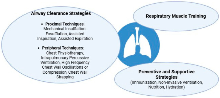 Figure 1: Main strategies for managing cough insufficiency and airway clearance in pediatric neuromuscular disorders (NMDs). Therapeutic modalities are categorized by target mechanism: assisted inspiration (air-stacking, lung volume recruitment via bag-valve-mask or mechanical insufflation to reach maximum insufflation capacity), assisted expiration (manually assisted cough, thoracic-abdominal thrusts, mechanical exsufflation), and combined mechanical insufflation-exsufflation (MI-E / CoughAssist) alongside non-invasive positive pressure ventilation (NIV) to prevent atelectasis, respiratory muscle fatigue, and recurrent aspiration pneumonia. Sourced from Randazzese et al., Children 2025; 12(10):1377; DOI: 10.3390/children12101377; PMCID: PMC12564417 under CC BY 4.0 Open Access.
4. Invasive Tracheostomy Indications & Diaphragmatic Pacing
- Tracheostomy Indications:
- Severe bulbar dysfunction with unmanageable salivary aspiration, recurrent laryngospasm, or failure of glottic closure preventing non-invasive airway clearance.
- Failure of NIV to control hypercapnia or oxygenation despite optimal interface and pressure titration.
- Continuous $24\text{-hour}$ ventilatory dependence in infants or young children unable to tolerate non-invasive interfaces or mouthpiece ventilation.
- Intolerance of facial interfaces due to severe facial deformities or skin necrosis.
- Diaphragmatic Pacing in CCHS:
- Implantation of phrenic nerve or direct diaphragmatic intramuscular electrodes connected to an external radiofrequency transmitter.
- Pacing provides rhythmic diaphragmatic contraction, freeing older children with CCHS from daytime mechanical ventilation and significantly improving mobility and developmental independence. Pacing requires an intact lower motor neuron pathway (normal C3–C5 anterior horn cells and phrenic nerves) and is contraindicated in primary neuropathies (e.g., SMA, GBS).
Bedside Protocols & Management Algorithms
Acute Decompensation Protocol in the Pediatric ICU / ER
When a child with chronic neuromuscular disease presents with acute respiratory distress:
flowchart TD
    classDef emerg fill:#8b0000,stroke:#ff4b5c,stroke-width:2px,color:#fff;
    classDef action fill:#1b4d3e,stroke:#2ecc71,stroke-width:2px,color:#fff;
    classDef warn fill:#d35400,stroke:#f39c12,stroke-width:2px,color:#fff;

    A["Acute Distress / Fever / Secretion Retention in Pediatric NMD"]:::emerg --> B["Immediate Clinical & Gas Assessment<br/>(Check Blood Gas, EtCO2, CXR — Do NOT just apply O2)"]:::warn

    B --> C["Aggressive Airway Clearance<br/>(MIE: +35/-35 cmH2O q1-2h with Inhaled Saline / Albuterol)"]:::action
    B --> D["Immediate Positive Pressure Support<br/>(BiPAP with High IPAP 16-20, EPAP 5, Backup Rate)"]:::action

    C --> E{"Clinical Response?<br/>(Clearance of atelectasis, normalization of PaCO2, PaO2)"}
    D --> E

    E -- "Stabilized" --> F["Continue PICU Non-Invasive Bundle<br/>Wean back to baseline settings slowly"]:::action
    E -- "Failure / Exhaustion / Severe Bulbar Failure" --> G["Elective Endotracheal Intubation<br/>(Avoid Succinylcholine! Use Video Laryngoscopy)"]:::emerg
Succinylcholine is strictly contraindicated in children with Duchenne Muscular Dystrophy, SMA, and other active myopathies due to the risk of life-threatening hyperkalemic cardiac arrest and malignant hyperthermia-like rhabdomyolysis triggered by massive potassium efflux from unstable extrajunctional acetylcholine receptors. If emergency intubation is required, use Rocuronium with Sugammadex readily available for reversal.
Key Clinical Matrix: Comparative Summary of Management
| Clinical Entity | Primary Pathophysiology | Daytime vs Nighttime Support | First-Line Airway Clearance | Special Hazards & Nuances |
|---|---|---|---|---|
| SMA Type 1 | Denervated intercostals; spared diaphragm; bell chest | Non-invasive BiPAP (often $>16\text{--}24\text{ h/day}$) | MIE ($+30/-30\text{ cmH}_2\text{O}$) via mask | Paradoxical breathing; disease-modifying therapies (Nusinersen, Risdiplam, Zolgensma) modify trajectory. |
| Duchenne (DMD) | Progressive diaphragmatic & abdominal myofiber loss | Nocturnal BiPAP in teens; daytime MPV in 20s | MIE + Air Stacking ($PCF < 270\text{ L/min}$) | Succinylcholine contraindicated; dilated cardiomyopathy monitoring mandatory. |
| CCHS (PHOX2B) | Absent central chemoreceptor sensitivity ($CO_2/O_2$) | Nocturnal BiPAP/Tracheostomy; 24h support in severe PARM/NPARM | Standard clearance (lungs and cough muscles are intact) | Unmonitored oxygen causes fatal apnea; diaphragmatic pacing indicated. |
| Guillain-Barré | Inflammatory demyelination / axonal loss | Acute BiPAP or ETT mechanical ventilation | MIE + In-line suctioning | Autonomic instability (labile BP/HR); serial bedside FVC/MIP monitoring. |
| Infant Botulism | Presynaptic ACh release blockade | Intubated mechanical ventilation vs BiPAP | Gentle suctioning, position drainage | Avoid aminoglycosides (worsen neuromuscular blockade); administer BabyBIG early. |
Key Takeaways & Summary
- Pathophysiological Core: Respiratory failure in pediatric neuromuscular diseases and central hypoventilation is a failure of ventilatory drive and mechanics (pump failure) rather than intrinsic gas-exchange parenchymal pathology.
- Micro-Atelectasis & Compliance: Chronic shallow breathing leads to micro-atelectasis, chest wall ankylosis, and severely reduced lung-thorax compliance, dramatically increasing the elastic work of breathing and causing nocturnal REM hypercapnia.
- Cough Flow Thresholds: An effective cough requires adequate inspiration, glottic closure, and vigorous abdominal contraction. A Peak Cough Flow ($PCF < 160\text{ L/min}$) indicates critical cough failure requiring mechanical insufflation-exsufflation (MIE / CoughAssist).
- Non-Invasive Strategy: BiPAP in Spontaneous/Timed (S/T) mode or AVAPS with an age-appropriate mandatory backup rate is essential to prevent hypercapnia during sleep.
- Supplemental Oxygen Hazard: Oxygen monotherapy without positive pressure support eliminates the hypoxic ventilatory drive, leading to acute hypercapnic coma and death.
- Pharmacological Safety: Succinylcholine is absolutely contraindicated in muscular dystrophies and denervating diseases due to fatal hyperkalemic arrest.