Mechanical ventilation in pediatric patients with congenital heart disease (CHD) represents one of the most demanding disciplines in critical care medicine. Unlike adult respiratory failure or pediatric acute respiratory distress syndrome (PARDS) where alveolar recruitment and oxygenation are paramount, post-operative pediatric cardiac ventilation requires continuous, dynamic balancing of cardiopulmonary interactions. The mechanical ventilator acts as an active hemodynamic modulator, directly manipulating right ventricular (RV) and left ventricular (LV) loading conditions, pulmonary vascular resistance ($PVR$), systemic vascular resistance ($SVR$), and shunt fractions.
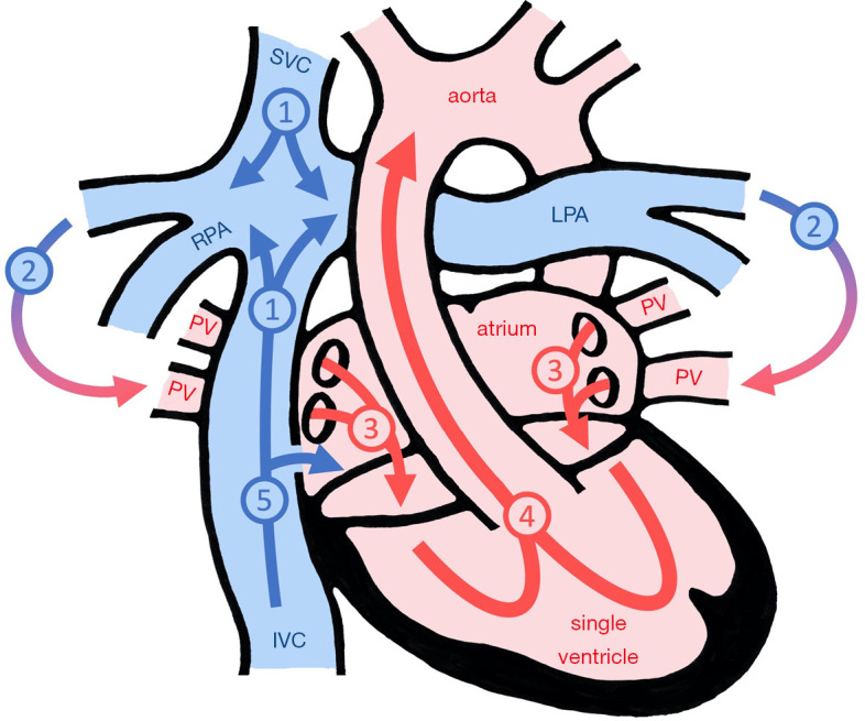 Figure 1: Determinants of pulmonary blood flow and vascular resistance in univentricular and cavopulmonary circulations. In the absence of a dedicated subpulmonary ventricle, pulmonary blood flow is passively driven across the pulmonary vascular bed by the transpulmonary pressure gradient ($\text{TPG} = PAP - LAP$). Positive intrathoracic pressure, alveolar overdistension, and microvascular remodeling directly heighten pulmonary vascular resistance ($PVR$), impairing ventricular filling and depressing cardiac index. Sourced from Becker et al., Cardiovasc Diagn Ther 2021; DOI: 10.21037/cdt-20-431.
Cardiopulmonary Hemodynamics in Pediatric Heart Disease
Positive pressure ventilation (PPV) fundamentally alters thoracic intracavity pressures, shifting transmural distending pressures across the cardiac chambers, great veins, and pulmonary vasculature.
Impact of Positive Pressure Ventilation and PEEP on Ventricular Loading
The cardiac effects of PPV and positive end-expiratory pressure (PEEP) diverge sharply between the right and left ventricles:
- Right Ventricular Preload: Increasing intrathoracic pressure ($P_{it}$) elevates mean systemic filling pressure ($MSFP$) but reduces the systemic venous return gradient ($\Delta P_{vr} = MSFP - CVP$). When right atrial pressure ($RAP$) rises secondary to elevated mean airway pressure ($mPaw$), venous return from the superior and inferior vena cavae diminishes, reducing RV end-diastolic volume.
- Right Ventricular Afterload: RV afterload is directly governed by transmural pulmonary arterial pressure and $PVR$. Alveolar distension increases transpulmonary pressure ($P_{tp} = P_{alv} - P_{pl}$)$, compressing alveolar microvessels and sharply elevating RV systolic wall stress. In patients with pre-existing RV dysfunction or pulmonary hypertension, excessive $mPaw$ triggers acute RV dilation, tricuspid regurgitation, and leftward interventricular septal shift (reversed Bernheim effect), impairing LV diastolic filling.
- Left Ventricular Preload: In a two-ventricle circulation with intact anatomy, LV preload depends on RV stroke volume and pulmonary venous transit. Reductions in RV output or severe compression of the pulmonary vascular bed secondarily decrease LV end-diastolic volume.
- Left Ventricular Afterload: LV afterload is determined by LV transmural systolic pressure ($P_{tm,LV} = P_{LV} - P_{it}$). By elevating $P_{it}$, PPV decreases LV transmural systolic pressure, acting as an effective mechanical unloading mechanism that augments LV stroke volume in systolic heart failure.
flowchart TD
    subgraph PPV_Effects["Positive Pressure Ventilation & Elevated PEEP"]
        direction TB
        A["Elevated Mean Airway Pressure (mPaw)"] --> B["Increased Intrathoracic Pressure (Pit)"]
        A --> C["Alveolar Expansion & Variable Lung Volume"]
    end

    subgraph RV_Branch["Right Ventricular Impact"]
        direction TB
        B --> D["Decreased CVP-to-MSFP Gradient\n(Reduced RV Preload)"]
        C --> E["Compression of Alveolar Capillaries\n(Increased RV Afterload)"]
        D & E --> F["Decreased RV Ejection & RV Dilation"]
        F --> G["Leftward Interventricular Septal Shift\n(Reversed Bernheim Effect)"]
    end

    subgraph LV_Branch["Left Ventricular Impact"]
        direction TB
        B --> H["Decreased Transmural LV Systolic Wall Stress\n(Reduced LV Afterload)"]
        G --> I["Impaired LV Diastolic Filling\n(Decreased LV Compliance)"]
        F --> J["Reduced Pulmonary Venous Return\n(Reduced LV Preload)"]
        H & I & J --> K["Modulated Cardiac Output\n(Net Dependent on Ventricle & Shunt)"]
    end

    style PPV_Effects fill:#1e293b,stroke:#3b82f6,stroke-width:2px,color:#fff
    style RV_Branch fill:#1e293b,stroke:#ef4444,stroke-width:2px,color:#fff
    style LV_Branch fill:#1e293b,stroke:#10b981,stroke-width:2px,color:#fff
The U-Shaped Relationship of Pulmonary Vascular Resistance and Lung Volume
Pulmonary vascular resistance is not a static property; it changes dynamically across lung volume states. The pulmonary vascular bed is divided into two anatomically distinct vascular compartments:
- Alveolar Vessels: Microscopic capillaries located within the interalveolar septa. These vessels are exposed to alveolar pressure ($P_{alv}$). As lung volume increases toward total lung capacity (TLC), alveolar distension stretches and compresses capillaries, increasing alveolar vessel resistance.
- Extra-Alveolar Vessels: Larger pulmonary arteries and veins traversing the bronchovascular sheaths. These vessels are exposed to pleural and interstitial pressures. At low lung volumes near residual volume (RV) or in atelectasis, loss of parenchymal radial traction and hypoxic pulmonary vasoconstriction cause extra-alveolar vessel collapse and high resistance.
Total PVR = Resistance(Alveolar Vessels) + Resistance(Extra-Alveolar Vessels)
The composite curve of $PVR$ plotted against lung volume is U-shaped, reaching its absolute physiological minimum at Functional Residual Capacity (FRC).
- At Low Lung Volumes (Atelectasis / FRC Deficit): Extra-alveolar vessels collapse due to loss of radial tethering traction, combined with localized hypoxic pulmonary vasoconstriction (HPV) and hypercapnic acidosis, driving $PVR$ upward.
- At High Lung Volumes (Alveolar Overdistension / High PEEP / Volutrauma): Alveolar capillaries are longitudinally stretched and compressed against distended alveolar walls, driving $PVR$ upward.
- Clinical Objective: Titrate PEEP and tidal volume ($V_T$) to establish ventilation precisely at normal FRC ($6-8\text{ mL/kg}$ without derecruitment or hyperinflation), minimizing total RV afterload.
Single Ventricle Physiology and Staged Palliative Ventilation
Children with functional single ventricle lesions (e.g., Hypoplastic Left Heart Syndrome [HLHS], Tricuspid Atresia, Double Inlet Left Ventricle [DILV]) undergo three-stage surgical palliation. Each stage presents unique respiratory-hemodynamic interdependencies.
Stage 1: Norwood, Sano, or Blalock-Taussig-Thomas (BTT) Shunt
In Stage 1 physiology, a single systemic ventricle (most commonly an anatomical RV in HLHS) pumps blood in parallel to both the systemic circulation ($Q_s$) and the pulmonary circulation ($Q_p$) via a systemic-to-pulmonary artery shunt (modified BTT shunt) or a right ventricle-to-pulmonary artery conduit (Sano shunt).
flowchart LR
    SV["Single Ventricle (SV)"] --> NeoAo["Neoaorta / Systemic Ejection"]
    NeoAo --> Qs["Systemic Vascular Bed (Qs)\nResistance = SVR"]
    NeoAo --> Shunt["mBTT Shunt / Sano Conduit"]
    Shunt --> Qp["Pulmonary Vascular Bed (Qp)\nResistance = PVR"]
    Qs --> RA["Right Atrium"]
    Qp --> LA["Common / Left Atrium"]
    RA & LA --> SV

    style SV fill:#dc2626,stroke:#fff,stroke-width:2px,color:#fff
    style Qp fill:#2563eb,stroke:#fff,stroke-width:2px,color:#fff
    style Qs fill:#16a34a,stroke:#fff,stroke-width:2px,color:#fff
Under parallel circulation, the distribution of blood flow is governed by the hydraulic resistance ratio between the pulmonary and systemic circuits:
$$\frac{Q_p}{Q_s} = \frac{\text{SVR}}{\text{PVR}} = \frac{SaO_2 - SvO_2}{SpvO_2 - SpaO_2}$$Where:
- $SaO_2$: Systemic arterial oxygen saturation (measured via arterial line or pulse oximetry, target $75-85\%$)
- $SvO_2$: Mixed venous oxygen saturation (measured from superior vena cava / pre-cardiac atrium, target $50-60\%$)
- $SpvO_2$: Pulmonary venous oxygen saturation (assumed $\approx 98-100\%$ in normal lungs)
- $SpaO_2$: Pulmonary arterial oxygen saturation (equal to mixed systemic venous saturation in common mixing)
Balancing Qp:Qs at the Bedside
The goal of mechanical ventilation in Stage 1 is maintaining $Q_p/Q_s \approx 0.8-1.2$:
- Pulmonary Overcirculation ($Q_p/Q_s \gg 1$, $SpO_2 > 88-90\%$):
- Mechanism: Low $PVR$ relative to $SVR$ diverts ventricular stroke volume away from the systemic and coronary beds into the compliant pulmonary vasculature.
- Clinical Sequelae: Diastolic run-off through the shunt reduces systemic diastolic arterial pressure ($DBP < 30-35\text{ mmHg}$), precipitating coronary hypoperfusion / steal, severe metabolic acidosis, oliguria, and necrotizing enterocolitis.
- Ventilator Interventions: Increase $PVR$ by lowering $FiO_2$ to $0.21$ (or using subatmospheric $FiO_2$ $0.17-0.19$ with medical nitrogen titration in specialized centers), instituting permissive hypercapnia ($PaCO_2 45-55\text{ mmHg}$, target pH $7.32-7.38$), avoiding high tidal volumes, and maintaining moderate PEEP ($4-5\text{ cmH}_2\text{O}$).
- Systemic Hypoxemia ($Q_p/Q_s \ll 1$, $SpO_2 < 70-75\%$):
- Mechanism: High $PVR$ or acute shunt thrombosis/stenosis limits pulmonary blood flow.
- Clinical Sequelae: Profound systemic cyanosis, tissue hypoxia, and lactic acidosis.
- Ventilator Interventions: Lower $PVR$ by increasing $FiO_2$ ($0.40-0.60$), increasing minute ventilation to normalize $PaCO_2$ ($35-40\text{ mmHg}$, mild alkalosis pH $7.45$), optimizing lung recruitment to FRC, and eliminating atelectasis.
An oxygen saturation ($SpO_2$) of $95\%$ in a post-operative Stage 1 Norwood neonate is not a sign of superior oxygenation; it is an imminent cardiac emergency indicating massive pulmonary overcirculation, systemic hypoperfusion, and coronary ischemia. Immediate therapeutic hypoventilation and $FiO_2$ reduction to $0.21$ are mandatory.
flowchart TD
    A["Post-Op Stage 1 Norwood / Sano Evaluation"] --> B{"Assess Arterial Saturation (SpO2) & Hemodynamics"}

    B -- "SpO2 > 88% | Wide Pulse Pressure | DBP < 35 mmHg" --> C["Pulmonary Overcirculation (Qp/Qs >> 1)"]
    C --> C1["Decrease FiO2 to 0.21 (Consider Subambient FiO2 0.18-0.20)"]
    C --> C2["Permissive Hypercapnia: Titrate PaCO2 48-55 mmHg (pH 7.30-7.35)"]
    C --> C3["Optimize PEEP (4-5 cmH2O) to prevent atelectrauma without overdistending"]
    C --> C4["Increase SVR / Systemic Tone (Vasopressin / Epinephrine)"]

    B -- "SpO2 75-85% | Lactate Normal | Mixed Venous SvO2 55-65%" --> D["Balanced Circulation (Qp/Qs ~ 1.0)"]
    D --> D1["Maintain Current Ventilator Strategy"]
    D --> D2["Lung-protective VT (6-8 mL/kg), PEEP 4-5 cmH2O"]

    B -- "SpO2 < 72% | Severe Cyanosis | Narrow Pulse Pressure" --> E["Pulmonary Hypoperfusion (Qp/Qs << 1)"]
    E --> E1["Increase FiO2 (0.40-0.80) to Induce Pulmonary Vasodilation"]
    E --> E2["Hyperventilate to Target PaCO2 32-38 mmHg (pH 7.45-7.50)"]
    E --> E3["Recruit Collapsed Alveolar Units to Restore FRC"]
    E --> E4["Rule Out Shunt Thrombosis (Urgent Echocardiogram / Heparin Bolus)"]

    style C fill:#7f1d1d,stroke:#ef4444,stroke-width:2px,color:#fff
    style D fill:#14532d,stroke:#22c55e,stroke-width:2px,color:#fff
    style E fill:#1e3a8a,stroke:#3b82f6,stroke-width:2px,color:#fff
Stage 2: Bidirectional Glenn / Superior Cavopulmonary Anastomosis
Performed at $3-6\text{ months}$ of age, the Bidirectional Glenn procedure disconnects the superior vena cava (SVC) from the right atrium and anastomoses it directly to the right pulmonary artery (RPA).
Passive Cavopulmonary Physiology
- The SVC supplies passive, non-pulsatile blood flow to the lungs, driven entirely by upper body central venous pressure ($CVP_{SVC} \approx 12-16\text{ mmHg}$) draining toward left atrial pressure ($LAP \approx 5-8\text{ mmHg}$).
- Mean Airway Pressure Sensitivity: Because no subpulmonary ventricle exists to generate pulsatile forward driving pressure, any increase in intrathoracic pressure or mean airway pressure ($mPaw$) directly opposes SVC venous return.
- An increase in $mPaw$ of just $3-5\text{ cmH}_2\text{O}$ can reduce pulmonary blood flow by $30-50\%$, causing severe upper body venous congestion (facial edema, elevated intracranial pressure) and profound hypoxemia.
Ventilator Management Protocol for Glenn Circulation
- Minimize Mean Airway Pressure ($mPaw$):
- Use short inspiratory times ($T_i \approx 0.5-0.6\text{ s}$).
- Set low PEEP ($3-5\text{ cmH}_2\text{O}$).
- Utilize pressure-controlled or PRVC modes with rapid decelerating flow profiles.
- Early Extubation Strategy:
- The restoration of spontaneous, negative-pressure ventilation acts as an "extracardiac thoracic pump", pulling venous blood from the SVC through the pulmonary vascular bed during inspiration.
- Target extubation within $4-8\text{ hours}$ post-operatively once bleeding is controlled, hypothermia resolved, and airway reflexes restored.
Stage 3: Fontan Circulation / Total Cavopulmonary Connection
Completed at $2-4\text{ years}$ of age, the Fontan operation connects the inferior vena cava (IVC) to the pulmonary arteries via an extracardiac conduit or lateral tunnel, achieving complete separation of systemic venous and arterial circuits.
flowchart TD
    subgraph Systemic_Venous_Return["Passive Total Cavopulmonary Circuit"]
        SVC["SVC Return (Upper Body)"] --> Glenn["Glenn Anastomosis"]
        IVC["IVC Return (Lower Body)"] --> Conduit["Extracardiac Conduit / Lateral Tunnel"]
        Glenn & Conduit --> PA["Pulmonary Arteries (Passive Non-Pulsatile Flow)"]
    end

    subgraph Pulmonary_Passage["Transpulmonary Gradient (TPG)"]
        PA --> Bed["Pulmonary Capillary Bed\n(Dependent on minimal PVR)"]
        Bed --> PV["Pulmonary Veins"]
        PV --> LA["Single/Common Atrium (LAP)"]
    end

    LA --> Ventricle["Systemic Single Ventricle"]
    Ventricle --> Aorta["Aorta -> Systemic Cardiac Output"]

    style Systemic_Venous_Return fill:#1e293b,stroke:#3b82f6,stroke-width:2px,color:#fff
    style Pulmonary_Passage fill:#1e293b,stroke:#10b981,stroke-width:2px,color:#fff
Transpulmonary Gradient Derivation
In the Fontan circuit, total cardiac output ($CO$) is strictly determined by passive flow across the pulmonary vascular bed:
$$\text{TPG} = PAP_{\text{mean}} - LAP_{\text{mean}}$$$$CO = \frac{\text{TPG}}{\text{PVR}} = \frac{PAP_{\text{mean}} - LAP_{\text{mean}}}{\text{PVR}}$$Where:
- $\text{TPG}$: Transpulmonary pressure gradient (normal $5-8\text{ mmHg}$)
- $PAP_{\text{mean}}$: Mean pulmonary artery pressure (equivalent to Fontan circuit pressure / CVP, ideally $< 15\text{ mmHg}$)
- $LAP_{\text{mean}}$: Left atrial pressure / single ventricular end-diastolic pressure (ideally $< 8-10\text{ mmHg}$)
- PVR is the Master Gatekeeper: Because the single ventricle cannot compensate for increased afterload on the pulmonary circuit, any rise in $PVR$ directly reduces ventricular filling, causing an immediate, linear drop in stroke volume and cardiac index.
- Avoid High PEEP ($> 5\text{ cmH}_2\text{O}$): Elevated PEEP compresses pulmonary capillaries and raises intrathoracic pressure, destroying the critical $5-8\text{ mmHg}$ $\text{TPG}$.
- Extubate Early: Transition to spontaneous breathing instantly lowers mean intrathoracic pressure, augments passive pulmonary perfusion, and increases cardiac output.
Post-Operative Cardiac Lesion Specifics
| Congenital Lesion / Surgical Procedure | Dominant Pathophysiological Vulnerability | Ventilator Strategy & Target Parameters | Hemodynamic & Pharmacologic Adjuncts |
|---|---|---|---|
| Transposition of the Great Arteries (TGA) (Arterial Switch Operation / Jatene) | Coronary artery reimplantation ischemia, acute LV dysfunction, pulmonary artery band distortion | • $V_T: 6-8\text{ mL/kg}$ • PEEP: $4-6\text{ cmH}_2\text{O}$ • $PaCO_2: 38-42\text{ mmHg}$ • $FiO_2$: Titrate to $SpO_2 > 95\%$ | • LV afterload reduction (Milrinone, Nicardipine) • Continuous ST-segment monitoring for coronary kinking • Maintain adequate coronary perfusion pressure ($DBP > 40$) |
| Tetralogy of Fallot (ToF) Repair (Transannular Patch / RVOT Reconstruction) | Junctional Ectopic Tachycardia (JET), severe free pulmonary regurgitation, restrictive RV "stiff" physiology | • Short $T_i$ ($0.5-0.6\text{ s}$) • Low-to-moderate PEEP ($4-5\text{ cmH}_2\text{O}$) • Avoid hyperventilation-induced alkalosis if JET is present | • Active cooling to $35.5-36.5^\circ\text{C}$ for JET • Atrioventricular sequential (DDD) pacing • Inodilator support (Milrinone/Epinephrine) • Adequate RV preload loading |
| Left-to-Right Shunts (VSD, AVSD, Truncus) (Complete Intracardiac Repair) | Reactive pulmonary vascular bed, pre-existing pulmonary overcirculation, acute post-op Pulmonary Hypertensive Crisis | • Avoid hypoxemia ($SpO_2 > 95\%$) • Mild hypocapnia ($PaCO_2 32-36\text{ mmHg}$, pH $7.45-7.50$) • Sedation/paralysis during unstable phase | • Inhaled Nitric Oxide (iNO $20\text{ ppm}$) • Milrinone / Sildenafil • Deep analgosedation (Dexmedetomidine, Fentanyl) |
| Truncus Arteriosus / Interrupted Aortic Arch | Complex combined LV/RV dysfunction, rapid vascular reactivity, airway compression from dilated neo-PA | • High-flow synchronization • Maintain FRC without bronchial compression • Low driving pressure ($\Delta P < 14\text{ cmH}_2\text{O}$) | • Inhaled prostacyclin or iNO • Aggressive afterload reduction |
Acute Pulmonary Hypertensive (PHTN) Crisis Protocol
Post-operative pulmonary hypertensive crisis is a life-threatening complication in pediatric cardiac surgery, most commonly occurring after repair of large left-to-right shunts (large VSD, complete AV canal, truncus arteriosus) or in infants with pre-existing elevated $PVR$.
Pathophysiological Cascade of a PHTN Crisis
An acute stimulus (e.g., endotracheal suctioning, agitation, hypoxia, acidosis, hypothermia) triggers intense pulmonary vasoconstriction:
- Pulmonary artery pressure abruptly exceeds systemic arterial pressure ($PAP > SAP$).
- Acute RV afterload mismatch leads to acute RV dilation and free tricuspid regurgitation.
- The interventricular septum shifts into the LV cavity, compressing the LV and abruptly dropping LV filling.
- Severe systemic hypotension, coronary hypoperfusion, and ventricular fibrillation/asystole ensue within seconds.
flowchart TD
    Trigger["Trigger: Agitation / Hypoxia / Acidosis / Suctioning"] --> Vasoconstriction["Intense Pulmonary Vasoconstriction"]
    Vasoconstriction --> PAP_Surge["PAP Surpasses Systemic Arterial Pressure (PAP > SAP)"]
    PAP_Surge --> RV_Failure["Acute RV Dilation & RV Ischemia"]
    RV_Failure --> Septal_Shift["Interventricular Septal Shift into LV"]
    Septal_Shift --> LV_Collapse["Loss of LV Preload & Low Cardiac Output"]
    LV_Collapse --> Hypotension["Profound Systemic Hypotension & Coronary Ischemia"]
    Hypotension --> Arrest["Cardiovascular Collapse / Cardiac Arrest"]

    style Trigger fill:#7f1d1d,stroke:#ef4444,stroke-width:2px,color:#fff
    style PAP_Surge fill:#7f1d1d,stroke:#ef4444,stroke-width:2px,color:#fff
    style Arrest fill:#450a0a,stroke:#dc2626,stroke-width:3px,color:#fff
Emergency Resuscitation and Management Protocol
Upon detection of acute systemic hypotension, rising PAP/CVP, and desaturation:
- Immediate Hyperoxygenation: Increase $FiO_2$ immediately to $1.0$. Oxygen is a potent, direct pulmonary vasodilator.
- Emergency Hyperventilation: Hand-ventilate or adjust ventilator settings to induce acute hypocarbic alkalosis:
- Target $PaCO_2: 30-34\text{ mmHg}$
- Target Arterial $\text{pH}: 7.50-7.55$ (administer Sodium Bicarbonate $1-2\text{ mEq/kg}$ IV if metabolic acidosis is present).
- Initiate / Escalate Inhaled Vasodilators:
- Start Inhaled Nitric Oxide (iNO) at $20\text{ ppm}$ directly into the inspiratory limb.
- If iNO is unavailable, deliver Inhaled Epoprostenol / Prostacyclin ($10-50\text{ ng/kg/min}$ or nebulized Iloprost).
- Deepen Sedation & Abolish Spontaneous Triggering:
- Bolus Fentanyl ($2-5\text{ mcg/kg}$) or Morphine.
- Administer immediate neuromuscular blockade (e.g., Rocuronium $1\text{ mg/kg}$ or Vecuronium $0.1\text{ mg/kg}$) to eliminate cough reflexes, airway resistance spikes, and metabolic oxygen consumption.
- Support Systemic Arterial Pressure:
- Bolus Epinephrine ($0.1-1.0\text{ mcg/kg}$) or Vasopressin ($0.0003-0.002\text{ units/kg/min}$) to keep systemic vascular resistance greater than pulmonary vascular resistance ($SAP > PAP$), preserving coronary perfusion.
- Inotropic and Lusitropic Support:
- Infuse Milrinone ($0.375-0.75\text{ mcg/kg/min}$) to enhance RV inotropy and induce pulmonary vasodilation (ensure adequate volume loading to counteract peripheral vasodilation).
- Mechanical Circulatory Support (ECLS / VA-ECMO):
- If refractory to maximal medical therapy and $PAP \ge SAP$ with persistent cardiac arrest or intractable shock, immediately initiate emergency cannulation for Veno-Arterial Extracorporeal Membrane Oxygenation (VA-ECMO).
Bedside Troubleshooting and Red Flags
- Sudden SpO2 Drop in Stage 1 Shunt: Rule out acute shunt thrombosis immediately. Check shunt murmur on auscultation (disappearance of continuous murmur is diagnostic of shunt occlusion), bolus heparin, and perform urgent echocardiography.
- Narrowing Pulse Pressure with Elevated CVP in Glenn / Fontan: Indicates acute failure of passive cavopulmonary transit due to high $mPaw$, pulmonary parenchymal collapse, or elevated left atrial pressure from single ventricle diastolic failure.
- Accelerated Narrow-Complex Tachycardia post-ToF Repair: Suspicion for Junctional Ectopic Tachycardia (JET). Discontinue hyperventilation alkalosis, cool patient to $36.0^\circ\text{C}$, optimize magnesium levels ($> 2.0\text{ mg/dL}$), initiate Dexmedetomidine / Amiodarone, and establish AV sequential pacing.
Key Takeaways and Summary
- Cardiopulmonary Interdependence: In pediatric cardiac surgery, mechanical ventilation is a potent hemodynamic tool. Mean airway pressure ($mPaw$) directly determines RV preload, RV afterload, and cavopulmonary blood transit.
- The FRC "Sweet Spot": Total $PVR$ is minimal at FRC. Both atelectasis (via extra-alveolar vessel collapse and HPV) and overdistension (via alveolar capillary compression) increase RV afterload and must be avoided.
- Stage 1 Norwood Principle: Balance $Q_p/Q_s$ at $\approx 1.0$ ($SpO_2 75-85\%$). Avoid hyperoxia and hypocarbia, which cause lethal pulmonary overcirculation and coronary steal.
- Glenn and Fontan Principles: Passive pulmonary blood flow requires low mean airway pressure, minimal PEEP ($3-5\text{ cmH}_2\text{O}$), and early liberation to negative-pressure spontaneous breathing.
- Pulmonary Hypertensive Crisis: Manage immediately with $100\%$ $FiO_2$, hyperventilation to $\text{pH } 7.50-7.55$, iNO $20\text{ ppm}$, neuromuscular blockade, and systemic vasopressors to maintain $SAP > PAP$.