Acute viral bronchiolitis is the leading cause of lower respiratory tract infection, acute hypoxemic-hypercapnic respiratory failure, and intensive care admission among infants under 12 months of age worldwide. While the vast majority of cases exhibit a self-limiting course manageable with supportive hydration and supplemental oxygen, a subset of patients—particularly neonates, former preterm infants, and infants with hemodynamically significant congenital heart disease, bronchopulmonary dysplasia (BPD), neuromuscular disorders, or immunodeficiency—develop severe critical bronchiolitis.
Managing severe bronchiolitis in the Pediatric Intensive Care Unit (PICU) presents unique physiological challenges. Unlike homogenous parenchymal lung diseases or pure central airway obstructions, bronchiolitis generates a complex, spatially heterogeneous mixed obstructive-restrictive mechanical phenotype characterized by wide regional variance in respiratory time constants ($\\tau$), extensive dynamic airway collapse, micro-atelectasis, severe ventilation-perfusion ($\\dot{V}/\\dot{Q}$) mismatch, and elevated risk of dynamic hyperinflation (intrinsic PEEP).
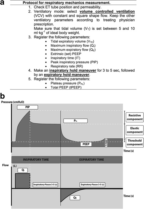 Figure 1: Bedside assessment of respiratory mechanics and partitioning of working pressure in an infant with severe viral bronchiolitis receiving controlled mechanical ventilation. (a) Measurement protocol illustrating constant-flow volume-controlled ventilation during end-inspiratory and end-expiratory occlusions. (b) Scalar waveforms showing decomposition of peak inspiratory pressure ($PIP$) into ohmic resistive pressure ($P_{res} = PIP - P_1$), viscoelastic tissue stress relaxation ($P_1 - P_2/P_{plat}$), elastic driving pressure ($P_{el} = P_2 - tPEEP$), and total intrinsic positive end-expiratory pressure ($tPEEP - PEEP$). Source: Cruces et al., BMC Pulm Med, 2017; DOI: 10.1186/s12890-017-0475-6.
Viral Etiology and Molecular Pathophysiology
Acute bronchiolitis is triggered by trophic viral pathogens that infect the pseudostratified ciliated columnar epithelium of the distal bronchiolar tree (internal lumen diameter $< 1\\text{ mm}$, lacking cartilaginous support).
Primary Pathogens and Cytopathology
- Respiratory Syncytial Virus (RSV, Orthopneumovirus): Responsible for $60-80\\%$ of PICU admissions. RSV utilizes its surface Fusion ($F$) and Attachment ($G$) glycoproteins to infect apical ciliated epithelial cells. Viral replication induces cell swelling, loss of functional ciliary axonemes (ciliary denudation), syncytia formation (large multinucleated syncytial syncytioplasms), and extensive apoptotic/necrotic shedding into the airway lumen.
- Human Rhinovirus (HRV, predominantly HRV-C): Highly associated with severe bronchospasm, family history of atopy, and wheezing phenotypes in infants $> 6$ months.
- Human Metapneumovirus (hMPV): Produces clinical courses nearly identical to RSV, with marked lower airway edema and dynamic airflow limitation.
- Viral Co-infections & Secondary Bacterial Colonization: Viral-viral (e.g., RSV + HRV) or viral-bacterial co-infections (most commonly Moraxella catarrhalis, Streptococcus pneumoniae, or Haemophilus influenzae) dramatically amplify peribronchiolar inflammation, duration of mechanical ventilation, and secondary pediatric ARDS (PARDS).
flowchart TD
    A["Viral Inoculation (RSV, Rhinovirus, hMPV)"] --> B["Infection of Bronchiolar Ciliated Epithelium"]
    B --> C["Epithelial Cytonecrosis & Ciliary Denudation"]
    B --> D["Pro-Inflammatory Chemokine Surge (IL-6, IL-8, TNF-a)"]

    D --> E["Neutrophilic & Lymphocytic Peribronchiolar Infiltration"]
    D --> F["Submucosal Microvascular Hyperpermeability & Edema"]

    C --> G["Sloughed Epithelial Debris + Dense Fibrin/Mucus Plugs"]
    F --> H["Circumferential Small-Airway Lumen Narrowing (r ↓)"]

    G --> I["Heterogeneous Airway Luminal Occlusion"]
    H --> I

    I --> J["High Resistance (Raw ↑↑) & Ball-Valve Obstruction"]
    I --> K["Complete Distal Resorption Micro-Atelectasis"]

    J --> L["Prolonged Time Constants (τ_long) & Dynamic Gas Trapping (PEEPi)"]
    K --> M["Short Time Constants (τ_short), Low Compliance (Crs ↓) & Shunt"]

    L --> N["Dual Heterogeneous Mechanical Phenotype & V/Q Mismatch"]
    M --> N
Micro-Anatomical Cascade of Airway Obstruction
Poiseuille's law dictates that laminar airway resistance ($R_{aw}$) is inversely proportional to the fourth power of the internal radius:
$$R_{aw} = \\frac{8 \\cdot \\eta \\cdot l}{\\pi \\cdot r^4}$$In an infant where baseline bronchiolar internal diameter is merely $1.0-1.5\\text{ mm}$, a $0.5\\text{ mm}$ circumferential accumulation of submucosal edema, cellular debris, and dense mucus reduces luminal radius by $50\\%$, increasing frictional airway resistance by a factor of $2^4 = 16$-fold ($1,600\\%$ increase in resistive work of breathing).
- Intraluminal Mucociliary Failure: Massive sloughing of dead ciliated epithelial cells combines with fibrin-rich inflammatory exudate and neutrophil extracellular traps (NETs), forming thick, tenacious intraluminal casts.
- Peribronchiolar Lymphocytic Infiltration: Dense cuffing of lymphocytes, plasma cells, and degranulating neutrophils surrounds small conducting airways, destroying the surrounding radial alveolar tethering that normally maintains bronchiolar patency during passive exhalation.
- Submucosal Edema & Microvascular Leak: Proinflammatory cytokines ($IL-1\\beta$, $IL-6$, $IL-8$, $TNF-\\alpha$, $MIP-1\\alpha$) trigger intense capillary leakage and localized engorgement within the delicate bronchiolar submucosa.
- Secondary Surfactant Dysfunction: Severe inflammatory alveolar exudate containing albumin, fibrin monomer, and cholesterol inhibits pulmonary surfactant biophysical activity. Furthermore, direct injury to alveolar Type II pneumocytes leads to quantitative surfactant deficiency, precipitating progressive regional micro-atelectasis and alveolar instability.
Dual Mechanical Phenotype and Time-Constant Heterogeneity
The defining pathophysiological hallmark of critical bronchiolitis is extreme inter-regional ventilation heterogeneity. The infant lung behaves as a multi-compartmental parallel network with wildly disparate regional mechanical properties.
graph TD
    subgraph Heterogeneous Lung Compartments
        A["Obstructed Compartment (Mucus Plugs / Narrowed Bronchioles)"]
        B["Atelectatic / Collapsed Compartment (Secondary Surfactant Loss)"]
    end

    A --> C["High Resistance (Raw ↑↑)<br>Normal / Preserved Compliance (C)"]
    B --> D["Normal / Mildly Elevated Resistance (Raw)<br>Low Compliance (C ↓↓)"]

    C --> E["Prolonged Expiratory Time Constant (τ_long > 0.6-1.0 s)"]
    D --> F["Short Expiratory Time Constant (τ_short < 0.15-0.25 s)"]

    E --> G["Dynamic Air Trapping & Auto-PEEP (PEEPi)<br>Expiratory Flow Truncation"]
    F --> H["Rapid Emptying & Cyclic Alveolar Collapse<br>Intense Intrapulmonary Shunt"]

    G --> I["Pendelluft Gas Transfer between Asynchronous Alveoli<br>Regional Volutrauma & Dynamic Shear Stress"]
    H --> I
Mathematical Formulation of Respiratory Time Constants
The expiratory time constant ($\\tau_{rs}$) represents the physical duration required for the lungs to passively eliminate $63.2\\%$ of a delivered tidal volume:
$$\\tau_{rs} = R_{rs} \\times C_{rs}$$Where:
- $\\tau_{rs}$ = Respiratory system time constant ($\\text{seconds}$)
- $R_{rs}$ = Total respiratory system resistance ($\\text{cmH}_2\\text{O}/(\\text{L}/\\text{s})$)
- $C_{rs}$ = Total respiratory system static compliance ($\\text{mL}/\\text{cmH}_2\\text{O}$ or $\\text{L}/\\text{cmH}_2\\text{O}$)
For passive exhalation, the expired lung volume as a function of expiratory time ($t$) is modeled exponentially:
$$V(t) = V_T \\cdot e^{-t / \\tau_{rs}}$$Complete alveolar emptying ($> 95-99.3\\%$ volume clearance) requires $3$ to $5$ full time constants:
$$t_{emptying} \\ge 3 \\text{ to } 5 \\times \\tau_{rs}$$- Obstructed Segments ($\\tau_{long}$): Resistance is severely elevated ($R_{aw} = 60-150\\text{ cmH}_2\\text{O}/(\\text{L}/\\text{s})$), while parenchymal compliance remains relatively preserved ($C_{rs} \\approx 1.0-1.5\\text{ mL}/\\text{cmH}_2\\text{O}$). Thus, $\\tau_{long} = 0.08 \\times 1.2 \\approx 0.8-1.5\\text{ s}$. Complete exhalation requires an expiratory time ($T_e$) of $3 \\times 1.0\\text{ s} = 3.0\\text{ s}$. If $T_e$ is set to $< 1.5\\text{ s}$, gas remains dynamically trapped behind floppy, dynamic-collapsed bronchioles, generating intrinsic positive end-expiratory pressure ($PEEP_i$).
- Micro-Atelectatic Segments ($\\tau_{short}$): Non-compliant, collapsed alveoli exhibit dramatically reduced compliance ($C_{rs} \\approx 0.2-0.4\\text{ mL}/\\text{cmH}_2\\text{O}$) with near-normal resistance ($R_{aw} \\approx 25\\text{ cmH}_2\\text{O}/(\\text{L}/\\text{s})$), yielding $\\tau_{short} \\approx 0.08-0.15\\text{ s}$. These units fill and empty almost instantaneously, remaining vulnerable to cyclic derecruitment and shear-induced atelectrauma.
Pendelluft Phenomenon and Gas Trapping
During both spontaneous efforts and positive-pressure mechanical breaths, gas redistributes asynchronously between parallel lung units with unequal time constants without contributing to fresh gas exchange:
- At the initiation of inspiration, fast-filling units ($\\tau_{short}$) receive tidal gas rapidly.
- As fast units reach peak pressure, gas continues to slowly inflate high-resistance slow units ($\\tau_{long}$).
- At end-inspiration or the onset of expiration, before external exhalation begins, gas shunts internally from high-pressure fast units into low-pressure slow units (Pendelluft gas exchange), exacerbating regional overdistension and hypercapnia.
Clinical Indications and Escalation Hierarchy
The clinical spectrum of severe infant bronchiolitis progresses from upper airway rhinitis to tachypnea, severe subcostal/intercostal retractions, grunting, nasal flaring, and ultimately diaphragmatic fatigue, apnea, and respiratory failure.
flowchart TD
    A["Infant with Severe Viral Bronchiolitis in Respiratory Distress"] --> B{"Apnea, Severe Lethargy, or Cardiac Arrest?"}
    B -- Yes --> C["Immediate Endotracheal Intubation & Invasive MV"]
    B -- No --> D["Trial of Non-Invasive Support: HFNC (1.5 - 2.0 L/kg/min)"]

    D --> E{"Clinical Response at 60-120 min?<br>(Work of Breathing, RR, FiO2 ≤ 0.40)"}
    E -- Favorable --> F["Continue HFNC & Gradual Weaning"]
    E -- Failure / Deterioration --> G["Escalate to Nasal CPAP (6-8 cmH2O) or BiPAP"]

    G --> H{"Target pH ≥ 7.22, PaCO2 Stabilizing,<br>SpO2 ≥ 90%, No Apnea?"}
    H -- Success --> I["Maintain CPAP/BiPAP & Airway Clearance"]
    H -- Refractory Exhaustion / Severe Acidosis --> C

    C --> J["Invasive Lung-Protective Mechanical Ventilation Protocol"]
    J --> K{"Refractory Hypoxemia / Hypercapnia<br>(OI ≥ 20-25 or PALICC-2 Severe PARDS)?"}
    K -- Yes --> L["PICU Advanced Rescue: Surfactant Lavage / Inhaled NO / VV-ECMO Evaluation"]
    K -- Controlled --> M["Permissive Hypercapnia & Progressive Weaning Ladder"]
Absolute and Relative Indications for Invasive Mechanical Ventilation
Apnea is a primary, life-threatening manifestation of viral bronchiolitis in young infants ($< 2$ months of age or ex-premature infants $< 44$ weeks post-menstrual age). RSV infects sensory unmyelinated bronchopulmonary C-fibers, triggering exaggerated laryngeal chemoreflexes and substance P-mediated central blunting of medullary respiratory drive. A single episode of prolonged, unprovoked apnea with bradycardia in an infant with bronchiolitis warrants high-vigilance PICU monitoring and a very low threshold for invasive endotracheal intubation.
- Refractory Apnea: Central or obstructive apneas associated with bradycardia ($HR < 80\\text{ bpm}$) and cyanosis unresponsive to non-invasive tactile stimulation or CPAP.
- Impending Respiratory Muscle Exhaustion: Clinical deterioration marked by paradoxical thoracoabdominal respirations ("see-saw breathing"), silent chest on auscultation (loss of wheeze due to minimal tidal flow), obtundation, or sudden bradycardia.
- Severe Uncompensated Respiratory Acidosis: Arterial or capillary blood gas demonstrating $pH < 7.15-7.20$ with progressive hypercapnia ($PaCO_2 > 70-80\\text{ mmHg}$) and clinical exhaustion.
- Refractory Hypoxemic Respiratory Failure: Inability to maintain target oxygen saturations ($SpO_2 \\ge 90\\%$) despite non-invasive CPAP/BiPAP with $FiO_2 > 0.60-0.70$ ($PaO_2/FiO_2 \\le 150-200$).
Invasive Mechanical Ventilation Strategy
Mechanical ventilation in severe bronchiolitis requires balancing two conflicting mechanical targets: providing adequate expiratory time for prolonged obstructed units while delivering sufficient distending pressure (PEEP) to stent dynamic small airways and recruit collapsed atelectatic zones.
Mode Selection
- Pressure-Controlled Synchronized Intermittent Mandatory Ventilation (PC-SIMV) + Pressure Support (PS) or Pressure-Controlled Assist/Control (PC-A/C):
- Physiological Rationale: Delivers a decelerating inspiratory flow waveform that rapidly delivers flow to low-resistance units while tapering flow toward end-inspiration, minimizing peak dynamic shear stress in heterogeneous infant airways. Fixed inspiratory pressures ensure that peak alveolar distension is capped regardless of regional mucus flux.
- PRVC / Volume Guarantee (PC-VG):
- Physiological Rationale: Automatically modulates inspiratory working pressure on a breath-by-breath basis to guarantee a preset tidal volume ($V_T$) while retaining decelerating flow dynamics.
- Caution: In the presence of acute airway resistance changes from thick mucus plugs, the ventilator will automatically increase peak pressure up to the safety limit ($P_{max} - 5\\text{ cmH}_2\\text{O}$), requiring strict bedside monitoring.
Initial Ventilator Settings Protocol
flowchart LR
    A["Intubated Infant with Severe Bronchiolitis"] --> B["Mode: PC-A/C or PC-SIMV + PS (Decelerating Flow)"]
    B --> C["Tidal Volume: 6 - 8 mL/kg (Monitor Exhaled Vte)"]
    B --> D["Respiratory Rate: 20 - 30 bpm (Avoid Tachypnea)"]
    B --> E["Inspiratory Time (Ti): 0.5 - 0.7 s"]
    B --> F["Expiratory Time (Te): ≥ 1.5 - 2.5 s (I:E = 1:2.5 to 1:3.5)"]
    B --> G["PEEP: 5 - 8 cmH2O (Airway Stenting & Atelectasis Prevention)"]
    B --> H["Target FiO2: Titrate to SpO2 90 - 95%"]

    C & D & E & F & G & H --> I["Continuous Bedside Scalar Monitoring: Flow-Time & PEEPi"]
| Ventilator Parameter | Initial Recommended Setting | Physiological Justification | Bedside Titration / Safety Limits |
|---|---|---|---|
| Mode | PC-A/C, PC-SIMV+PS, or PRVC / VG | Decelerating flow profile accommodates heterogeneous time constants | Ensure adequate synchronization; set PS flow trigger sensitive without auto-cycling ($0.5-1.0\\text{ L/min}$). |
| Tidal Volume ($V_T$) | $6-8\\text{ mL/kg}$ PBW | Normal physiological infant $V_T$; avoids regional volutrauma | Monitor exhaled tidal volume ($V_{Te}$) continuously at the airway flow sensor. |
| Inspiratory Time ($T_i$) | $0.5-0.7\\text{ s}$ | Sufficient to overcome resistive-inertial forces and deliver volume | Adjust according to the inspiratory flow scalar; flow should decelerate to near zero. |
| Respiratory Rate ($f$) | $20-30\\text{ breaths/min}$ (Infants) | Lower than normal physiological infant rates to ensure prolonged $T_e$ | Avoid high ventilator rates ($> 35-40\\text{ bpm}$); prevents dynamic breath-stacking. |
| Expiratory Time ($T_e$) | $\\ge 1.5-2.5\\text{ s}$ ($I:E = 1:2.5 \\text{ to } 1:3.5$) | Allows slow units ($3-5 \\times \\tau_{long}$) to empty completely | Inspect flow-time scalar: expiratory flow MUST reach the zero baseline before next breath. |
| PEEP | $5-8\\text{ cmH}_2\\text{O}$ | Pneumatic stent for floppy small airways; recruits surfactant-deficient zones | Avoid excessive PEEP ($> 9-10\\text{ cmH}_2\\text{O}$) which worsens dynamic hyperinflation and RV afterload. |
| Driving Pressure ($\\Delta P$) | $\\Delta P \\le 14-15\\text{ cmH}_2\\text{O}$ | $\\Delta P = P_{plat} - PEEP_{total}$; key determinant of mechanical strain | Minimize driving pressure while tolerating permissive hypercapnia. |
| $FiO_2$ | Titrate to $SpO_2\\ 90-95\\%$ | Prevents hyperoxic absorption micro-atelectasis and oxygen toxicity | Target $PaO_2 = 60-80\\text{ mmHg}$ ($SpO_2\\ 88-92\\%$ acceptable in severe PARDS). |
PEEP Management: The Pneumatic Stenting Paradigm
In bronchiolitis, small cartilaginous support is absent, and peribronchiolar radial traction is lost due to inflammation. During unassisted passive exhalation, the positive pleural pressure compresses small airways, inducing dynamic downstream collapse and gas trapping:
$$P_{airway} < P_{pleural} \\implies \\text{Airway Closure}$$Application of moderate extrinsic PEEP ($5-8\\text{ cmH}_2\\text{O}$) raises the intraluminal pressure above the critical closing pressure ($P_{crit}$) throughout expiration:
$$PEEP_{extrinsic} > P_{crit}$$This acts as an internal pneumatic stent, preventing dynamic airway collapse, reducing downstream expiratory resistance, accelerating overall expiratory emptying, and decreasing the difference between extrinsic and intrinsic PEEP ($PEEP_i - PEEP_{ext}$).
graph LR
    subgraph Zero or Inadequate Extrinsic PEEP (0-3 cmH2O)
        A["Intraluminal Pressure Drops during Expiration"] --> B["Extraluminal Pleural Forces Exceed Luminal Pressure"]
        B --> C["Dynamic Bronchiolar Collapse & Premature Airway Closure"]
        C --> D["Severe Gas Trapping, High PEEPi, & Regional Atelectasis"]
    end

    subgraph Optimal Extrinsic PEEP (5-8 cmH2O)
        E["Continuous Positive Distending Pressure Maintained"] --> F["Intraluminal Pressure Exceeds Closing Pressure (P > Pcrit)"]
        F --> G["Pneumatic Stenting of Floppy Bronchiolar Walls"]
        G --> H["Unimpeded Expiratory Flow, Reduced PEEPi, & Stable FRC"]
    end
In severe obstructive phenotypes, setting excessive extrinsic PEEP ($> 9-10\\text{ cmH}_2\\text{O}$) can push lung units beyond the upper inflection point onto the flat, non-compliant portion of the pressure-volume curve. This leads to alveolar overdistension, increased dead-space ventilation ($V_D/V_T$), pulmonary vascular resistance elevation, right ventricular outflow impedance, and risk of barotrauma (pneumothorax/pneumomediastinum). Continuous bedside monitoring of intrinsic PEEP via end-expiratory occlusion is essential.
Permissive Hypercapnia Protocol
In severe viral bronchiolitis, attempting to normalize $PaCO_2$ ($35-45\\text{ mmHg}$) by escalating minute ventilation ($V_E = V_T \\times f$) causes dangerous dynamic hyperinflation, elevated mechanical power, and barotrauma.
- Permissive Hypercapnia Target: Maintain arterial $pH \\ge 7.20-7.25$ with $PaCO_2$ typically between $60-80\\text{ mmHg}$ (and occasionally $80-90\\text{ mmHg}$ if renal bicarbonate compensation preserves $pH > 7.18$).
- Contraindications to Permissive Hypercapnia: Severe pulmonary hypertension, right ventricular failure with cor pulmonale, hemodynamic instability, or coexisting acute hypoxic-ischemic encephalopathy/intracranial hypertension.
Non-Invasive Support and Rescue Therapies
graph TD
    A["Non-Invasive and Adjunctive Support Arsenal"]

    A --> B["High-Flow Nasal Cannula (HFNC)"]
    A --> C["Continuous Positive Airway Pressure (CPAP)"]
    A --> D["Heliox (Helium-Oxygen Mixture)"]
    A --> E["Inhaled Hypertonic Saline (3%)"]
    A --> F["Exogenous Surfactant Replacement"]
    A --> G["Veno-Venous Extracorporeal Membrane Oxygenation (VV-ECMO)"]

    B --> B1["Flow 1.5 - 2.0 L/kg/min<br>Washes out nasopharyngeal dead space<br>Delivers 2 - 4 cmH2O PEEP"]
    C --> C1["Nasal / Full-Face Mask CPAP 5 - 8 cmH2O<br>Decreases inspiratory work of breathing<br>Stents upper and lower dynamic airways"]
    D --> D1["Density-lowering 70:30 or 60:40 mixture<br>Converts turbulent flow to laminar flow<br>Reduces Reynolds number & resistive WOB"]
    E --> E1["Osmotic hydration of dehydrated mucus<br>Accelerates mucociliary clearance<br>Administer with bronchodilator if bronchospastic"]
    F --> F1["Indicated in secondary surfactant degradation / PARDS<br>Endotracheal bolus or bronchoalveolar lavage<br>Improves compliance & oxygenation index"]
    G --> G1["PALICC-2 Severe PARDS Criteria<br>Oxygenation Index (OI) ≥ 40 or refractory hypercapnic arrest<br>Complete lung rest & gas exchange support"]
High-Flow Nasal Cannula (HFNC) & CPAP Mechanics
- HFNC Mechanism of Action:
- Delivers heated ($37^\\circ\\text{C}$), fully humidified ($100\\%$ relative humidity, $44\\text{ mg } H_2O/L$) gas at flow rates of $1.5-2.0\\text{ L/kg/min}$ (up to $20-30\\text{ L/min}$ in older infants).
- Dead-Space Washout: Purges anatomical nasopharyngeal dead space of expired carbon dioxide ($CO_2$), creating an upper airway reservoir of fresh gas that significantly enhances alveolar ventilation efficiency.
- Distending Pharyngeal Pressure: Generates flow-dependent positive airway pressure ($2-4\\text{ cmH}_2\\text{O}$ with mouth closed), stenting upper pharyngeal collapsibility and lowering diaphragmatic work of breathing.
- Nasal CPAP (5–8 $\\text{cmH}_2\\text{O}$):
- Indicated when HFNC fails to alleviate moderate-to-severe retractions or when recurrent apneas occur.
- Generates stable, sealed distending airway pressure that counteracts intrinsic PEEP and improves functional residual capacity (FRC).
Heliox Therapy
According to fluid dynamics, the Reynolds number ($Re$), which dictates whether gas flow is laminar ($Re < 2000$) or turbulent ($Re > 4000$), is directly proportional to gas density ($\\rho$):
$$Re = \\frac{\\rho \\cdot v \\cdot d}{\\mu}$$Where $\\rho$ = gas density, $v$ = velocity, $d$ = airway diameter, and $\\mu$ = gas dynamic viscosity.
In turbulent flow through obstructed, narrowed bronchioles, the driving pressure gradient ($\\Delta P$) required to generate flow ($\\dot{V}$) is density-dependent:
$$\\Delta P = k_1 \\cdot \\mu \\cdot \\dot{V} + k_2 \\cdot \\rho \\cdot \\dot{V}^2$$Heliox ($70\\%\\text{ He} : 30\\%\\text{ O}_2$ or $60\\%\\text{ He} : 40\\%\\text{ O}_2$) has a density approximately one-third that of atmospheric nitrogen-oxygen gas ($\\rho_{Heliox} \\approx 0.43\\text{ g/L}$ vs $\\rho_{Air} \\approx 1.20\\text{ g/L}$). By drastically lowering density, Heliox converts turbulent flow into smooth laminar flow, decreasing resistive pressure drops across obstructed bronchioles and reducing patient work of breathing by $30-50\\%$. Note: Heliox efficacy diminishes rapidly when $FiO_2 > 0.40$ is required.
Pharmacological & Adjunctive Mucociliary Clearances
- Inhaled 3% Hypertonic Saline: Creates an osmotic gradient across the damaged bronchiolar epithelium, drawing water into the dehydrated mucus gel layer, reducing mucus viscoelasticity, breaking ionic bonds in mucus polymer networks, and stimulating ciliary beat frequency.
- Bronchodilators (Trial Basis Only): Routine beta-2 agonists (albuterol) and epinephrine are not universally recommended by AAP/NICE guidelines due to lack of alpha-smooth muscle hypertrophy in young infants $< 6$ months. However, in ventilated infants with high baseline resistance or atopic phenotypes, a documented therapeutic trial (measuring post-aerosol expiratory resistance on the ventilator) can identify clinical responders.
- Surfactant Lavage / Replacement: In severe cases progressing to secondary PARDS with profound alveolar collapse and compliance loss ($C_{rs} < 0.4\\text{ mL}/\\text{cmH}_2\\text{O}$), exogenous surfactant instillation ($100\\text{ mg/kg}$ Poractant alfa) restores alveolar interfacial surface tension, improves $P/F$ ratio, and accelerates mechanical liberation.
Bedside Monitoring, Troubleshooting, and Complications
In infants mechanically ventilated for viral bronchiolitis, sudden catastrophic deterioration (acute desaturation, bradycardia, severe loss of delivered tidal volume in PC mode, or peak pressure alarm spikes in VC mode) is caused by an endotracheal tube (ETT) or mainstem bronchus mucus plug until proven otherwise:
- Disconnect from the Ventilator: Immediately attach a self-inflating manual resuscitation bag with $100\\%\\text{ O}_2$ and a PEEP valve.
- Assess Chest Rise and Resistance: If manual bag ventilation encounters high resistance with absent chest rise, suspect complete lumen occlusion.
- Deep Endotracheal Suctioning: Pass an appropriately sized in-line suction catheter with $0.5-1.0\\text{ mL}$ sterile normal saline instillation.
- Emergency ETT Exchange: If suction catheter fails to pass beyond the ETT tip or resistance remains absolute, perform immediate re-intubation over an airway exchange catheter or direct laryngoscopy.
Bedside Monitoring Matrix
| Clinical Parameter | Target Objective | Bedside Monitoring Modality | Actionable Threshold / Intervention |
|---|---|---|---|
| Expiratory Flow Scalar | Complete decay to zero baseline before next trigger | Continuous ventilator graphics screen | If expiratory flow is truncated: decrease respiratory rate ($f$), increase $T_e$, or optimize PEEP. |
| Intrinsic PEEP ($PEEP_i$) | $PEEP_i < 3-5\\text{ cmH}_2\\text{O}$ | End-expiratory occlusion hold ($2-3\\text{ s}$) in paralyzed or deeply sedated infant | If $PEEP_i > 5\\text{ cmH}_2\\text{O}$: lengthen expiratory time ($T_e$) and decrease respiratory frequency. |
| Static Compliance ($C_{stat}$) | $> 0.6-1.0\\text{ mL}/(\\text{cmH}_2\\text{O} \\cdot \\text{kg})$ | $C_{stat} = \\frac{V_{Te}}{P_{plat} - PEEP_{total}}$ via inspiratory hold | If $C_{stat} < 0.5\\text{ mL}/(\\text{cmH}_2\\text{O} \\cdot \\text{kg})$: evaluate for atelectasis or secondary PARDS; titrate PEEP. |
| Airway Resistance ($R_{aw}$) | $< 30-50\\text{ cmH}_2\\text{O}/(\\text{L}/\\text{s})$ | $R_{insp} = \\frac{PIP - P_{plat}}{\\dot{V}_{insp}}$ during constant-flow VC | If $R_{aw} > 60-80$: perform endotracheal suctioning, clear condensation, and consider trial bronchodilator. |
| Oxygenation Index (OI) | $OI < 8-10$ | $OI = \\frac{MAP \\times FiO_2 \\times 100}{PaO_2}$ | If $OI > 15-20$: escalate PARDS management (proning, surfactant, inhaled nitric oxide, or ECMO). |
When severe bronchiolitis induces extensive bilateral infiltrates with parenchymal consolidation and severe hypoxemia not fully explained by heart failure, it meets the Second Pediatric Acute Lung Injury Consensus Conference (PALICC-2) definition for PARDS:
- Non-Invasive PARDS: Full-face mask / nasal CPAP $\\ge 5\\text{ cmH}_2\\text{O}$ with $SpO_2/FiO_2 \\le 221$ ($SpO_2 \\le 97\\%$) or $PaO_2/FiO_2 \\le 300$.
- Invasive Mild PARDS: $4.0 \\le OI < 8.0$ or $5.0 \\le OSI < 7.5$.
- Invasive Moderate PARDS: $8.0 \\le OI < 16.0$ or $7.5 \\le OSI < 12.3$.
- Invasive Severe PARDS: $OI \\ge 16.0$ or $OSI \\ge 12.3$. Refractory cases with $OI \\ge 40$ or uncompensated hypercapnic arrest are candidates for Veno-Venous Extracorporeal Membrane Oxygenation (VV-ECMO).
Liberation and Extubation Protocol
Liberation from mechanical ventilation in bronchiolitis requires objective evidence of resolved small-airway obstruction, cleared intraluminal secretions, and restored respiratory muscle endurance.
flowchart TD
    A["Daily Morning Liberation Screen"] --> B{"Criteria Met?<br>1. FiO2 ≤ 0.40 & PEEP ≤ 6 cmH2O<br>2. Stable Hemodynamics & Minimal Sedation<br>3. PaCO2 45-55 mmHg with Normal pH<br>4. Secretions Manageable & Cough Reflex Intact"}

    B -- No --> C["Continue Lung-Protective Ventilation & Secretion Clearance"]
    B -- Yes --> D["Perform Spontaneous Breathing Trial (SBT) for 30-60 min"]

    D --> E["SBT on CPAP 5 cmH2O / PS ≤ 5-8 cmH2O or T-Piece"]
    E --> F{"SBT Tolerance Assessment:<br>- RR within age-appropriate limits<br>- No severe subcostal retractions or nasal flaring<br>- SpO2 ≥ 92% on FiO2 ≤ 0.40<br>- ETT Air-Leak Test > 15-20% at P_cuff < 20-25 cmH2O"}

    F -- Fails SBT --> G["Resume Supportive Mechanical Ventilation & Reassess in 24h"]
    F -- Passes SBT --> H["Extubate Directly to Prophylactic Post-Extubation HFNC or CPAP"]

    H --> I["Post-Extubation Monitoring & Airway Lavage"]
Post-Extubation Respiratory Support Bundle
- Elective Transition to Non-Invasive Support: Extubating directly to HFNC ($1.5-2.0\\text{ L/kg/min}$) or nasal CPAP ($5-7\\text{ cmH}_2\\text{O}$) prevents post-extubation upper airway collapse, maintains functional residual capacity, and dramatically reduces re-intubation rates in infants $< 6$ months.
- Croup / Laryngeal Edema Prophylaxis: If an endotracheal cuff leak is absent at airway pressures $> 20-25\\text{ cmH}_2\\text{O}$, administer intravenous dexamethasone ($0.5\\text{ mg/kg}$, max $10\\text{ mg}$) $4-6$ hours prior to extubation.
- Targeted Suctioning and Analgesia Weaning: Gradually taper opioids and benzodiazepines to prevent withdrawal-induced tachypnea while providing gentle nasopharyngeal suctioning to maintain upper airway patency.
Key Takeaways and Clinical Pearls
- Heterogeneous Mixed Mechanics: Critical bronchiolitis is characterized by dual time-constant dynamics—slow, high-resistance obstructed units ($\\tau_{long}$) juxtaposed against fast, low-compliance atelectatic units ($\\tau_{short}$).
- Protect Against Dynamic Hyperinflation: Set moderate tidal volumes ($6-8\\text{ mL/kg}$), age-appropriate low respiratory rates ($20-30\\text{ bpm}$), and prolonged expiratory times ($T_e \\ge 1.5-2.5\\text{ s}$) to permit complete alveolar emptying.
- PEEP as a Pneumatic Stent: Maintain PEEP at $5-8\\text{ cmH}_2\\text{O}$ to prevent dynamic downstream collapse of floppy bronchioles, accelerate expiratory emptying, and stabilize functional residual capacity.
- Practice Permissive Hypercapnia: Prioritize lung protection and barotrauma prevention over normocapnia; tolerate $pH \\ge 7.20$ with $PaCO_2\\ 60-80\\text{ mmHg}$.
- Vigilance for Acute Mucus Plugs: Sudden loss of tidal volume or severe desaturation demands immediate manual bagging, inline saline suctioning, and urgent tube verification.