Status asthmaticus in the pediatric intensive care unit (PICU) represents a medical emergency characterized by refractory lower airway obstruction that fails to respond to initial standard bronchodilator and systemic corticosteroid therapy. In severe, near-fatal asthma, the triad of intense smooth muscle spasm, extensive submucosal edema, and viscous intraluminal mucus plugging produces marked expiratory flow limitation, massive airway resistance ($R_{aw}$), and profound dynamic hyperinflation.
When mechanical ventilation becomes unavoidable, the intensivist faces a high-stakes physiological paradox: positive pressure ventilation in the setting of severe airflow obstruction carries an extreme risk of barotrauma, tension pneumothorax, and life-threatening hemodynamic collapse. Successful management demands a paradigm shift from traditional normocapnic ventilation toward controlled hypoventilation with permissive hypercapnia, precise matching of expiratory time to respiratory mechanics, and timely deployment of targeted rescue adjuncts.
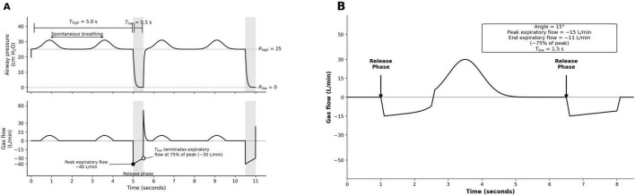 Figure 1: Ventilator flow waveforms contrasting normal respiratory mechanics with severe asthma during positive pressure ventilation. In panel (A), normal lung mechanics exhibit a brisk peak expiratory flow ($-40\text{ L/min}$) with a steep expiratory decay angle ($\sim 45^\circ$), allowing complete alveolar gas evacuation in a short expiratory phase ($T_e = 0.5\text{ s}$). In panel (B), severe airflow obstruction and dynamic hyperinflation result in a severely depressed peak expiratory flow ($-15\text{ L/min}$) and a flat, prolonged decay trajectory ($\sim 15^\circ$), illustrating the marked lengthening of the expiratory time constant ($\tau_E$) and persistent end-expiratory flow truncation (auto-PEEP). Adapted from Jain et al., Front Med (Lausanne) 2026;13:1901959. DOI: 10.3389/fmed.2026.1901959 (Open Access / CC BY 4.0).
Pathophysiological Cascade: Resistance, Time Constants, and Dynamic Hyperinflation
The fundamental biomechanical disturbance in acute severe pediatric asthma is severe expiratory flow limitation caused by the concentric narrowing and obstruction of medium and small conducting airways ($< 2\text{ mm}$ internal diameter).
   Allergic / Viral Trigger
              │
              ▼
   ┌─────────────────────────────────────────────────────────┐
   │  Airway Hyperreactivity & Type 2 Inflammatory Cascade    │
   │  • Smooth muscle bronchospasm                           │
   │  • Submucosal edema & microvascular leakage             │
   │  • Tenacious mucus hypersecretion & plug formation      │
   └─────────────────────────────────────────────────────────┘
              │
              ▼
   ┌─────────────────────────────────────────────────────────┐
   │  Marked Increase in Airway Resistance (Raw ∝ 1/r⁴)      │
   └─────────────────────────────────────────────────────────┘
              │
              ▼
   ┌─────────────────────────────────────────────────────────┐
   │  Prolonged Expiratory Time Constant (τE = Raw × Crs)     │
   │  • Inadequate expiratory time (Te < 3–5 × τE)           │
   │  • End-expiratory alveolar gas trapping                 │
   └─────────────────────────────────────────────────────────┘
              │
              ▼
   ┌─────────────────────────────────────────────────────────┐
   │  Dynamic Hyperinflation & Intrinsic PEEP (PEEPi)        │
   │  • Alveolar overdistension & static compliance loss     │
   │  • Massive intrathoracic pressure elevation             │
   └─────────────────────────────────────────────────────────┘
              │
              ▼
   ┌─────────────────────────────────────────────────────────┐
   │  Cardiopulmonary Collapse & Barotrauma Risk             │
   │  • Decreased venous return & right ventricular strain   │
   │  • Pneumothorax / Pneumomediastinum                     │
   └─────────────────────────────────────────────────────────┘
The Expiratory Time Constant and Airway Resistance Derivations
Under laminar flow conditions governed by the Hagen-Poiseuille relationship, resistance to gas flow is inversely proportional to the fourth power of the airway internal radius:
$$R_{aw} = \frac{8 \eta L}{\pi r^4}$$Where:
- $\eta$ = dynamic gas viscosity ($\text{Pa}\cdot\text{s}$)
- $L$ = conducting airway tube length ($\text{m}$)
- $r$ = internal airway radius ($\text{m}$)
In young children whose basal airway calibers are inherently narrow (e.g., $4\text{ mm}$ in a toddler vs. $8\text{ mm}$ in an adolescent), a $1\text{ mm}$ concentric reduction in luminal radius from mucosal edema and spasm increases airway resistance by a factor of $16$ ($1600\%$), compared to only a $3.16$-fold increase in an older child.
The rate of passive alveolar emptying is determined by the Expiratory Time Constant ($\tau_E$), which represents the mathematical product of total airway resistance and respiratory system compliance:
$$\tau_E = R_{aw} \times C_{rs}$$Where:
- $\tau_E$ = expiratory time constant ($\text{seconds}$)
- $R_{aw}$ = airway resistance ($\text{cmH}_2\text{O}\cdot\text{s/L}$)
- $C_{rs}$ = static respiratory system compliance ($\text{L/cmH}_2\text{O}$)
During passive expiration driven by elastic lung recoil, the volume of gas remaining in the alveoli at time $t$ after opening the expiratory valve follows an exponential decay:
$$V(t) = V_T \cdot e^{-t / \tau_E}$$| Time Interval Elapsed | Theoretical Alveolar Volume Emptied | Alveolar Volume Trapped |
|---|---|---|
| $1 \times \tau_E$ | $63.2\%$ | $36.8\%$ |
| $2 \times \tau_E$ | $86.5\%$ | $13.5\%$ |
| $3 \times \tau_E$ | $95.0\%$ | $5.0\%$ |
| $4 \times \tau_E$ | $98.2\%$ | $1.8\%$ |
| $5 \times \tau_E$ | $99.3\%$ | $0.7\%$ |
In healthy, intubated pediatric patients, normal $R_{aw}$ is $10\text{--}20\text{ cmH}_2\text{O}\cdot\text{s/L}$ and $C_{rs}$ is approximately $0.02\text{ L/cmH}_2\text{O}$ ($20\text{ mL/cmH}_2\text{O}$), yielding a baseline $\tau_E$ of $0.2\text{--}0.4\text{ seconds}$. A complete exhalation requiring $4\text{--}5 \times \tau_E$ is comfortably achieved within $1.0\text{--}1.5\text{ seconds}$.
In severe pediatric status asthmaticus, $R_{aw}$ commonly surges to $60\text{--}120+\text{ cmH}_2\text{O}\cdot\text{s/L}$. Even with preserved parenchyma, $\tau_E$ lengthens to $1.2\text{--}2.0+\text{ seconds}$. Consequently, full passive exhalation requires an expiratory time ($T_e$) of $5\text{ to }10\text{ seconds}$. If the ventilator rate is set too high (e.g., $25\text{ breaths/min}$ with $T_e = 1.6\text{ s}$), each subsequent breath is delivered before prior alveolar exhalation finishes, creating progressive gas accumulation termed dynamic hyperinflation (or "breath stacking").
  Flow (L/min)
    ^
 +40|        ┌─────┐                 ┌─────┐
    |        │     │                 │     │
    |________│     │_________________│     │_________  Inspiration (Ti)
   0|──────────────────────────────────────────────── Time
    |        \                       \
    |         \                       \
    |          \                       \
    |           \                       \
 -20|            \_________              \_________    Expiratory Flow Truncation
    |                      |                       |   (Auto-PEEP / Dynamic Hyperinflation)
    v                      ^                       ^
                 Next breath delivered        Next breath delivered
                 BEFORE flow reaches ZERO     BEFORE flow reaches ZERO
Hemodynamic Collapse Mechanisms and Intubation Hazards
The initiation of invasive positive pressure ventilation in severe pediatric status asthmaticus carries a high mortality risk due to catastrophic cardiopulmonary interactions.
flowchart TD
    A["Dynamic Hyperinflation (Massive Alveolar Gas Trapping)"] --> B["Severe Rise in Mean Intrathoracic Pressure (ITP)"]
    A --> C["Alveolar Overdistension (Zone 1 West Physiology)"]

    B --> D["Compression of Great Veins & Right Atrium"]
    D --> E["Severe Reduction in Systemic Venous Return (RV Preload)"]

    C --> F["Extrinsic Compression of Intra-Alveolar Pulmonary Capillaries"]
    F --> G["Acute Surge in Pulmonary Vascular Resistance (PVR)"]
    G --> H["Acute Right Ventricular Afterload & Dilation"]

    H --> I["Ventricular Interdependence: Leftward Septal Shift (Reversed Bernheim)"]
    E --> J["Decreased RV Stroke Volume"]

    I --> K["Impaired Left Ventricular Filling & Compliance"]
    J --> L["Reduced LV Preload & Stroke Volume"]

    K --> M["Precipitous Fall in Cardiac Output"]
    L --> M

    M --> N["Post-Intubation Circulatory Arrest / PEA"]
Pathophysiological Determinants of Shock in Ventilated Asthma
- Impaired Systemic Venous Return (Preload Failure): As trapped alveolar gas elevates mean intrathoracic pressure ($ITP$), the pressure gradient for venous return from the extrathoracic systemic veins ($\Delta P_{VR} = P_{msf} - P_{ra}$) is diminished.
- Right Ventricular Afterload Mismatch: Alveolar overdistension stretches the pulmonary microvasculature, converting vast areas of the lung into West Zone 1 condition ($P_{alveolar} > P_{arterial} > P_{venous}$). This elevates pulmonary vascular resistance ($PVR$), precipitating acute cor pulmonale.
- Ventricular Interdependence: RV dilation within the non-distensible pediatric pericardium shifts the interventricular septum leftward into the left ventricular cavity (reversed Bernheim effect), restricting LV diastolic filling, reducing stroke volume, and triggering profound hypotension.
- Blunted Sympathetic Tone from Induction: Patients with status asthmaticus survive pre-intubation through high endogenous catecholamine secretion. Pharmacologic induction agents (e.g., propofol, benzodiazepines) and neuromuscular blockers ablate this sympathetic compensation, causing immediate vascular collapse.
If a ventilated asthmatic child develops sudden pulseless electrical activity (PEA), bradycardia, or severe hypotension immediately following intubation:
- DISCONNECT THE VENTILATOR CIRCUIT AT THE ETT: Immediately break the connection to ambient air.
- MANUALLY DECOMPRESS THE THORAX: Apply bilateral, firm chest compressions to expel trapped air through the ETT until expiratory flow cessation is observed.
- ADMINISTER RAPID ISOTONIC CRYSTALLOID BOLUSES ($20\text{ mL/kg}$): Restore right ventricular preload against high intrathoracic pressures.
- RULE OUT TENSION PNEUMOTHORAX: Perform immediate bedside lung ultrasound or bilateral needle thoracostomy if chest decompression does not restore spontaneous circulation.
Mechanical Ventilation Strategies in Severe Pediatric Asthma
Mechanical ventilation in status asthmaticus does not heal bronchial inflammation; its sole goal is to maintain safe oxygenation and viable alveolar ventilation while buying time for systemic bronchodilators and anti-inflammatory therapies to restore airway caliber.
Controlled Hypoventilation and Permissive Hypercapnia
Attempting to normalize $PaCO_2$ in severe pediatric asthma requires high minute ventilation, which accelerates dynamic hyperinflation and leads to barotrauma. Modern ventilation relies on controlled hypoventilation with permissive hypercapnia.
$$\dot{V}_A = \frac{\dot{V}_{CO_2}}{PaCO_2} \cdot (1 - V_D / V_T)$$- Permissive Arterial Targets: Accept $PaCO_2$ between $60\text{--}90\text{ mmHg}$ (and occasionally $> 100\text{ mmHg}$ if tolerated), provided arterial $pH \ge 7.15\text{--}7.20$.
- Renal Bicarbonate Compensation: Over $24\text{--}48\text{ hours}$, renal retention of $HCO_3^-$ buffers respiratory acidosis. Routine infusion of sodium bicarbonate is contraindicated as it produces dissolved carbonic acid, increasing the $CO_2$ burden: $$HCO_3^- + H^+ \rightleftharpoons H_2CO_3 \rightleftharpoons H_2O + CO_2 \uparrow$$
- Absolute Contraindications to Permissive Hypercapnia:
- Traumatic brain injury, intracranial hypertension, or cerebral edema ($CO_2$-induced cerebral vasodilation increases ICP).
- Severe preexisting pulmonary hypertension or right-to-left intracardiac shunts (acidosis increases $PVR$).
- Uncompensated severe myocardial dysfunction.
Ventilator Parameters and Initial Initialization Matrix
| Ventilator Variable | Recommended PICU Target | Physiological Rationale & Safety Threshold |
|---|---|---|
| Ventilator Mode | VC-CMV (Constant Square Flow) | Ensures constant inspiratory flow to allow clean static plateau pressure ($P_{plat}$) and airway resistance measurements. Pressure Control (PC) is an acceptable alternative if $T_i$ is kept short. |
| Tidal Volume ($V_T$) | $6\text{--}8\text{ mL/kg}$ PBW | Avoids alveolar overdistension. Extremely low volumes ($< 5\text{ mL/kg}$) increase dead-space ratio ($V_D / V_T$); high volumes ($> 8\text{ mL/kg}$) worsen gas trapping. |
| Respiratory Rate ($f$) | $10\text{--}16\text{ breaths/min}$ (School-age/Adolescent) $14\text{--}20\text{ breaths/min}$ (Infant/Toddler) | Deliberately slow respiratory rate to maximize total cycle time ($T_{tot} = 60 / f$), reserving sufficient time for expiration. |
| Inspiratory Time ($T_i$) | $0.5\text{--}0.8\text{ seconds}$ | Short $T_i$ maximizes the time window remaining for passive exhalation ($T_e = T_{tot} - T_i$). |
| Inspiratory Flow ($\dot{V}_{insp}$) | $40\text{--}80\text{ L/min}$ (Square Waveform) | High peak inspiratory flow rapidly delivers the tidal volume, shortening $T_i$. |
| $I:E$ Ratio | $1:3\text{ to }1:5$ (or wider) | Ensures expiratory time ($T_e$) is at least $3\text{ to }5 \times \tau_E$ ($T_e \ge 3.5\text{--}5.0\text{ s}$). |
| Extrinsic PEEP ($PEEP_e$) | $0\text{--}5\text{ cmH}_2\text{O}$ (Passive / Paralyzed) $\le 80\% \times PEEP_i$ (Spontaneously Triggering) | In passive patients, applied PEEP adds directly to intrinsic PEEP, increasing total intrathoracic pressure. In triggering patients, low PEEP reduces triggering effort. |
| $FiO_2$ | Titrate to $SpO_2\ 90\text{--}94\%$ | Pure asthma is an obstructive ventilatory failure, not an alveolar diffusion defect. Severe refractory hypoxemia indicates atelectasis, pneumothorax, or pneumonia. |
Respiratory Mechanics Monitoring: Peak vs. Plateau Pressure
In volume-controlled ventilation with a constant square flow, total airway pressure is divided into resistive and elastic components via an end-inspiratory pause maneuver.
   Airway Pressure (Paw)
     ^
     |             /|
Ppeak|            / |  <── Resistive Pressure Drop: ΔPres = Raw · Flow
     |           /  |
     |          /   |................
Pplat|         /    :               | <── Elastic Recoil Pressure: ΔPel = VT / Crs
     |        /     :               |     (Maintained during zero flow hold)
     |       /      :               |
PEEP |______/_______:_______________|________
     0      <───────Ti──────────────>
            Constant Flow   Zero-Flow
                            Pause (0.5s)
Static vs. Dynamic Airway Pressure Formulas
- Resistive Pressure Drop ($\Delta P_{res}$): $$\Delta P_{res} = P_{peak} - P_{plat} = R_{aw} \times \dot{V}$$
- Elastic Driving Pressure ($\Delta P_{elastic}$): $$\Delta P = P_{plat} - PEEP_{tot} = \frac{V_T}{C_{rs}}$$
- Static Plateau Pressure Safety Limit: $$P_{plat} < 28\text{--}30\text{ cmH}_2\text{O}$$
In severe asthma, peak inspiratory pressure ($P_{peak}$) often reaches alarmingly high values ($50\text{--}70\text{ cmH}_2\text{O}$). This elevated pressure reflects the resistive dissipation required to force gas through the narrow endotracheal tube and bronchospastic central airways.
Because this pressure drops along the conducting tree, the delicate alveoli are protected from this peak, provided that $P_{plat} < 30\text{ cmH}_2\text{O}$.
- High $P_{peak}$ + Normal $P_{plat}$ ($\le 25\text{ cmH}_2\text{O}$): Severe airway resistance / bronchospasm. The lung is protected from barotrauma.
- High $P_{peak}$ + High $P_{plat}$ ($> 30\text{ cmH}_2\text{O}$): Severe dynamic hyperinflation (massive auto-PEEP) or acute pneumothorax. Immediate reduction in respiratory rate or chest decompression is required.
Quantifying Intrinsic PEEP ($PEEP_i$) at the Bedside
Intrinsic PEEP ($PEEP_i$) is the positive alveolar pressure present at the end of expiration resulting from incomplete alveolar emptying:
$$PEEP_i = P_{exp,hold} - PEEP_{extrinsic}$$ Paw (cmH2O)
^
20| ┌───┐
| │ │
10| │ │
| │ │ ┌───────────────────────┐
| │ │ │ Alveolar Equilibration│
5|───────────────────┘ └───────────────┘ (End-Expiratory │ = Total PEEP (PEEPtot)
| Hold Maneuver) │
0| └───────────────────────
+───────────────────────────────────────────────────────────────────────────────────────>
<─────── Exp Hold ──────>
To measure $PEEP_i$ accurately:
- Ensure the patient is passive (sedated $\pm$ paralyzed) to avoid active expiratory muscle recruitment.
- Perform an end-expiratory occlusion maneuver for $1.5\text{--}3.0\text{ seconds}$.
- As the expiratory valve remains closed, trapped alveolar gas equilibrates throughout the ventilator circuit, revealing the total PEEP ($PEEP_{tot}$).
The Low PEEP Dilemma: Setting Extrinsic PEEP in Asthma
The application of extrinsic PEEP ($PEEP_e$) in status asthmaticus is guided by the patient's respiratory drive and underlying airway collapse mechanics:
flowchart TD
    A["Patient with Severe Status Asthmaticus"] --> B{"Patient Condition"}

    B -->|"Passive / Paralyzed / Deeply Sedated"| C["No Active Triggering Efforts"]
    C --> D["Set PEEP_e = 0 to 3 cmH2O (ZEEP / Low PEEP)"]
    D --> E["Avoid Cascade Hyperinflation: Any added PEEP_e directly adds to PEEP_i"]

    B -->|"Spontaneously Breathing / Triggering"| F["Dynamic Airway Collapse & High WOB"]
    F --> G["Measure Intrinsic PEEP (PEEP_i)"]
    G --> H["Apply PEEP_e = 60% to 80% of PEEPi (Water-Fall Effect)"]
    H --> I["Reduces Inspiratory Trigger Threshold & Alleviates Ineffective Efforts"]
The Waterfall Effect in Spontaneous Breathing
In spontaneously triggering patients, dynamic small-airway compression acts as a mechanical "waterfall." To trigger a ventilator breath, the patient's diaphragm must generate enough negative pleural pressure to overcome the intrinsic positive alveolar pressure:
$$\Delta P_{trigger} = PEEP_i + \text{Ventilator Trigger Sensitivity}$$By setting $PEEP_e$ to approximately $60\text{--}80\%$ of $PEEP_i$ (without exceeding $PEEP_i$), the clinician elevates the downstream circuit baseline without increasing upstream alveolar volume, reducing trigger work of breathing and resolving ineffective triggering efforts. In a fully paralyzed patient, this waterfall benefit does not apply; added $PEEP_e$ simply increases total intrathoracic pressure.
Pharmacological Titration and Rescue Adjuncts
Medical management must be escalated alongside mechanical support to resolve the underlying bronchoconstriction.
flowchart TD
    A["Invasive Mechanical Ventilation Established"] --> B["First-Line Continuous Pharmacotherapy"]
    B --> B1["Continuous Nebulized Albuterol (0.5 mg/kg/h)"]
    B --> B2["IV Methylprednisolone (1-2 mg/kg q6h)"]
    B --> B3["IV Magnesium Sulfate (50-75 mg/kg over 30-60 min)"]

    B --> C{"Refractory Severe Bronchospasm & High Pplat?"}

    C -->|"Yes"| D["Second-Line Systemic Infusions"]
    D --> D1["IV Terbutaline Infusion (0.1-10 mcg/kg/min)"]
    D --> D2["IV Epinephrine Infusion (0.05-0.5 mcg/kg/min)"]
    D --> D3["IV Ketamine Infusion (1-3 mg/kg/h)"]

    D --> E{"Persistent Severe Acidosis (pH < 7.10) / Barotrauma?"}

    E -->|"Yes"| F["Third-Line & Rescue Modalities"]
    F --> F1["Inhaled Volatile Anesthetics (Isoflurane / Sevoflurane via AnaConDa)"]
    F --> F2["Heliox (70:30 or 80:20 Mixture)"]
    F --> F3["Veno-Venous Extracorporeal Membrane Oxygenation (VV-ECMO)"]
Systemic and Inhaled Pharmacotherapy Matrix
| Therapeutic Agent | PICU Dosage & Administration | Mechanism of Action | Monitoring & Toxicity Concerns |
|---|---|---|---|
| Albuterol (Salbutamol) | Continuous nebulization: $0.5\text{ mg/kg/h}$ (max $20\text{ mg/h}$) via vibrating mesh nebulizer inline | $\beta_2$-adrenoreceptor agonism $\rightarrow$ adenylyl cyclase activation $\rightarrow$ intracellular cAMP $\uparrow \rightarrow$ smooth muscle relaxation | Tachycardia, hypokalemia, tremor, type B lactic acidosis (elevated lactate from $\beta_2$ glycolysis stimulation). |
| Methylprednisolone | $1\text{--}2\text{ mg/kg IV}$ loading dose, then $1\text{ mg/kg IV q6h}$ | Glucocorticoid receptor binding $\rightarrow$ inhibition of inflammatory transcription factors (NF-$\kappa$B), upregulation of $\beta_2$-receptor density | Hyperglycemia, acute steroid myopathy (especially when co-administered with prolonged neuromuscular blockade). |
| Magnesium Sulfate | $50\text{--}75\text{ mg/kg IV}$ (max $2.5\text{ g}$) infused over $30\text{--}60\text{ min}$, followed by continuous infusion at $20\text{--}40\text{ mg/kg/h}$ | Voltage-gated calcium channel blockade in airway smooth muscle; inhibits acetylcholine release at motor endplates | Hypotension, flushing, loss of deep tendon reflexes, respiratory depression at serum $Mg^{2+} > 3.5\text{ mmol/L}$ ($> 8.5\text{ mg/dL}$). |
| Terbutaline | Loading dose: $2\text{--}10\text{ mcg/kg IV}$ over $10\text{ min}$, then continuous infusion $0.1\text{--}10.0\text{ mcg/kg/min}$ | Systemic $\beta_2$-agonist delivery to peripheral occluded airways inaccessible by inhaled aerosols | Tachyarrhythmias, myocardial ischemia (monitor troponin/ECG), severe hypokalemia, hypotension from peripheral vasodilation. |
| Ketamine | Loading dose: $1\text{--}2\text{ mg/kg IV}$, followed by continuous infusion $1\text{--}3\text{ mg/kg/h}$ | Non-competitive NMDA receptor antagonist; blocks catecholamine reuptake $\rightarrow$ endogenous catecholamine-mediated bronchodilation | Hypertension, hypersalivation (mitigate with glycopyrrolate), emergence delirium, bronchorrhea. |
| Epinephrine (Adrenaline) | Continuous infusion: $0.05\text{--}0.5\text{ mcg/kg/min IV}$ | Mixed $\alpha_1, \beta_1, \beta_2$ agonist. $\alpha_1$ vasoconstriction reduces mucosal edema; $\beta_2$ relaxes smooth muscle | Hypertension, tachycardia, ventricular arrhythmias, severe metabolic lactic acidosis. |
Advanced Rescue Modalities in Refractory Status Asthmaticus
1. Volatile Inhalational Anesthetics (Isoflurane and Sevoflurane)
Volatile halogenated anesthetics exert potent, direct, dose-dependent relaxation of airway smooth muscle via multiple pathways:
- Inhibition of voltage-dependent L-type $Ca^{2+}$ channels in bronchial smooth muscle.
- Depletion of sarcoplasmic reticulum intracellular calcium stores via IP3 pathway inhibition.
- Direct attenuation of vagally mediated airway reflexes.
Delivery in the PICU is achieved using dedicated miniature anesthetic conserving devices (e.g., AnaConDa, Sedaconda) placed in the inspiratory limb of standard intensive care ventilators.
- Titration: Titrated to an end-tidal volatile concentration of $0.5\text{--}1.5\text{ MAC}$.
- Safety Monitoring: Scavenging systems are mandatory to prevent PICU ambient contamination. Clinicians must monitor for peripheral vasodilation-induced hypotension, nephrotoxicity (fluoride ion accumulation with prolonged sevoflurane), and malignant hyperthermia.
2. Helium-Oxygen Gas Mixtures (Heliox)
Heliox is a medical gas mixture composed of helium and oxygen (typically formulated as $80:20$ or $70:30\ \text{He:O}_2$).
Fluid flow transitions from smooth laminar streamlines to turbulent eddy currents when the dimensionless Reynolds Number ($Re$) exceeds a critical threshold ($Re > 2000\text{--}4000$):
$$Re = \frac{\rho \cdot v \cdot D}{\eta}$$Where:
- $\rho$ = gas density ($\text{kg/m}^3$)
- $v$ = mean linear flow velocity ($\text{m/s}$)
- $D$ = airway diameter ($\text{m}$)
- $\eta$ = dynamic gas viscosity ($\text{Pa}\cdot\text{s}$)
Helium is an inert noble gas with a density approximately one-seventh that of nitrogen ($\rho_{He} = 0.179\text{ kg/m}^3$ vs. $\rho_{N_2} = 1.25\text{ kg/m}^3$). Replacing nitrogen with helium reduces total gas mixture density by up to $66\%$, lowering $Re$ below critical thresholds in narrowed airways:
- Converts turbulent flow regimes into smooth laminar flow, reducing resistive pressure drop ($\Delta P = K_2 \rho \dot{V}^2$).
- Improves peripheral deposition of nebulized bronchodilator aerosols into distal obstructed lung segments.
Clinical Limitation: Heliox requires $FiO_2 \le 0.30\text{--}0.40$ to retain its low-density physical advantages; high oxygen requirements render Heliox ineffective.
3. Veno-Venous Extracorporeal Membrane Oxygenation (VV-ECMO)
When severe dynamic hyperinflation, unmanageable plateau pressures ($P_{plat} > 32\text{--}35\text{ cmH}_2\text{O}$), intractable respiratory acidosis ($pH < 7.00$), or tension pneumothorax prevent safe mechanical ventilation, VV-ECMO serves as a bridge to recovery.
- Cannulation Strategy: Single dual-lumen bicaval cannula (e.g., Avalon) in the right internal jugular vein or two-site femoral-jugular cannulation.
- Ultra-Protective "Rest" Ventilator Settings: Once the extracorporeal circuit takes over carbon dioxide clearance, the mechanical ventilator is transitioned to lung-rest parameters:
- Respiratory rate: $4\text{--}8\text{ breaths/min}$
- Tidal volume: $2\text{--}4\text{ mL/kg}$ PBW
- Peak inspiratory pressure: $< 20\text{--}25\text{ cmH}_2\text{O}$
- $PEEP_e$: $0\text{--}5\text{ cmH}_2\text{O}$
- $FiO_2$: $0.30\text{--}0.40$
Bedside Troubleshooting and Acute Complications
                 SUDDEN CLINICAL DETERIORATION IN VENTILATED ASTHMATIC
                                         │
                                         ▼
                           DISCONNECT FROM VENTILATOR
                           (Manual Chest Compression)
                                         │
                    ┌────────────────────┴────────────────────┐
                    ▼                                         ▼
         HEMODYNAMICS RESTORED                       HYPOTENSION PERSISTS
         (Dynamic Air-Trapping)                               │
                    │                                         ▼
    • Prolong Expiratory Time (↓ Rate)               PERFORM IMMEDIATE POCUS
    • Decrease Tidal Volume (VT)                     / CLINICAL EVALUATION
    • Suction Mucus Plugs                                     │
                                        ┌─────────────────────┼─────────────────────┐
                                        ▼                     ▼                     ▼
                               TENSION PNEUMOTHORAX     ETT OBSTRUCTION       MASSIVE ATELECTASIS
                               • Absent lung sliding    • Unable to pass       • Unilateral whiteout
                               • Tracheal deviation       suction catheter     • Volume loss
                                        │                     │                     │
                                        ▼                     ▼                     ▼
                               Needle Decompression   Extubate / Re-intubate  Therapeutic
                               & Chest Tube Insertion  over bougie             Bronchoscopy
The "DOPES" Algorithm Adapted for Status Asthmaticus
- D - Dislodgement: Accidental extubation or right mainstem endobronchial intubation. In small pediatric airways, selective right mainstem intubation leads to unilateral hyperinflation, tension pneumothorax, and left lung atelectasis. Verify symmetric breath sounds and depth markings.
- O - Obstruction: Complete lumen occlusion from inspissated mucus plugs or kinking of the flexible endotracheal tube. Attempt to pass a soft inline suction catheter immediately; if obstructed, immediately exchange the ETT over an airway exchange catheter.
- P - Pneumothorax: Ruptured subpleural blebs from excessive alveolar distending pressure produce tension pneumothorax. Perform immediate ultrasound-guided needle decompression followed by tube thoracostomy.
- E - Equipment Failure: Ventilator valve malfunction or closed expiratory port. Disconnect the patient and bag manually using a dedicated bag-valve-mask circuit with a manometer.
- S - Stacking (Breath Stacking / Dynamic Hyperinflation): The primary mechanism of failure. Relieved by disconnecting the circuit and allowing prolonged passive exhalation with manual thoracic compression.
Liberation and Extubation Protocol
Weaning the pediatric patient with status asthmaticus from invasive mechanical ventilation begins as soon as airway resistance improves and dynamic hyperinflation resolves.
Extubation Readiness Criteria
- Resolution of Dynamic Obstruction: Expiratory flow scalar smoothly returns to the zero-flow baseline well before the initiation of the subsequent breath.
- Normalized Mechanics:
- Static plateau pressure: $P_{plat} < 20\text{ cmH}_2\text{O}$
- Resistive gradient: $\Delta P_{res} = P_{peak} - P_{plat} < 10\text{--}15\text{ cmH}_2\text{O}$ at standard flow rates
- Measured intrinsic PEEP: $PEEP_i < 3\text{--}5\text{ cmH}_2\text{O}$
- Blood Gas Normalization: Stable spontaneous acid-base balance with $pH > 7.30$ and $PaCO_2 < 45\text{--}50\text{ mmHg}$ on minimal ventilatory support.
- Cuff Leak Assessment: Presence of an audible peritubal air leak upon cuff deflation at $Paw \le 20\text{--}25\text{ cmH}_2\text{O}$, confirming absence of severe subglottic or laryngeal edema.
- Neurological Competence: Patient is awake, exhibits strong protective airway reflexes (cough and gag), and maintains adequate spontaneous inspiratory efforts.
Post-Extubation Management Strategy
- Prophylactic High-Flow Nasal Cannula (HFNC) or Bi-Level PAP: Immediate post-extubation application of heated humidified HFNC ($1.0\text{--}1.5\text{ L/kg/min}$) or non-invasive positive pressure ventilation (IPAP $10\text{--}14\text{ cmH}_2\text{O}$, EPAP $4\text{--}5\text{ cmH}_2\text{O}$) prevents early muscle fatigue.
- Continued Inhaled Aerosol Delivery: Maintain scheduled vibrating-mesh or jet-nebulized bronchodilators with systemic corticosteroids.
Key Takeaways
- Controlled Hypoventilation is Mandatory: The priority of mechanical ventilation in status asthmaticus is preventing alveolar overdistension and barotrauma. Accept permissive hypercapnia ($PaCO_2\ 60\text{--}90\text{ mmHg}$, $pH \ge 7.15\text{--}7.20$) rather than pursuing normocapnia.
- Expiratory Time is King: Set slow respiratory rates ($10\text{--}16\text{ bpm}$) and high inspiratory flows ($40\text{--}80\text{ L/min}$) to achieve prolonged expiratory times ($T_e \ge 3.5\text{--}5.0\text{ s}$, $I:E\ 1:3\text{ to }1:5$).
- Plateau Pressure Protects Alveoli: High peak inspiratory pressures reflect conducting airway resistance. Maintain static plateau pressure $P_{plat} < 30\text{ cmH}_2\text{O}$ to protect the alveolar compartment.
- Immediate Action for Acute Collapse: If acute hemodynamic collapse occurs following intubation, disconnect the ventilator immediately and perform manual chest decompression to exhaust trapped alveolar gas.
- Escalate Medical Therapies: Utilize continuous bronchodilators, systemic corticosteroids, IV magnesium, and continuous IV $\beta_2$-agonists/ketamine. For refractory disease, consider volatile anesthetics, Heliox, and VV-ECMO.