This closing chapter synthesises the seventeen that precede it. The earlier notes describe neonatal respiratory physiology and mechanical strategy in isolation; here the work is retrieval — a deteriorating infant, an incomplete history, and a clock that is already running.
Each vignette gives a short stem, poses the bedside decision, then answers it with a rationale and a pointer to the parent chapter. The Spotter Gallery distils five recurring patterns from neonatal ventilator graphics, blood gases and radiographs, and the closing table maps every case to its diagnosis, its principle and its parent chapter.
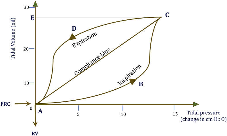 Figure 1: The pressure–volume loop is the bedside signature of compliance and of the work of breathing; the lower inflection marks recruitment, the upper inflection overdistension, so the safest tidal breath sits on the linear compliance line between them. (Source: Chakkarapani AA, Adappa R, Mohammad Ali SK, Gupta S, Soni NB, Chicoine L, Hummler HD. International Journal of Pediatrics & Adolescent Medicine 2020. DOI: 10.1016/j.ijpam.2020.03.003).
Case 1: Progression on CPAP — Early Rescue Surfactant or Patience
A 27+2-week, 980 g infant on bubble CPAP 7 cmH2O has risen from FiO2 0.21 to 0.35 over four hours, with mild recession, a lung-ultrasound score of 10 and a gas of pH 7.28, PaCO2 52 mmHg, PaO2 58 mmHg.
Question: Give early rescue surfactant by LISA/MIST, or continue CPAP and reassess?
Give surfactant by LISA/MIST with CPAP maintained. Below 28 weeks the trigger is a rising FiO2 at or above 0.30 on CPAP of at least 6 cmH2O, and a high lung-ultrasound score predicts CPAP failure. Delay permits atelectrauma, air leak and the inflammatory priming of bronchopulmonary dysplasia, and thin-catheter administration avoids the intubation the technique was designed to prevent (Chapter 03), while waiting allows the injury Chapter 02 describes.
A rapidly rising oxygen requirement in a very immature infant is the signal; a 27-week lung tolerates delay far less well than a 30-week one.
Case 2: Volume Guarantee, a Large Leak and an Unexpected Hypocarbia
A 28-week infant on day 3 is on pressure-controlled volume guarantee with a set tidal volume of 5 mL/kg. PIP has climbed to the ceiling of 26 cmH2O, the measured leak is 55%, and the gas shows pH 7.48 and PaCO2 28 mmHg. The team proposes raising the ceiling.
where $V_i$ and $V_e$ are inspired and expired tidal volume (mL).
Question: What is the mechanism of the hypocarbia, and should the ceiling be raised?
No. With an uncuffed tube, a leak above 40–50% makes the expired signal unreliable, so the controller raises PIP to chase volume it cannot measure; because the leak is flow-dependent, the higher pressure enlarges it, producing a spiral of rising pressure with unchanged measured volume. The inspired volume actually delivered is over-generous, and since $P_{aCO_2} \propto 1/\dot V_A$, hypocarbia follows. Treat the leak: reposition or upsize the tube, reduce neck extension, suction, drain or replace the flow sensor, and lower the set tidal volume towards 4 mL/kg with a reduced rate.
A PaCO2 below 35 mmHg, and especially below 30 mmHg, causes cerebral vasoconstriction and is an antecedent of periventricular leukomalacia and bronchopulmonary dysplasia. A "volume not reached" alarm at the ceiling is therefore often a safety success (Chapter 04).
Case 3: PPHN with a Raised Oxygenation Index
A term infant with meconium aspiration has a mean airway pressure of 16 cmH2O, FiO2 1.0 and PaO2 48 mmHg, giving an oxygenation index near 33. There is a pre- to post-ductal gradient and echocardiographic pulmonary hypertension with right-to-left ductal shunting; lung volume, blood pressure and sedation are optimised.
where MAP is mean airway pressure (cmH2O), FiO2 the inspired oxygen fraction and PaO2 the arterial oxygen tension (mmHg). An OI above 15–20 prompts consideration of vasodilators, above about 25 justifies escalation, and a sustained $\geq 40$ over four hours is the classic ECMO trigger.
Question: When and how do you start and wean inhaled nitric oxide?
Start inhaled nitric oxide at 20 ppm, the dose validated in the pivotal trials; the trigger is an OI above about 25 despite a recruited lung and an adequate systemic pressure head, which this infant meets. Reassess vasoreactivity after 30–60 minutes: a PaO2 rise of at least 20 mmHg, or an OI fall of at least 20%, identifies a responder. Wean FiO2 before the drug, in 1 ppm steps every 1–4 hours. Measure methaemoglobin 4–8 hours after starting and daily, and keep nitrogen dioxide below about 3 ppm (Chapter 06).
Rebound pulmonary hypertension is most likely at low doses and in the first hours after withdrawal. Restart at the last effective dose and add a phosphodiesterase-5 inhibitor before attempting withdrawal again.
Case 4: Meconium Aspiration with Air Leak
A term infant with meconium aspiration needs PIP 30 cmH2O. The radiograph shows hyperinflation with reticular and cystic lucencies; minutes later the infant desaturates and bradycardiates, the left chest is hyperlucent and transilluminates brightly.
Question: What ventilation changes are required, and in what order?
The final event is a tension pneumothorax: needle decompression immediately, without waiting for a radiograph, then a chest drain. The preceding picture is pulmonary interstitial emphysema, the signal to reduce airway pressures. Then ventilate gently — lower PIP and MAP, shorten inspiratory time, lengthen expiratory time to avoid trapping, keep PEEP at 5–7 cmH2O to stent obstructed airways, and accept permissive hypercapnia. When PIP exceeds 25–28 cmH2O, HFOV is a rational rescue (Chapter 07).
Each escalation raises transpulmonary pressure and the risk of further air leak. Ask what else the rising pressure is treating.
Case 5: Congenital Diaphragmatic Hernia after Face-Mask Ventilation
An infant with an antenatally diagnosed left CDH received three minutes of face-mask ventilation in the delivery room. The abdomen is distended, breath sounds are absent on the left, saturations are 70% with bradycardia, and ventilation is difficult.
Question: What are your immediate priorities?
Stop mask ventilation and intubate early, ideally with a T-piece resuscitator. Place a large-bore 8–10 Fr orogastric tube on continuous low suction. Use a pre-ductal oximeter and target SpO2 85–95%, avoiding hyperoxia. Ventilate gently: PIP at or below about 25 cmH2O, PEEP 3–5 cmH2O, tidal volumes 4–6 mL/kg, accepting PaCO2 45–65 mmHg and pH of at least about 7.20. The defect is not the emergency; the physiology is (Chapter 10).
Mask pressure inflates the stomach and herniated bowel, worsening lung compression and impeding venous return. Suspect a contralateral pneumothorax in any abrupt deterioration.
Case 6: Apnoea of Prematurity Blocking Extubation
A 25+4-week, 750 g infant ventilated for respiratory distress syndrome is on PIP 16 cmH2O, PEEP 5 cmH2O, rate 20/min and FiO2 0.25, but has had frequent apnoeas with bradycardia and desaturation over 24 hours. Caffeine is established and haemoglobin is 95 g/L.
Question: Is the infant ready for extubation, and onto what support?
Not yet: a high pre-extubation apnoea burden is among the strongest predictors of extubation failure. Correct the reversible factors first — transfuse the anaemia, treat a significant ductus and sepsis, confirm nutrition, and continue caffeine without interruption. Perform a spontaneous breathing trial and a sedation hold. Extubate high-risk ELBW infants onto synchronised nasal IPPV with CPAP of at least 6 cmH2O rather than high-flow nasal cannula, whose pressure is unmeasured, and consider non-invasive NAVA if synchrony remains limiting. Reintubate promptly if apnoea or acidosis is refractory (Chapter 12).
Prolonged stimulation and recurrent desaturation restart lung injury and worsen outcomes.
Case 7: Bronchopulmonary Dysplasia at 36 Weeks
An infant born at 24+1 weeks remains invasively ventilated at 36 weeks' postmenstrual age with FiO2 0.35, heterogeneous time constants and a large dead space, on the volume-targeted settings chosen at birth.
Question: What ventilatory strategy should now be used?
Established BPD is not the stiff, surfactant-deficient lung of RDS. Shift to larger tidal volumes of 6–8.5 mL/kg, slower rates of 15–25 breaths/min, longer expiratory times and higher PEEP of 7–10 cmH2O to stent collapsible airways, while avoiding overdistension of a simplified lung. Screen for BPD-associated pulmonary hypertension, the strongest predictor of death, by echocardiography; treat persistent hypertension with sildenafil and reserve inhaled nitric oxide for acute deterioration. Use diuretics selectively, attend to nutrition, and consider a low-dose DART-style corticosteroid only in ventilator-dependent infants with the trade-off documented (Chapter 11).
Case 8: Tracheoesophageal Fistula with Gastric Distension
A term infant with type C oesophageal atresia and a distal tracheoesophageal fistula is ventilated; over an hour PIP has risen for an unchanged tidal volume, carbon dioxide clearance is poor, and the abdomen is distended and tympanitic.
Question: What is the mechanism, and what is the order of management?
This is fistula steal. The trachea divides each breath between lung and fistula in parallel, and because resistance falls with the fourth power of radius, a 1–2 mm fistula commands much of each breath; the distended stomach then splints the diaphragm and the spiral reinforces itself. The order is fixed: first position the tube tip distal to the fistula but above the carina, removing the communication from the circuit; second, lower the driving pressure, keeping PIP at about 20–25 cmH2O with a short inspiratory time and preserved spontaneous breathing; third, decompress the stomach with a large-bore tube or gastrostomy on open drainage. Titrate PEEP cautiously at 4–5 cmH2O (Chapter 14).
A tense, enlarging abdomen with hypotension and falling saturations means gas in the peritoneum: stop positive-pressure ventilation where possible, decompress the stomach, and call for surgery.
Case 9: Therapeutic Hypothermia with Settings Set for 37 °C
A term asphyxiated infant is undergoing hypothermia at 33.5 °C. Ventilator settings are unchanged since admission; the gas shows pH 7.52 and PaCO2 27 mmHg, and cerebral oximetry is low.
Question: What has gone wrong, and how do you correct it?
Hypothermia lowers carbon dioxide production by roughly 6–7% per degree Celsius, so settings appropriate at 37.0 °C over-ventilate at 33.5 °C. Cerebral blood flow changes by roughly 3–4% per mmHg, approximated by $CBF \approx CBF_{0}\left[1 + k\left(PaCO_2 - 40\right)\right]$ with $k \approx 0.03\text{–}0.04$ per mmHg; a fall from 40 to 30 mmHg therefore cuts cerebral blood flow by about 30%, at the moment the injured brain is most vulnerable to ischaemia. Reduce the rate and tidal volume to target PaCO2 35–45 mmHg. Interpret gases uncorrected (alpha-stat), target pre-ductal SpO2 94–98%, and never hyperventilate for pulmonary hypertension (Chapter 15).
Hypocapnia worsens neurodevelopmental outcome. Treat pulmonary hypertension pharmacologically and defend systemic blood pressure instead.
Case 10: Pulmonary Haemorrhage on the Ventilator
A 26-week infant on day 3 with a haemodynamically significant patent ductus arteriosus and recent surfactant develops sudden blood-stained, frothy endotracheal secretions, desaturation, pallor and a rising FiO2 requirement.
Question: What are the immediate measures, including PEEP and ductal closure?
This is pulmonary haemorrhage from rupture of the immature airspace–capillary barrier. Secure the airway with gentle suction, use FiO2 1.0 and raise PEEP or MAP by 2–3 cmH2O to 8–12 cmH2O to tamponade capillaries without overdistension. Correct coagulopathy deliberately: platelets below $50 \times 10^{9}/\text{L}$ with active bleeding, fresh frozen plasma for a prolonged prothrombin or partial thromboplastin time, vitamin K, fibrinogen above 1.0 g/L, and haemoglobin above 120 g/L. Obtain targeted echocardiography, then close a significant ductus with ibuprofen, indomethacin or paracetamol once stabilised. Give post-haemorrhage surfactant for secondary inactivation, avoid fluid boluses, and minimise handling (Chapter 16).
In any high-risk preterm infant with respiratory distress syndrome, a significant ductus, growth restriction or recent surfactant, treat a sudden unexplained deterioration as pulmonary haemorrhage until proven otherwise.
Spotter Gallery
Each spotter compresses a longer reasoning process into one observation and one action.
Spotter 1: The pressure–volume loop
An inspiratory limb that flattens or "beaks" at the top, or a lower inflection with a concave initial segment, marks the limits of safe ventilation: the upper inflection signals overdistension of the most compliant units, while the lower inflection marks the pressure at which collapsed units recruit. Reduce tidal volume or PIP and reassess the driving pressure $\Delta P = P_{plat} - PEEP$; raise PEEP if a lower inflection is present (Figure 1).
Spotter 2: The capnography waveform
An end-tidal carbon dioxide tracing falling abruptly to near zero with desaturation means the airway, not the lung, has failed: tube dislodgement, obstruction, or massive air leak. Verify tube position and patency, transilluminate, and decompress a tension pneumothorax before adjusting the ventilator.
Spotter 3: The blood-gas pattern of over-delivery
pH 7.48, PaCO2 28 mmHg and a rising PaO2 on volume guarantee with a large uncuffed-tube leak is a controller fooled, not a lung improved; the over-generous inspired volume risks periventricular leukomalacia and bronchopulmonary dysplasia. Reduce the set tidal volume and rate, inspect the tube and sensor, and never raise the ceiling.
Spotter 4: The hyperlucent hemithorax
A hyperlucent hemithorax with absent lung markings, mediastinal shift and a distended abdomen, with sudden desaturation and bradycardia, is a tension pneumothorax. Perform immediate needle decompression, then a chest drain — imaging must never delay it.
Spotter 5: The pre- to post-ductal gradient
A pre-ductal (right hand) to post-ductal (left foot) saturation gradient above roughly 10–15%, with labile hypoxaemia disproportionate to the radiographic disease, indicates right-to-left ductal shunting from PPHN. Confirm by echocardiography, optimise lung volume and blood pressure, and start inhaled nitric oxide when the oxygenation index exceeds about 25.
flowchart TD
    A["Ventilated neonate deteriorates abruptly"] --> B{"Symmetrical chest wall movement?"}
    B -->|"No"| C{"Bright transillumination glow?"}
    C -->|"Yes"| D["Tension pneumothorax: immediate needle decompression"]
    C -->|"No"| E["Check ETT depth, patency and leak"]
    B -->|"Yes"| F{"Pre- to post-ductal SpO2 gradient above 10 percent?"}
    F -->|"Yes"| G["PPHN: optimise recruitment and blood pressure, then iNO"]
    F -->|"No"| H["Rising PIP at fixed Vt: obstruction, leak or falling compliance"]
Summary Table
| Vignette | Diagnosis | Key ventilator principle | Parent chapter |
|---|---|---|---|
| 1 | Preterm RDS progressing on CPAP | Early rescue surfactant by LISA/MIST at the gestational-age FiO2 threshold | Chapter 02, Chapter 03 |
| 2 | Volume guarantee with a large uncuffed-tube leak | A leak above 40–50% makes the expired signal unreliable; treat the leak, not the ceiling | Chapter 04 |
| 3 | PPHN with an oxygenation index above 25 | Optimise lung volume and systemic pressure, then iNO 20 ppm; wean in 1 ppm steps | Chapter 06 |
| 4 | Meconium aspiration with tension pneumothorax and PIE | Decompress before imaging; lower PIP and MAP, lengthen expiratory time | Chapter 07 |
| 5 | Congenital diaphragmatic hernia after face-mask ventilation | No mask ventilation; early intubation, gastric decompression, gentle ventilation | Chapter 10 |
| 6 | Apnoea of prematurity preventing extubation in an ELBW infant | Correct reversible factors, then extubate onto NIPPV with CPAP at least 6 cmH2O | Chapter 12 |
| 7 | Bronchopulmonary dysplasia at 36 weeks | Larger tidal volumes, slower rates, higher PEEP; screen and treat BPD-PH | Chapter 11 |
| 8 | Tracheoesophageal fistula with gastric distension | Exclude the fistula by tube position, reduce driving pressure, decompress the stomach | Chapter 14 |
| 9 | Hypoxic-ischaemic encephalopathy during hypothermia | Cooling lowers carbon dioxide production; reduce minute ventilation for normocapnia | Chapter 15 |
| 10 | Pulmonary haemorrhage with a significant ductus | Tamponade with PEEP 8–12 cmH2O, correct coagulopathy, then close the duct | Chapter 16 |
Key Takeaways
- The ventilator is a diagnostic instrument: a rising pressure, falling volume or unexplained gas localises the fault to the tube, the lung or the circulation.
- Escalating oxygen requirement on effective CPAP in preterm RDS indicates early rescue surfactant by LISA/MIST, not patience.
- Under volume guarantee the target is a ceiling delivered within a strict pressure limit; a large uncuffed-tube leak makes the expired signal unreliable and can generate hypocarbia.
- Air leak is treated by reducing transpulmonary pressure; tension pneumothorax by immediate needle decompression before imaging.
- Gastric distension links congenital diaphragmatic hernia and tracheoesophageal fistula; in both, decompression and low driving pressures precede escalation.
- Apnoea of prematurity is a control failure, not a parenchymal one; caffeine, correction of reversible factors and the right interface make extubation succeed.
- Established bronchopulmonary dysplasia needs a different paradigm from RDS, and its pulmonary hypertension should be sought actively as the strongest predictor of death.
- Therapeutic hypothermia lowers carbon dioxide production, so unchanged settings produce hypocapnia; target normocapnia and never hyperventilate the asphyxiated newborn.
- Pulmonary haemorrhage is time-critical: tamponading mean airway pressure, haemostatic correction and echocardiography-guided ductal management proceed in parallel.