Neonatal pulmonary haemorrhage (PH) is the acute extravasation of blood from the pulmonary microcirculation into the alveolar spaces and interstitium, recognised as frank blood or heavily blood-stained, often frothy secretions aspirated from the endotracheal tube (ETT). In an infant supported non-invasively it may announce itself less clearly — as blood in the posterior pharynx or gastric aspirate, or as an abrupt, unexplained escalation in oxygen requirement. PH is not a single disease but a final common pathway: a competent yet immature capillary bed is exposed to a haemodynamic, inflammatory and mechanical load it cannot withstand, the airspace–capillary barrier fails, and the alveoli fill with haemorrhagic pulmonary oedema. It is typically acute and occurs most often between 48 and 96 hours of life, during the transitional period when pulmonary vascular resistance (PVR) is falling, ductal shunting is most dynamic and surfactant has recently been given.
Three failures converge. Gas exchange collapses as alveoli flood with blood, which also inactivates surfactant and worsens compliance; circulating volume and oxygen-carrying capacity fall, producing hypovolaemic shock in the severest cases; and the same haemodynamic instability threatens the brain, so severe intraventricular haemorrhage (IVH) frequently coexists. Management is therefore three simultaneous tasks — secure the airway without provoking further bleeding, tamponade the capillaries and restore haemostasis, and correct the underlying haemodynamic and coagulation derangement — executed with minimal handling and deliberate restraint.
Definition, Recognition and the Diagnostic Threshold
The defining event and its timing
The hallmark is the sudden appearance of blood or blood-stained secretions from the airway of a previously stable neonate, with a rapid rise in oxygen and ventilatory requirement; coarse breath sounds, reduced air entry, pallor, cyanosis, bradycardia and hypotension follow as alveolar flooding progresses. Operationally, pulmonary haemorrhage is bleeding that is clinically evident but compensated, whereas massive pulmonary haemorrhage (MPH) causes respiratory failure, haemodynamic collapse or death. Timing is characteristic: PH clusters in the first week of life, peaking between 48 and 96 hours, and is rare after the first fortnight. In infants on non-invasive support, look for blood in the posterior pharynx or gastric aspirate.
Distinguishing true haemorrhage from its mimics
Not every bloody airway secretion is pulmonary haemorrhage, and mislabelling a mimic generates both unnecessary intervention and false reassurance.
- Traumatic intubation or airway injury bleeds immediately at instrumentation, is localised and self-limiting, and lacks the diffuse radiological flooding and abrupt gas-exchange collapse of true PH.
- A bleeding diathesis such as thrombocytopenia, vitamin K deficiency or disseminated intravascular coagulation (DIC) presents as generalised oozing from puncture sites, the umbilicus or the gut; it often coexists with and aggravates PH rather than excluding it.
- Upper-airway or gastrointestinal bleeding, including swallowed maternal blood and epistaxis, can mimic haemoptysis but spares the lower airway.
- Pulmonary oedema may produce pink, frothy ETT fluid without a primary breach of the vascular wall.
In a preterm infant with respiratory distress syndrome (RDS), a haemodynamically significant patent ductus arteriosus (hsPDA), fetal growth restriction (FGR) or recent surfactant, any sudden unexplained deterioration in oxygenation or perfusion should be treated as pulmonary haemorrhage until proven otherwise. Delay in recognition, not the volume of the bleed, is the commonest avoidable contributor to death.
Epidemiology and the Diseases It Complicates
Incidence and mortality
Reported incidence depends on the definition used and the intensity of care. Population estimates range from 1 to 12 per 1,000 live births, rising to about 12% in very low birth weight (VLBW, <1500 g) infants and beyond 20% below 1000 g. In some series PH accounts for roughly 10% of deaths among VLBW infants, and the attributable mortality of the most severe form reaches 50–57%. Centres using high-frequency ventilation and early targeted haemodynamic management report lower rates, and male infants are affected more often than females.
The conditions that set the stage
PH is best understood as a complication of prematurity and of the very treatments that keep these infants alive. Each contributor can be assigned a point of failure in the airspace–capillary unit, which clarifies which intervention is likely to help.
| Domain | Contributing factor | Principal mechanism |
|---|---|---|
| Pulmonary | RDS and surfactant deficiency | High surface tension, poor compliance, high mean airway pressure and epithelial barrier breakdown |
| Pulmonary | Surfactant administration | Sudden fall in PVR and rise in pulmonary blood flow against fragile capillaries |
| Pulmonary | Mechanical ventilation | Volutrauma, barotrauma and oxygen toxicity disrupting endothelium |
| Cardiovascular | Haemodynamically significant PDA | Left-to-right shunting and pulmonary overcirculation ($Q_p/Q_s$ elevation) |
| Cardiovascular | Left ventricular diastolic dysfunction, restrictive foramen ovale | Post-capillary pulmonary venous congestion and capillary stress |
| Cardiovascular | Afterload surges from vasopressors | Disrupted stress–velocity relationship with rising left atrial pressure |
| Haematological | Thrombocytopenia and platelet dysfunction | Failure of primary haemostasis in a fragile capillary bed |
| Haematological | Coagulopathy, vitamin K deficiency, DIC | Impaired clot formation and secondary bleeding |
| Infective | Sepsis, especially Gram-negative | Endothelial injury, cytokine-driven permeability and DIC |
| Perinatal | Prematurity below 30 weeks, VLBW/ELBW, FGR/SGA, asphyxia, hypothermia | Immature vessels, reduced clotting factor synthesis, platelet dysfunction |
| Iatrogenic | Excessive tidal volume, aggressive fluid boluses | Capillary rupture and fluid overload with venous congestion |
Pathophysiology of Capillary Stress Failure
Fragile vessels and the stress–failure threshold
The preterm pulmonary capillary bed is structurally unprepared for postnatal life. Capillary walls are thin, invested with few supporting pericytes, and possess an immature basement membrane; autoregulation of pulmonary blood flow and capillary recruitment is poorly developed. Wall stress in such a vessel is governed by the Laplace relation:
$$T = \Delta P \times r$$where $T$ = circumferential wall tension ($\text{dyn}\cdot\text{cm}^{-1}$); $\Delta P$ = transmural distending pressure ($\text{mmHg}$); and $r$ = capillary radius ($\text{cm}$). Because the immature capillary dilates passively under a high-flow, low-resistance state, $r$ and $\Delta P$ rise together, so wall tension increases as their product. This squared dependence on the haemodynamic load explains why the transitional period is precisely when the barrier ruptures.
Surfactant, compliance and the haemodynamic surge
Surfactant replacement is one of the great successes of neonatology, yet it transiently destabilises the pulmonary circulation. As surface tension falls, compliance improves and PVR drops abruptly, so pulmonary blood flow rises sharply — especially with an unrestrictive ductus arteriosus. The result is a sudden increase in capillary recruitment and flow through vessels never exposed to such a load, superimposed on mechanical ventilation at airway pressures now too high for the newly compliant lung. Endothelial and epithelial integrity is lost and the alveolar space fills with red cells and proteinaceous fluid. This explains the characteristic pattern in which haemorrhage follows closely on surfactant administration in the most immature infants.
Post-capillary congestion and left ventricular immaturity
Elevated capillary pressure is not only a problem of precapillary flow. The immature left ventricle is relatively stiff, afterload-intolerant and dependent on adequate diastolic filling; FGR, twin-to-twin transfusion and perinatal asphyxia leave it with reduced compliance. When afterload rises abruptly — for example with vasopressor use — the stress–velocity relationship is disturbed, left atrial pressure rises, and pulmonary venous and capillary pressures follow. The transpulmonary gradient falls, promoting post-capillary congestion. This is directly analogous to the cerebral vulnerability that produces IVH, and it is why a bleeding infant may be harmed rather than helped by indiscriminate vasopressor escalation.
Net movement of fluid across the pulmonary capillary wall is described by the Starling equation:
$$Q_f = K_f \left[ (P_c - P_i) - \sigma(\pi_c - \pi_i) \right]$$where $Q_f$ = net transcapillary fluid flux ($\text{mL}\cdot\text{min}^{-1}$); $K_f$ = filtration coefficient, reflecting hydraulic permeability and surface area ($\text{mL}\cdot\text{min}^{-1}\cdot\text{mmHg}^{-1}$); $P_c$ = pulmonary capillary hydrostatic pressure ($\text{mmHg}$); $P_i$ = interstitial hydrostatic pressure ($\text{mmHg}$); $\sigma$ = protein reflection coefficient (dimensionless, 0–1); $\pi_c$ = capillary colloid osmotic pressure ($\text{mmHg}$); and $\pi_i$ = interstitial colloid osmotic pressure ($\text{mmHg}$). Oedema accumulates when the net filtration pressure is positive and exceeds lymphatic clearance.
Inflammation, sepsis and the coagulation cascade
In overwhelming sepsis, endotoxin activates mononuclear phagocytes and triggers a cytokine cascade that recruits neutrophils into the lung, releases proteases and reactive oxygen species, and dismantles intercellular junctions. The same process consumes platelets and clotting factors, producing DIC. The synergy is critical: increased permeability, coagulopathy, thrombocytopenia and fragile capillaries together turn a minor haemodynamic perturbation into a catastrophic bleed. Intra-alveolar blood then inactivates surfactant, perpetuating atelectasis, inflammation and further capillary stress.
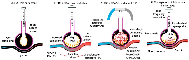 Figure 1: The dynamic interplay of surfactant therapy, ductal shunting and left ventricular dysfunction that ruptures the immature airspace–capillary barrier, and the tamponading strategies that oppose it. (Source: Sahussarungsi S, Lapointe A, Villeneuve A, et al. Biomedicines 2025;13(7):1744. DOI: 10.3390/biomedicines13071744).
Immediate Resuscitation and Ventilatory Management
Secure the airway and clear it gently
The first priority is a patent, secured airway. Blood and clots must be cleared by gentle, brief suctioning sufficient to restore the lumen and permit ventilation; repeated disconnection, deep suctioning and vigorous catheterisation cause mucosal trauma and may provoke further haemorrhage. Where the ETT is occluded by clot, or the infant is not intubated and is deteriorating, intubation should be performed with the largest appropriate tube to allow both ventilation and future suction. An apnoeic, bradycardic or profoundly hypoxaemic infant should be hand-ventilated while the team organises definitive management.
Raise mean airway pressure to tamponade
The central ventilatory intervention is an increase in mean airway pressure (MAP), which compresses the bleeding alveolar capillaries and provides mechanical tamponade. On conventional ventilation this is achieved by raising positive end-expiratory pressure (PEEP) — typically to 8–12 cmH$_2$O — while accepting the associated rise in MAP; on high-frequency oscillatory ventilation (HFOV) it is achieved by titrating MAP upwards. Raising MAP also recruits flooded alveoli and improves oxygenation. The strategy must respect the ceiling imposed by the immature lung: excessive MAP overdistends alveoli, compresses capillaries elsewhere, impairs venous return and raises intracranial pressure, increasing the risk of IVH.
- Call for help and start a timer; assign a scribe and a drug calculator.
- Increase $FiO_2$ to 1.0 and raise PEEP or MAP by 2–3 cmH$_2$O immediately.
- Suction gently to clear clot and confirm tube patency.
- Send an urgent full blood count, coagulation profile, fibrinogen and blood gas; cross-match.
- Give vitamin K and prepare platelets, fresh frozen plasma (FFP) and packed red cells.
- Perform targeted neonatal echocardiography to define shunt, ventricular function and pulmonary pressures.
- Maintain normothermia, normoglycaemia and adequate perfusion with minimal handling.
Ventilator targets and the ceiling of safe pressure
Once haemostasis begins, the aim shifts to lung-protective ventilation at the lowest MAP that sustains oxygenation. Working targets are a pre-ductal $SpO_2$ of 91–95% and a $PaCO_2$ of 40–50 mmHg, avoiding both hypoxaemia and hypocarbia; tidal volumes of 4–6 mL/kg limit volutrauma. There is no benefit from sustained hyperoxia, so $FiO_2$ should be weaned promptly once bleeding slows and MAP can be reduced. HFOV is a reasonable option when oxygenation remains inadequate despite high MAP, because it sustains a high mean airway pressure with a small tidal amplitude.
Minimal handling, thermoregulation and perfusion
Every unnecessary stimulation — weighing, repositioning, loud alarms, repeated suctioning — can raise systemic and pulmonary vascular pressures and restart bleeding. Care should be clustered, sedation optimised, and the infant kept normothermic, since hypothermia impairs platelet function and aggravates coagulopathy. Perfusion is judged by capillary refill, lactate, urine output and blood pressure, with the caveat that a normal blood pressure does not guarantee adequate systemic flow in a vasoconstricted infant.
Haemostatic, Pharmacological and Haemodynamic Therapy
Coagulopathy and thrombocytopenia
Bleeding is rarely isolated to the lung, so the coagulation system must be corrected deliberately rather than reflexively. Platelets should be transfused for active pulmonary bleeding when the count falls below $50 \times 10^9/\text{L}$; lower thresholds apply when bleeding is less severe, and decisions are individualised according to trajectory, concurrent coagulopathy and haemodynamic stability. Fresh frozen plasma is indicated for clinically significant bleeding with prolonged prothrombin time (PT) or activated partial thromboplastin time (aPTT) relative to gestational and postnatal norms. Vitamin K corrects deficiency, particularly in growth-restricted and asphyxiated infants with immature hepatic synthesis of clotting factors, and fibrinogen should be maintained above $1.0\ \text{g}/\text{L}$. For ventilated preterm infants with active bleeding, haemoglobin is conventionally kept above $120\ \text{g}/\text{L}$ to preserve oxygen delivery.
Endotracheal epinephrine
In life-threatening or refractory bleeding, diluted epinephrine instilled at the airway may induce localised pulmonary vasoconstriction and reduce haemorrhage. The best evidence favours delivery via a catheter advanced to the tip of the ETT rather than through the ETT connector: 1:10,000 epinephrine, $0.3$–$1.0\ \text{mL}/\text{kg}$ ($0.03$–$0.1\ \text{mg}/\text{kg}$), has been associated with markedly improved survival in one series. The manoeuvre is not standard therapy and risks mucosal ischaemia, necrosis, arrhythmia and systemic hypertension, so it is reserved for catastrophic bleeding as a temporising measure.
Surfactant replacement once bleeding is controlled
Because intra-alveolar blood inactivates surfactant, worsening compliance and atelectasis after the acute event often reflect secondary surfactant deficiency rather than ongoing haemorrhage. Administering a further dose once bleeding has stabilised improves recruitment and gas exchange; cohort data describe substantial reductions in oxygenation and ventilatory indices within an hour, without apparent excess of bronchopulmonary dysplasia or mortality. Surfactant is therefore a treatment for the aftermath of PH, not only a trigger of it.
Closing the haemodynamically significant ductus
When echocardiography demonstrates a large left-to-right ductal shunt with systemic steal or pulmonary overcirculation, closure reduces the flow load on the fragile capillary bed. Cyclo-oxygenase inhibitors (ibuprofen or indomethacin) or paracetamol may be used, though evidence that early closure prevents PH is mixed: prophylactic indomethacin reduced early severe PH in one trial but not overall, and contemporary trials of expectant ductal management report low PH rates. Ductal closure is therefore one component of haemodynamic optimisation rather than a definitive cure, and the temptation to treat every ductus aggressively should be resisted.
Volume, inotropes and the cardiovascular phenotype
Haemorrhage may cause true hypovolaemia, but the immature left ventricle tolerates rapid volume loading poorly, so boluses should be small, cautious and guided by echocardiography. When perfusion is inadequate, inotropes are preferable to repeated fluid challenges, although afterload surges from vasopressors can worsen post-capillary congestion. The correct choice depends on the identified phenotype — hypovolaemia, left ventricular dysfunction, pulmonary overcirculation or persistent pulmonary hypertension — which is why targeted neonatal echocardiography should be obtained early rather than deferred.
A Structured Approach to the Acute Event
The sequence below emphasises that tamponade, haemostatic correction and haemodynamic assessment proceed in parallel rather than in series, and that interventions are continuously retitrated.
graph TD
    A["Sudden bloody ETT secretions, desaturation or collapse<br/>in a high-risk neonate"] --> B["Call for help; declare pulmonary haemorrhage; start timer"]
    B --> C["FiO2 to 1.0; increase PEEP or MAP by 2-3 cmH2O"]
    C --> D["Gentle suction to clear clot; confirm ETT patency; secure airway"]
    D --> E{"Persistent bleeding or haemodynamic instability?"}
    E -->|"Yes"| F["Blood products: platelets, FFP, packed red cells, vitamin K;<br/>target fibrinogen above 1.0 g/L"]
    E -->|"No"| G["Continue lung-protective ventilation with<br/>tamponading MAP; avoid overdistension"]
    F --> H["Targeted neonatal echocardiography:<br/>shunt, LV function, pulmonary pressures"]
    G --> H
    H --> I{"Haemodynamically significant PDA?"}
    I -->|"Yes"| J["Consider ibuprofen, indomethacin or paracetamol;<br/>avoid indiscriminate fluid boluses"]
    I -->|"No"| K["Assess LV dysfunction, anaemia,<br/>sepsis and coagulopathy"]
    J --> L{"Life-threatening bleeding refractory to the above?"}
    K --> L
    L -->|"Yes"| M["Endotracheal epinephrine 1:10,000,<br/>0.3-1.0 mL/kg via catheter at ETT tip"]
    L -->|"No"| N["Post-haemorrhage surfactant if hypoxic<br/>respiratory failure from surfactant inactivation"]
    M --> N
    N --> O["Wean FiO2 and MAP as bleeding stops;<br/>minimal handling, normothermia, glucose, perfusion"]
| Priority | Action | Target or caveat |
|---|---|---|
| Airway | Gentle suction; confirm ETT patency; re-intubate if occluded | Clear clot without repeated disconnection |
| Oxygenation | $FiO_2$ to 1.0, then wean | Pre-ductal $SpO_2$ 91–95%; avoid sustained hyperoxia |
| Ventilation | PEEP to 8–12 cmH$_2$O or raise MAP on HFOV | Tamponade without overdistension or impaired venous return |
| Coagulation | Platelets, FFP, vitamin K | Platelets below $50 \times 10^9/\text{L}$ if actively bleeding; fibrinogen above $1.0\ \text{g}/\text{L}$ |
| Oxygen carriage | Packed red cell transfusion | Haemoglobin above $120\ \text{g}/\text{L}$ in ventilated actively bleeding infants |
| Haemodynamics | Echocardiography-guided therapy | Treat hsPDA, LV dysfunction or hypovolaemia selectively |
| Thermoregulation | Maintain 36.5–37.5 $^\circ\text{C}$ | Hypothermia impairs platelet function |
| Handling | Cluster care; minimal suction | Prevent re-bleeding and concurrent IVH |
Complications, Outcomes and Prevention
Complications and prognosis
The immediate hazards are exsanguination and hypoxic–ischaemic injury; the subsequent ones are those of any catastrophic neonatal event. Severe (grade 3–4) IVH is strongly associated with PH and may occur concurrently or shortly afterwards, and infants with PH are several times more likely to develop major IVH. Bronchopulmonary dysplasia, prolonged ventilation, air leak and nosocomial infection are common. Among survivors, severe PH is associated with a significantly increased risk of death or neurodevelopmental impairment, so PH marks a particularly vulnerable brain as well as a damaged lung.
Prevention in the at-risk infant
Prevention is largely the avoidance of iatrogenic harm in infants whose vessels are inherently fragile. A complete course of antenatal corticosteroids reduces the incidence and severity of PH by accelerating lung maturation and stabilising the pulmonary vasculature. Ventilation should scrupulously avoid excessive tidal volumes and airway pressures, since volutrauma and barotrauma are direct mechanisms of capillary rupture. Fluid balance should be conservative, avoiding rapid boluses that precipitate venous congestion, and transfusion practice should follow restrictive thresholds. Early recognition — a low threshold for investigating sudden deterioration in any high-risk infant — and prompt echocardiographic characterisation of the cardiovascular phenotype allow ductal and ventricular contributors to be addressed before the barrier fails. Prophylactic indomethacin may reduce early severe PH but not overall incidence, and is no substitute for these measures.
Key Takeaways
- Pulmonary haemorrhage is the acute rupture of the immature airspace–capillary barrier, presenting as bloody ETT secretions with abrupt deterioration, most often between 48 and 96 hours of life.
- Distinguish true PH from traumatic intubation, a generalised bleeding diathesis, upper-airway bleeding and pulmonary oedema; the mimics may coexist with PH and do not exclude it.
- PH complicates prematurity and RDS, surfactant administration, a haemodynamically significant PDA, mechanical ventilation, sepsis, coagulopathy and thrombocytopenia, and fetal growth restriction.
- Capillary stress failure is driven by a haemodynamic surge, high surface tension, inflammation and post-capillary congestion from a stiff immature left ventricle; the Starling relation and Laplace wall stress quantify these forces.
- Immediately secure the airway, suction gently, and increase mean airway pressure — PEEP of 8–12 cmH$_2$O or a higher MAP on HFOV — to tamponade the bleeding capillaries.
- Correct coagulopathy and thrombocytopenia deliberately: platelets below $50 \times 10^9/\text{L}$ with active bleeding, FFP for prolonged PT or aPTT, vitamin K, and fibrinogen above $1.0\ \text{g}/\text{L}$.
- Treat a haemodynamically significant ductus with a cyclo-oxygenase inhibitor or paracetamol where indicated, give post-haemorrhage surfactant for secondary surfactant inactivation, and tailor volume and inotropes to the echocardiographic phenotype.
- Prevention rests on antenatal corticosteroids, lung-protective ventilation, prudent fluid and transfusion practice, and early recognition with minimal handling in every at-risk infant.