Non-invasive respiratory support (NIRS) has displaced early intubation as the default initial strategy for most preterm infants with, or at risk of, respiratory distress syndrome (RDS). By sustaining end-expiratory lung volume, splinting the compliant pharynx, and unloading the diaphragm, NIRS attenuates the atelectrauma and volutrauma that initiate ventilator-induced lung injury (VILI) and so steers development away from bronchopulmonary dysplasia (BPD). It also preserves spontaneous breathing, avoids sedation and neuromuscular blockade, protects the immature glottis, and leaves surfactant deliverable by the minimally invasive LISA/MIST route described in the preceding chapter.
The modern NIRS armamentarium is best understood as a pressure source (bubble column, ventilator-derived constant pressure, or intermittent inflations) married to an interface (short binasal prongs, nasopharyngeal prong, nasal mask, or the leak-prone RAM cannula). Outcome depends as much on interface seal and delivered mean airway pressure as on the nominal mode label. This chapter covers the modality landscape, the physics of bubble-generated pressure, the interfaces that convert pressure into distending force, the anchoring trial evidence, and a practical escalation algorithm.
graph TD
    A["Pressure source"] --> B["Bubble column<br/>hydrostatic set-point"]
    A --> C["Ventilator-derived CPAP<br/>constant pressure"]
    A --> D["NIPPV / SNIPPV<br/>CPAP + intermittent inflations"]
    A --> E["HHHFNC / nHFO<br/>flow- or oscillator-derived"]
    B --> F["Interface"]
    C --> F
    D --> F
    E --> F
    F --> G["Short binasal prongs<br/>most effective seal"]
    F --> H["Nasopharyngeal prong / nasal mask"]
    F --> I["RAM cannula<br/>high leak, larger dead space"]
The Modality Landscape
Bubble CPAP
Bubble CPAP is the simplest and often the most reproducible pressure generator. A blended, heated, humidified gas stream is delivered to the infant while the expiratory limb is submerged to a measured depth in a water column; escaping bubbles hold circuit pressure at the hydrostatic set-point. It needs no pressure transducer or servo-control, is intrinsically fail-safe against over-pressurisation, and generates the characteristic bubbling oscillation. Its limitations are a fixed dependence on submersion depth, insensitivity to large leaks, and circuit noise.
Ventilator-Derived CPAP and NIPPV/SNIPPV
Ventilator-derived (constant-pressure) CPAP uses a servovalve or flow generator to hold a target pressure against variable leak; because it incorporates a trigger and an inflation valve, the same device can deliver nasal intermittent positive-pressure ventilation (NIPPV) — background CPAP punctuated by ventilator-delivered inflations. When those inflations are synchronised to spontaneous inspiratory effort by pneumotachograph, pressure capsule, or impedance, the mode is synchronised NIPPV (SNIPPV). Synchrony improves comfort and gas exchange and avoids inflation against active expiration, which risks gastric insufflation; the Cochrane evidence is nevertheless insufficient to mandate synchrony as an independent determinant of outcome.
Nasal High-Frequency Oscillation
Nasal high-frequency oscillation (nHFO) superimposes a small-amplitude oscillatory waveform on a nasal bias flow. It has been studied mainly as a rescue modality in infants failing CPAP, with mechanistic appeal in carbon-dioxide clearance and recruitment, but comparative data are sparse and it remains an investigational adjunct rather than a first-line modality.
Heated Humidified High-Flow Nasal Cannula and the RAM Cannula
Heated humidified high-flow nasal cannula (HHHFNC) delivers conditioned gas at 2–8 L/min through thin, tapered nares. Its mechanisms differ partly from CPAP: nasopharyngeal dead-space washout improves carbon-dioxide clearance, while the high flow generates a variable, leak-dependent distending pressure. HHHFNC is better tolerated and causes less nasal trauma, but delivered pressure is unpredictable and, as primary support, its treatment-failure rate exceeds CPAP's (see HIPSTER). The RAM cannula is a short binasal interface used for NIRS that forms no true seal; it behaves as a high-leak, dead-space-bearing conduit whose effective pressure is well below that of conventional short binasal prongs.
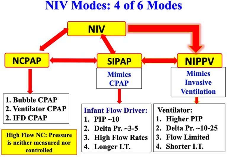 Figure 1: The principal non-invasive modalities for preterm infants differ by pressure source and interface, from bubble CPAP through NIPPV/SNIPPV and nasal high-frequency oscillation to heated humidified high-flow nasal cannula. (Source: Shi Y, Muniraman H, Biniwale M, Ramanathan R. Frontiers in Pediatrics 2020. DOI: 10.3389/fped.2020.00270).
Interfaces and Fixation
Short Binasal Prongs
Short binasal prongs are the reference interface for CPAP in preterm infants and, in the Cochrane review of CPAP devices, confer a lower intubation rate than single nasopharyngeal prongs. Prong tips sit just inside the nares, forming a partial seal that permits the intentional leak required for carbon-dioxide clearance and gastric venting. Correctly sized prongs should occlude roughly 60–80% of the nares; a prong that fills the naris completely abolishes the leak, raises nasal resistance, and predisposes to columella necrosis.
Nasopharyngeal Prongs and Nasal Masks
A single nasopharyngeal prong is advanced until its tip lies in the nasopharynx; it is easier to secure in an agitated infant but less effective and prone to secretion obstruction. Nasal masks suit difficult prong sizing or larger infants, distributing force over a wider area and reducing focal septal pressure, but are more prone to dislodgement and leak-related eye irritation.
The RAM Cannula and Interface Dead Space
The RAM cannula's absent seal means that a large fraction of circuit flow escapes and that the device contributes an additional dead-space burden. This is clinically relevant in two ways: the effective distending pressure is lower and less predictable than with prongs, and the extra dead space can blunt carbon-dioxide clearance. The RAM cannula is best reserved for situations in which the small-calibre nares cannot accommodate prongs — notably in the delivery room and in the smallest infants.
Fixation and Skin Protection
Interface fixation is a nursing-intensive intervention, not an afterthought. Prongs should be secured with a purpose-designed cap or bonnet so the vector of force is axial and the tips are not driven into the septum, with a hydrocolloid barrier over the columella and philtrum. The interface should be repositioned every 4–6 hours and the nares inspected each shift. Over-tightening, the commonest error, trades a marginal seal gain for a linear increase in mucosal ischaemia.
Increasing the seal by tightening the interface is not free. Nasal septal and columellar injury is a function of contact pressure and duration, not of measured airway pressure, and a snug interface that trades a small leak reduction for mucosal ischaemia is a poor bargain. Audit interfaces hourly, protect the skin, and never use pressure alone to overcome a poorly fitting interface.
The Physics of Bubble CPAP
The Hydrostatic Set-Point
In bubble CPAP the applied pressure is generated by a water column of depth $h$:
$$P_{CPAP} = \rho\, g\, h$$where $P_{CPAP}$ = applied airway pressure ($\text{cmH}_2\text{O}$); $\rho$ = density of water ($\approx 1{,}000\ \text{kg}\cdot\text{m}^{-3}$); $g$ = gravitational acceleration ($9.81\ \text{m}\cdot\text{s}^{-2}$); $h$ = submersion depth of the expiratory limb ($\text{m}$). Since $\rho g = 9{,}810\ \text{Pa}\cdot\text{m}^{-1}$ and $1\ \text{cmH}_2\text{O} \approx 98.07\ \text{Pa}$, the bedside identity is $P_{CPAP}[\text{cmH}_2\text{O}] \approx h[\text{cm}]$: each centimetre of submersion yields approximately one centimetre of water of distending pressure.
The set-point is therefore also a ceiling: once the column bubbles, higher bias flow does not raise mean pressure. This protects against barotrauma from a flow surge but means that raising flow cannot rescue a falling airway pressure — the clinician must correct the leak or deepen the submersion.
The Bubbling Oscillation
The escaping bubbles superimpose a small-amplitude, broadband pressure oscillation on the steady column pressure:
$$P_{aw}(t) = P_{CPAP} + \Delta P_{osc}\,\sin(2\pi f\, t)$$where $P_{aw}(t)$ = instantaneous airway opening pressure ($\text{cmH}_2\text{O}$); $\Delta P_{osc}$ = peak oscillatory amplitude ($\text{cmH}_2\text{O}$); $f$ = oscillation frequency ($\text{Hz}$); and $t$ = time ($\text{s}$). In vitro modelling shows the amplitude rises with increasing bias flow and again as compliance falls, with broadband frequency content from single-digit to low-tens-of-Hertz. The proposed benefit invokes stochastic resonance: a noisy signal superimposed on spontaneous effort raises the probability that a critically closed airway opens, so marginally recruited units are recruited and maintained. Preterm-lamb experiments support this, bubble CPAP giving higher oxygenation, greater oxygen uptake, lower alveolar protein, and less ventilation inhomogeneity than constant-pressure CPAP at matched flow. Clinical translation remains debated, and bubble CPAP is best regarded as a pressure-generation strategy rather than a proven outcome advantage over well-delivered ventilator CPAP.
Flow, Orifice Resistance and Leak
The pressure that actually reaches the alveolus is the set-point minus the losses incurred across the prongs and leak. For the turbulent, high-velocity flow through a prong orifice:
$$\dot{V} = C_d\, A \sqrt{\frac{2\,\Delta P}{\rho_{gas}}}$$where $\dot{V}$ = volumetric flow ($\text{L}\cdot\text{min}^{-1}$ or $\text{m}^{3}\cdot\text{s}^{-1}$); $C_d$ = discharge coefficient (dimensionless, $\approx 0.6\text{–}0.8$); $A$ = prong cross-sectional area ($\text{m}^{2}$); $\Delta P$ = pressure drop across the prong ($\text{Pa}$); and $\rho_{gas}$ = gas density ($\text{kg}\cdot\text{m}^{-3}$). The quadratic dependence means that forcing a higher flow through a fixed, too-small prong raises $\Delta P$ steeply and converts useful distending pressure into kinetic energy and noise.
Where flow is laminar — the narrow lumen of a thin catheter or a partially obstructed prong — the loss is described by the Hagen–Poiseuille relation:
$$\Delta P = \frac{8\,\mu\, L\, \dot{V}}{\pi\, r^{4}}$$with $\mu$ = dynamic viscosity ($\text{Pa}\cdot\text{s}$), $L$ = tube length ($\text{m}$), and $r$ = lumen radius ($\text{m}$). The fourth-power dependence on radius explains why a partial mucus obstruction so efficiently destroys delivered pressure and why prong calibre is a more powerful determinant of resistance than length.
Bernoulli's principle provides the governing intuition for the leak itself:
$$P + \tfrac{1}{2}\,\rho\, v^{2} + \rho\, g\, z = \text{constant}$$where $P$ = static pressure ($\text{Pa}$), $\rho$ = gas density ($\text{kg}\cdot\text{m}^{-3}$), $v$ = gas velocity ($\text{m}\cdot\text{s}^{-1}$), $g$ = gravitational acceleration ($\text{m}\cdot\text{s}^{-2}$), and $z$ = height ($\text{m}$). Gas that accelerates through a leak converts static into dynamic pressure, so the pressure sensed at the airway opening falls whenever the leak fraction rises. The leak fraction quantifies this competition:
$$\phi_{leak} = 1 - \frac{\dot{V}_{delivered}}{\dot{V}_{circuit}}$$where $\phi_{leak}$ = fraction of circuit flow lost at the interface (dimensionless); $\dot{V}_{delivered}$ = flow effective at maintaining airway pressure ($\text{L}\cdot\text{min}^{-1}$); and $\dot{V}_{circuit}$ = total bias flow delivered ($\text{L}\cdot\text{min}^{-1}$). A ventilator-derived CPAP generator compensates automatically for $\phi_{leak}$; a bubble circuit does not, which is why mouth leak, a displaced prong, or a RAM cannula can silently collapse effective pressure.
Comparative Matrix: CPAP, NIPPV/SNIPPV and HHHFNC
| Feature | CPAP (bubble or ventilator) | NIPPV / SNIPPV | HHHFNC |
|---|---|---|---|
| Mechanism | Continuous distending pressure holds functional residual capacity and splints the pharynx | Background CPAP plus ventilator-delivered inflations; synchronised in SNIPPV | Heated humidified high flow; dead-space washout with flow- and leak-dependent pressure |
| Typical settings | PEEP $5\text{–}8\ \text{cmH}_2\text{O}$; bias flow $6\text{–}8\ \text{L}\cdot\text{min}^{-1}$ | PEEP $5\text{–}6\ \text{cmH}_2\text{O}$; PIP $14\text{–}20\ \text{cmH}_2\text{O}$; rate $20\text{–}40\ \text{min}^{-1}$; $T_i$ $0.3\text{–}0.5\ \text{s}$ | Flow $2\text{–}8\ \text{L}\cdot\text{min}^{-1}$; $FiO_2$ titrated to target |
| Advantages | Simple, effective, most extensively validated | Lower extubation failure and reintubation than CPAP (Cochrane) | Comfort, less nasal trauma, easier feeding and handling |
| Drawbacks | Interface trauma, air leak, gastric distension | More gastric insufflation and air leak; needs a triggering-capable device | Unpredictable pressure; higher primary-support failure than CPAP (HIPSTER) |
| Best evidence | SUPPORT, COIN, Cochrane CPAP reviews | Cochrane NIPPV reviews (2023) | HIPSTER; Cochrane high-flow reviews |
The Evidence Base
Early CPAP versus Intubation and Surfactant
Two landmark randomised trials defined the role of early CPAP. The COIN trial (Morley et al., N Engl J Med 2008) randomised 610 infants of 25–28 weeks' gestation at 5 minutes of age to nasal CPAP or intubation and ventilation. The primary composite of death or BPD at 36 weeks was 33.9% with CPAP versus 38.9% with intubation (odds ratio 0.80, 95% CI 0.58–1.12; $p = 0.19$), a direction of benefit that did not reach significance. CPAP halved surfactant use, reduced ventilation days, and lowered the 28-day composite of death or oxygen requirement (odds ratio 0.63, 95% CI 0.46–0.88; $p = 0.006$), but the pneumothorax rate was 9% versus 3% ($p < 0.001$) — the signal that early CPAP trades a reduced need for ventilation against a higher air-leak burden.
The SUPPORT trial (Finer et al., N Engl J Med 2010) randomised 1,316 infants of 24$^{+0}$ to 27$^{+6}$ weeks to delivery-room CPAP versus intubation and surfactant with a protocol-driven limited-ventilation strategy. The primary outcome of death or BPD at 36 weeks was 47.8% with CPAP versus 51.0% with surfactant (relative risk 0.95, 95% CI 0.85–1.05) — again not significant. CPAP-treated infants required intubation and postnatal corticosteroids less often, required fewer days of mechanical ventilation, and were more likely to be alive and free of mechanical ventilation by day 7. Taken together, the trials support CPAP as a legitimate alternative to routine intubation, particularly when paired with early recourse to surfactant for infants who declare surfactant deficiency.
Early CPAP reduces the need for and duration of invasive ventilation, but it does not reliably reduce the composite of death or BPD, and it increases pneumothorax in some trials (COIN: 9% versus 3%). The correct conclusion is not that CPAP is harmless, but that it must be delivered with a competent interface, close air-leak surveillance, and a protocolised failure pathway.
Post-Extubation NIPPV versus CPAP
The 2023 Cochrane review of NIPPV versus NCPAP after extubation (Lemyre et al., 19 trials, 2,738 infants) found that NIPPV likely reduces respiratory failure (RR 0.75, 95% CI 0.67–0.84; NNTB 11) and reintubation (RR 0.78, 95% CI 0.70–0.87; NNTB 12), with a possible reduction in pulmonary air leak (RR 0.57, 95% CI 0.37–0.87), but little or no difference in gastrointestinal perforation, necrotising enterocolitis, chronic lung disease, or mortality. Ventilator-generated NIPPV outperformed bilevel devices for post-extubation respiratory failure (RR 0.49, 95% CI 0.40–0.62), and the authors noted that data in infants below 28 weeks remain sparse. For early NIPPV, the companion 2023 Cochrane review (17 trials, 1,958 infants) found a reduction in respiratory failure (RR 0.65, 95% CI 0.54–0.78) and in the need for endotracheal ventilation (RR 0.67, 95% CI 0.56–0.81), with a possible small reduction in chronic lung disease but no demonstrated mortality benefit.
HHHFNC versus CPAP
The HIPSTER trial (Roberts et al., N Engl J Med 2016) randomised 564 preterm infants of at least 28 weeks' gestation with early respiratory distress who had not received surfactant to HHHFNC or CPAP as primary support. The trial was stopped early by the data and safety monitoring committee: treatment failure within 72 hours occurred in 25.5% of the high-flow group versus 13.3% of the CPAP group (risk difference 12.3 percentage points, 95% CI 5.8–18.7) — the upper confidence bound crossed the pre-specified 10-percentage-point non-inferiority margin. Intubation rates did not differ significantly (15.5% versus 11.5%). HHHFNC is therefore a reasonable post-extubation and step-down modality, but not a superior primary-support strategy to CPAP.
Practical Settings, Failure Prediction and Escalation
Initial Settings and Monitoring
| Parameter | Recommendation | Rationale / Notes |
|---|---|---|
| CPAP pressure | $5\text{–}8\ \text{cmH}_2\text{O}$ | Start $6\text{–}7$; titrate to work of breathing, $FiO_2$, and $SpO_2$ |
| Bias flow | $6\text{–}8\ \text{L}\cdot\text{min}^{-1}$ | Higher flow increases oscillatory amplitude and leak noise |
| Gas conditioning | $37\ ^\circ\text{C}$, fully saturated | Prevents mucosal injury and secretion inspissation |
| $SpO_2$ target | $90\text{–}95\%$ (alarms $\approx 89\text{–}96\%$) | Avoid hyperoxia to limit retinopathy of prematurity risk |
| $FiO_2$ titration | Wean in $0.02\text{–}0.05$ steps | Lowest $FiO_2$ sustaining target $SpO_2$ |
| Interface | Short binasal prongs | Re-site every 4–6 h; inspect septum each shift |
| Gastric venting | Orogastric tube, open or intermittent suction | Prevents distension and splinting of the diaphragm |
Predictors of CPAP Failure
CPAP failure is best predicted by the rate of change in support requirement rather than any single threshold. Early warning signs include an $FiO_2$ requirement exceeding 0.30 within 2–3 hours despite optimised interface and pressure, a rising or persistently elevated $PaCO_2$ with respiratory acidosis ($pH < 7.25$), recurrent apnoea, increasing work of breathing, and progressive atelectasis. Lower gestational age, small-for-gestational-age status, absent antenatal corticosteroids, severe initial disease, and a large leak all raise the pre-test probability of failure. A high initial $FiO_2$ — reflecting shunt from unrecruited, surfactant-deficient lung — is particularly powerful and signals that the infant may need surfactant rather than more pressure.
graph TD
    A["Preterm infant on bubble CPAP<br/>5-8 cmH2O"] --> B{"Stabilising?<br/>SpO2 90-95%, FiO2 falling, pH > 7.25"}
    B -->|"Yes"| C["Continue; wean FiO2 then PEEP"]
    B -->|"No"| D["Optimise interface, position,<br/>secretion clearance, gastric venting"]
    D --> E{"FiO2 > 0.30 by 2-3 h,<br/>rising pCO2, or apnoea?"}
    E -->|"No"| C
    E -->|"Yes"| F["Escalate to NIPPV / SNIPPV<br/>PIP 14-20 cmH2O, rate 20-40/min, PEEP 5-6"]
    F --> G{"Improving within 1-2 h?"}
    G -->|"Yes"| H["Continue; consider de-escalation after 24-72 h"]
    G -->|"No"| I{"Surfactant deficiency likely?"}
    I -->|"Yes"| J["LISA / MIST on ongoing CPAP"]
    I -->|"No"| K["Intubate and ventilate invasively"]
    J --> L{"Improving after surfactant?"}
    L -->|"Yes"| C
    L -->|"No"| K
    H --> M["Step down to CPAP, then HHHFNC, then room air"]
Escalate without delay for $FiO_2 > 0.30$ by 2–3 hours, $FiO_2 > 0.40$ at any point, $pH < 7.20$ with $PaCO_2 > 60\text{ mmHg}$, recurrent apnoea requiring stimulation, or haemodynamic instability. A trial of NIPPV is appropriate before intubation, but LISA/MIST should be offered concurrently when surfactant deficiency is the dominant physiology; delaying surfactant while escalating pressure prolongs injurious support.
De-Escalation
De-escalation should be deliberate rather than reflexive. Once the infant is stable on $FiO_2 < 0.30$ with normal acid–base status, wean the $FiO_2$ first, then reduce PEEP in $1\ \text{cmH}_2\text{O}$ steps to a floor of about $5\ \text{cmH}_2\text{O}$. Stable infants may step down to HHHFNC, recognising that its pressure delivery is less predictable and that failure must be detected promptly. Feeding tolerance, nasal integrity, and growth trajectory legitimately govern how quickly the interface is removed.
Complications and Prevention
- Nasal septal and columellar trauma. The commonest and most preventable complication, arising from focal contact pressure and ischaemia. Prevention: correct prong size, axial fixation via a cap or bonnet, a hydrocolloid barrier, 4–6-hourly re-siting, and shift-wise inspection.
- Gastric distension. Nasal positive pressure and air-swallowing inflate the stomach, splint the diaphragm, and can trigger feeds intolerance or regurgitation. An orogastric tube, vented to air or on intermittent suction, is standard; persistent distension should prompt interface and synchrony review.
- Pulmonary air leak. Pneumothorax, pulmonary interstitial emphysema, and pneumomediastinum are the price of positive pressure in immature lung; COIN showed a threefold excess of pneumothorax with early CPAP. Keep a low threshold for transillumination and chest radiography when an infant deteriorates abruptly.
- Feeding intolerance and necrotising enterocolitis. NIRS is less disruptive to feeding than invasive ventilation, but gastric distension and unstable splanchnic perfusion justify cautious feed advancement, especially with NIPPV.
- Interface dead space. The RAM cannula, and to a lesser degree long nasopharyngeal prongs, add dead space that impairs carbon-dioxide clearance; prefer short binasal prongs whenever the nares permit.
When an infant on nominal CPAP is not responding, measure what is actually delivered rather than assuming the set-point. Confirm continuous bubbling at the correct depth, verify that the prong tips are visible in the nares, inspect for a gaping mouth leak, and ask whether the interface is the correct size. In bubble CPAP, restoring the seal and the submersion depth often resolves "CPAP failure" more effectively than escalating to NIPPV.
Prongs that fully obturate both nares abolish the intended leak, eliminate carbon-dioxide egress, raise nasal resistance, and rapidly produce ischaemic septal injury. Target 60–80% nares occlusion and treat any columellar blanching as an emergency requiring immediate interface change.
Key Takeaways
- Non-invasive support is first-line therapy for most preterm infants with or at risk of RDS; its benefit derives from maintaining end-expiratory lung volume while avoiding intubation-associated VILI.
- Bubble CPAP sets pressure hydrostatically — approximately $1\ \text{cmH}_2\text{O}$ per centimetre of expiratory-limb submersion — and superimposes a small-amplitude bubbling oscillation whose physiological value is plausible but not fully established.
- Interface quality determines delivered pressure: short binasal prongs are the most effective and best-evidenced interface, sized to occlude 60–80% of the nares, whereas the RAM cannula is leak-prone and adds dead space.
- Early CPAP reduces the need for and duration of mechanical ventilation but does not reliably reduce death or BPD, and increases pneumothorax in some trials (COIN 9% versus 3%); monitor closely for air leak.
- NIPPV and SNIPPV reduce post-extubation respiratory failure and reintubation compared with CPAP (Cochrane: RR 0.75 and 0.78 respectively), with ventilator-generated and synchronised delivery appearing advantageous.
- HHHFNC is better tolerated and useful for step-down care, but as primary support it failed non-inferiority against CPAP in HIPSTER (25.5% versus 13.3% treatment failure).
- A rising $FiO_2$ requirement — especially $FiO_2 > 0.30$ within 2–3 hours — is the most useful bedside predictor of CPAP failure and should trigger interface optimisation, escalation to NIPPV, and consideration of LISA/MIST before intubation.
- Nasal septal trauma and gastric distension are the signature complications of NIRS and are almost entirely preventable through correct sizing, axial fixation, skin protection, regular re-siting, and gastric venting.