The transition from intrauterine to extrauterine life is the most abrupt physiological event in human development. In minutes, a fluid-filled, high-resistance organ becomes an air-filled, low-resistance gas exchanger, while the placenta ceases to be the organ of gas exchange. Three processes dominate: clearance of fetal lung liquid, the first breath with establishment of a functional residual capacity (FRC), and an eight- to ten-fold fall in pulmonary vascular resistance (PVR) that redirects right ventricular output through the lungs.
Delay of any step produces the classic neonatal syndromes — transient tachypnoea of the newborn (TTN), respiratory distress syndrome (RDS) and persistent pulmonary hypertension of the newborn (PPHN) — and dictates how the delivery room is managed and why the preterm infant is exquisitely vulnerable.
The steps are interdependent. Liquid must be cleared before gas exchange is effective, and aeration and oxygenation must occur before PVR can fall. Interventions that invert this order — for example, clamping the cord before the lung is aerated — destabilise the transition.
Stages of Lung Development
Lung development spans five overlapping stages, from the fourth post-conception week to about eight years of age. The gestational window at which birth occurs determines both the gas-exchange surface available and the clinical syndrome that follows.
Embryonic and pseudoglandular stages
In the embryonic stage (4–7 post-conception weeks) the lung bud evaginates from the ventral foregut and divides into the trachea and main bronchi; disordered separation causes tracheo-oesophageal fistula. In the pseudoglandular stage (5–17 weeks), dichotomous branching generates all conducting airways down to the terminal bronchioles. Because airway number is fixed here, space-occupying lesions such as oligohydramnios or congenital diaphragmatic hernia cause irreversible pulmonary hypoplasia.
Canalicular, saccular and alveolar stages
The canalicular stage (16–26 weeks) is decisive: respiratory bronchioles and their capillaries appear, the epithelium thins, and type II pneumocytes begin synthesising surfactant near 24 weeks — the limit of viability. In the saccular stage (24–38 weeks) saccules enlarge and the air–blood barrier narrows. The alveolar stage begins near 36 weeks and continues for years; secondary septation multiplies alveoli from roughly 50 million at term to some 300 million by age eight. Preterm birth can permanently arrest alveolarisation, the structural basis of bronchopulmonary dysplasia.
| Stage | Gestational window (post-conception) | Defining events | Clinical correlate |
|---|---|---|---|
| Embryonic | 4–7 weeks | Lung bud from ventral foregut; trachea and main bronchi | Tracheo-oesophageal fistula |
| Pseudoglandular | 5–17 weeks | Branching morphogenesis; airways to terminal bronchioles | Airway number fixed; pulmonary hypoplasia |
| Canalicular | 16–26 weeks | Respiratory bronchioles and capillaries; surfactant ~24 weeks | Limit of viability; surfactant-deficient RDS |
| Saccular | 24–38 weeks | Enlarging saccules; thinning air–blood barrier | Preterm birth predisposes to BPD |
| Alveolar | 36 weeks – ~8 years | Secondary septation; alveolar multiplication | Alveolar growth; arrest in BPD |
Before 24 weeks there is no viable gas-exchange surface. Between 24 and 34 weeks the lung is structurally present but surfactant-deficient, producing RDS. At term the lung is competent, so failure reflects failed liquid clearance (TTN) or failed vascular relaxation (PPHN).
Fetal Lung Liquid and Its Clearance
The fetal lung is actively distended by a secreted liquid that provides the mechanical stimulus for growth; it is isosmotic and low in protein, distinguishing it from later pulmonary oedema.
Active chloride secretion in utero
Secretion is driven by apical chloride efflux through the cystic fibrosis transmembrane conductance regulator (CFTR) and calcium-activated chloride channels, with sodium and water following passively. The result is a luminal volume of approximately 20–30 mL/kg at term, renewed at 2–4 mL/kg/h. Because the process is energy-dependent, it can be reversed within minutes — the essential design feature of the perinatal transition.
The ion switch from secretion to absorption
At the onset of labour, a hormonal surge of catecholamines, glucocorticoids and thyroid hormones reprograms the epithelium to absorption. Apical epithelial sodium channels (ENaC) admit sodium into the type II pneumocyte, and the basolateral sodium–potassium ATPase (Na⁺/K⁺-ATPase) extrudes three sodium ions in exchange for two potassium ions into the interstitium. The vectorial sodium flux creates an osmotic gradient that draws water out through aquaporin channels (notably AQP5) and the paracellular route.
$$\text{Cl}^- \text{ secretion} \;\rightarrow\; \text{Na}^+ \text{ absorption} \quad (\text{ENaC} + \text{Na}^+/\text{K}^+\text{-ATPase})$$Secretory phenotype (fetal): apical Cl⁻ efflux (CFTR) with passive Na⁺ and water secretion. Absorptive phenotype (neonatal): apical Na⁺ influx via ENaC and basolateral extrusion by Na⁺/K⁺-ATPase (3 Na⁺ out : 2 K⁺ in per ATP); water follows osmotically. Catecholamines and glucocorticoids drive the switch and labour potentiates it.
Routes of lung liquid clearance
The "thoracic squeeze" of vaginal delivery removes only a small fraction, because distal airways collapse and limit expulsion. The dominant route is transepithelial absorption across the alveolar epithelium; the remainder is cleared by the pulmonary capillaries and lymphatics, which drain the interstitial fluid that the epithelium has extruded. Aeration then drives residual liquid inward as the air–liquid interface advances.
flowchart TD
    A["Fetal lung: apical Cl- secretion, liquid-filled lumen"] --> B["Labour: catecholamines and glucocorticoids"]
    B --> C["Upregulation of apical ENaC: Na+ enters the pneumocyte"]
    C --> D["Basolateral Na+/K+-ATPase: Na+ extruded, 3 Na+ per 2 K+"]
    D --> E["Osmotic gradient draws H2O via AQP5 and paracellular route"]
    E --> F["Fluid enters the interstitium"]
    F --> G["Cleared by pulmonary capillaries and lymphatics"]
    G --> H["Aeration advances the air-liquid interface and drives residual fluid inward"]
    H --> I["Air-filled alveoli; functional residual capacity established"]
Caesarean delivery before labour deprives the fetus of the catecholamine and glucocorticoid surge and of the mechanical effects of contractions; ENaC-mediated absorption is not upregulated, so the infant is born with a fluid-filled lung and is at markedly increased risk of TTN.
The First Breath and the Establishment of Functional Residual Capacity
The first breath must overcome the surface tension of the liquid lining the airways and the viscous and resistive impedance of the fluid-filled lung. Transpulmonary pressures during the first breath are correspondingly extreme:
$$P_{tp} \approx -40 \text{ to } -70 \text{ cmH}_2\text{O}$$where $P_{tp}$ is transpulmonary pressure (cmH₂O), the gradient between alveolar and pleural pressure. The pressure required to open a collapsed airway is governed by Laplace's law:
$$P = \frac{2\gamma}{r}$$in which $P$ is the distending pressure (dyn/cm²), $\gamma$ is the surface tension of the air–liquid interface (dyn/cm) and $r$ is the airway or alveolar radius (cm). Surfactant lowers $\gamma$ towards zero at low volumes, powerfully reducing the opening pressure; in surfactant deficiency, $\gamma$ is high, so atelectasis is favoured and the work of breathing rises sharply.
Resistance to gas flow is described by the Poiseuille relationship:
$$Q = \frac{\pi\,\Delta P\, r^{4}}{8\,\eta\, L}$$where $Q$ is flow (mL/s), $\Delta P$ is the pressure gradient (cmH₂O), $r$ is airway radius (cm), $\eta$ is gas viscosity (dyn·s/cm²) and $L$ is airway length (cm). The fourth-power dependence on radius explains why liquid filling or small reductions in calibre increase resistance dramatically, and why positive end-expiratory pressure is so effective.
The net result is retention of gas at end-expiration: an FRC of roughly 30 mL/kg, maintained by the balance of elastic recoil, post-inspiratory diaphragmatic braking, laryngeal closure and applied PEEP. Its mechanical correlate is respiratory system compliance:
$$C_{rs} = \frac{\Delta V}{\Delta P}$$where $C_{rs}$ is respiratory system compliance (mL/cmH₂O), $\Delta V$ is the change in lung volume (mL) and $\Delta P$ is the change in airway pressure (cmH₂O). FRC is the volume at which compliance is optimal; both atelectasis and overdistension reduce $C_{rs}$.
A single effective inflation both clears liquid and establishes FRC. If the first breaths are weak or unsupported, liquid persists, surfactant is inactivated by residual fluid, and a self-perpetuating cycle of atelectasis and respiratory failure begins — which is why the first minute carries disproportionate physiological weight.
Pulmonary Vascular Transition
In fetal life PVR is high because the pulmonary vessels are constricted by hypoxia and low pH, compressed by the fluid-filled lung, and invested with a thick muscular medial layer. Only about 10% of combined ventricular output passes through the lungs; the remainder is diverted through the ductus arteriosus (DA) and foramen ovale (FO), with the placenta performing gas exchange.
After aeration, PVR falls eight- to ten-fold within minutes and pulmonary blood flow rises to receive essentially the entire right ventricular output. This fall results from converging mechanisms: oxygenation relieves hypoxic pulmonary vasoconstriction; nitric oxide from endothelial nitric oxide synthase acts through cGMP; prostacyclin (PGI₂) acts through cAMP; and bradykinin, generated and degraded at the pulmonary endothelium, contributes to vasodilatation. Opening of potassium channels hyperpolarises smooth muscle, and lung expansion physically dilates the extra-alveolar vessels.
flowchart TD
    A["Fetal state: fluid-filled lungs and hypoxic vasoconstriction"] --> B["PVR high; most right ventricular output bypasses the lungs via the ductus arteriosus"]
    B --> C["Birth: lung aeration raises alveolar and arterial oxygen tension"]
    C --> D["Hypoxic vasoconstriction relieved"]
    C --> E["Endothelial nitric oxide and prostacyclin released"]
    C --> F["Bradykinin generated at the pulmonary endothelium"]
    D --> G["Smooth-muscle relaxation and potassium channel opening"]
    E --> G
    F --> G
    G --> H["PVR falls 8- to 10-fold within minutes"]
    H --> I["Pulmonary blood flow rises; the lung replaces the placenta as the source of left ventricular output"]
    I --> J["Functional closure of the ductus arteriosus; foramen ovale closes as left atrial pressure exceeds right atrial pressure"]
    J --> K["Anatomical ductal closure over days to weeks, forming the ligamentum arteriosum"]
The magnitude of the transition is quantified by the pulmonary vascular resistance equation:
$$PVR = \frac{PAP - LAP}{Q_p}$$where $PVR$ is pulmonary vascular resistance (Wood units, mmHg·min/L), $PAP$ is mean pulmonary artery pressure (mmHg), $LAP$ is left atrial pressure (mmHg) and $Q_p$ is pulmonary blood flow (L/min). Because $PVR$ is a ratio, it falls both as pressure decreases and as flow increases — after birth both occur together.
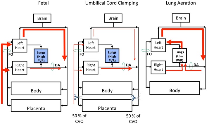 Figure 1: Lung aeration, not cord clamping alone, triggers the fall in pulmonary vascular resistance and reverses ductal shunt — the physiological rationale for ventilation before cord clamping. (Source: Bhatt S, Polglase GR, Wallace EM, Te Pas AB, Hooper SB. Frontiers in Pediatrics 2014. DOI: 10.3389/fped.2014.00113).
Closure of the ductus arteriosus and foramen ovale
The ductus arteriosus constricts within hours in term infants in response to the rise in arterial oxygen tension, a fall in circulating prostaglandin E₂ (its placental source is removed and the lung metabolises it) and reduced flow. Functional closure is followed by anatomical obliteration over days to weeks, leaving the ligamentum arteriosum; in preterm infants the ductus is less oxygen-responsive and more prostaglandin-sensitive, so patency persists. The foramen ovale closes functionally when pulmonary venous return raises left atrial pressure above right atrial pressure; anatomical sealing takes months to years, and a probe-patent foramen ovale persists in roughly a quarter of healthy adults.
If PVR remains elevated after birth, right-to-left shunting persists and the infant becomes profoundly hypoxaemic despite a structurally normal heart. Therapy is to recruit the lung and give pulmonary vasodilators such as inhaled nitric oxide: oxygen and lung volume are vasodilator therapies, not merely supportive care.
Delivery-Room Implications
The physiology translates directly into practice. The overriding aim is to establish effective ventilation and lung aeration early and gently, because aeration is the trigger for liquid clearance, FRC formation and the fall in PVR.
The golden minute, cord clamping and sustained inflation
The "golden minute" is the first minute of life, during which tone, breathing and heart rate are assessed and, if needed, effective positive-pressure ventilation is begun. Heart rate is the best single indicator of a successful transition and is best assessed by auscultation or electrocardiography rather than palpation.
Delayed cord clamping for at least 60 seconds in vigorous term and preterm infants allows placental transfusion to continue while the lung aerates; in preterm infants requiring support, ventilation before clamping is favoured. The rationale is precise: the fall in PVR and the establishment of pulmonary blood flow are aeration-dependent, so clamping before aeration removes placental preload before the lung can take over.
A sustained inflation (an initial inflation of 10–15 seconds) recruits the fluid-filled lung and establishes FRC, and is used in some preterm protocols, although evidence for improved long-term outcomes is mixed and excessive pressures must be avoided. Early CPAP or PEEP of 5–8 cmH₂O preserves end-expiratory volume, prevents atelectrauma and reduces the work of breathing. Oxygen is titrated to target saturations rather than given in excess, because hyperoxia generates reactive oxygen species, injures the immature lung and impairs the vascular transition.
Transient tachypnoea and persistent pulmonary hypertension
Two common disorders map onto this physiology. Transient tachypnoea of the newborn results from delayed clearance of fetal lung liquid, classically after elective caesarean section without labour; retained fluid reduces compliance and increases the work of breathing. Persistent pulmonary hypertension of the newborn results from failure of PVR to fall, leaving a fetal pattern of right-to-left shunting and severe hypoxaemia.
To interpret oxygenation in these infants, recall the alveolar gas equation:
$$P_AO_2 = F_iO_2\,(P_B - P_{H_2O}) - \frac{P_aCO_2}{R}$$where $P_AO_2$ is alveolar oxygen tension (mmHg), $F_iO_2$ is the inspired oxygen fraction (dimensionless), $P_B$ is barometric pressure (760 mmHg at sea level), $P_{H_2O}$ is saturated water vapour pressure (47 mmHg at 37 °C), $P_aCO_2$ is arterial carbon dioxide tension (mmHg) and $R$ is the respiratory exchange ratio (approximately 0.8). The equation shows that raising $F_iO_2$ raises alveolar oxygen tension linearly, whereas a rise in $P_aCO_2$ lowers it — so hypoventilation, not oxygen, is the correctable variable in many distressed neonates.
- Assess tone, breathing and heart rate within the first minute.
- Deliver an effective inflation: mask seal, airway patency, adequate pressure.
- Prefer CPAP/PEEP of 5–8 cmH₂O to maintain FRC.
- Delay cord clamping for at least 60 seconds when the infant is vigorous.
- Titrate oxygen to target saturations; avoid hypoxia and hyperoxia alike.
- Anticipate TTN after non-laboured caesarean section and PPHN when hypoxaemia is disproportionate to lung disease.
Key Takeaways
- Lung development proceeds through five stages — embryonic, pseudoglandular, canalicular, saccular and alveolar — with the canalicular stage (~24 weeks) marking both the appearance of surfactant and the limit of viability.
- The fetal lung is distended by 20–30 mL/kg of secreted liquid, produced by apical chloride secretion (CFTR) and renewed at 2–4 mL/kg/h.
- At birth, catecholamines, glucocorticoids and labour switch the epithelium to absorption: apical ENaC admits sodium, the basolateral Na⁺/K⁺-ATPase extrudes it, and water follows osmotically.
- Transepithelial absorption, not the thoracic squeeze, is the dominant route of lung liquid clearance; capillaries, lymphatics and aeration clear the remainder.
- The first breath generates transpulmonary pressures of about −40 to −70 cmH₂O to overcome surface tension (Laplace) and airway resistance (Poiseuille), establishing an FRC of roughly 30 mL/kg.
- Lung aeration drives an eight- to ten-fold fall in PVR via oxygenation, nitric oxide, prostacyclin and bradykinin, reversing ductal shunt and closing the foramen ovale.
- Delivery-room practice — delayed cord clamping, sustained inflation, early CPAP/PEEP and careful oxygen titration — exists to support this physiology, whose failure produces TTN and PPHN.