Mechanical ventilation is an indispensable, life-sustaining intervention in modern critical care. However, the presence of an artificial airway and exposure to positive-pressure ventilation subject critically ill patients to substantial risks of secondary iatrogenic complications. These adverse sequelae encompass infectious catastrophes (such as ventilator-associated tracheobronchitis and ventilator-associated pneumonia), mechanical and pulmonary parenchymal disruptions (barotrauma, volutrauma, dynamic hyperinflation), airway injury (cuff-induced ischemic necrosis, post-extubation subglottic stenosis), and systemic neuromusculoskeletal failure (ventilator-induced diaphragmatic dysfunction and critical illness neuromyopathy).
To optimize clinical outcomes, reduce attributable intensive care unit (ICU) length of stay, and minimize mortality, critical care medicine has evolved standardized surveillance frameworks—most notably the Centers for Disease Control and Prevention (CDC) National Healthcare Safety Network (NHSN) Ventilator-Associated Events (VAE) hierarchy—and multimodal, evidence-based preventive care bundles.
CDC Ventilator-Associated Events (VAE) Surveillance Hierarchy
Historically, the surveillance and epidemiologic tracking of pulmonary infections in mechanically ventilated patients relied on clinical criteria for Ventilator-Associated Pneumonia (VAP). However, conventional VAP definitions suffered from extreme subjectivity, poor inter-observer reliability, and high susceptibility to diagnostic manipulation (driven largely by variable interpretation of portable bedside chest radiographs). In 2013, the CDC NHSN replaced classic clinical VAP definitions for surveillance purposes with an objective, tiered construct known as Ventilator-Associated Events (VAE).
The VAE framework identifies sustained respiratory deterioration occurring after a baseline period of stability on invasive mechanical ventilation. It is structured into three progressive, hierarchical tiers:
[ Baseline Stability (>= 2 calendar days) ]
                     │
                     ▼
  ┌─────────────────────────────────────┐
  │ Tier 1: VAC                         │  (PEEP increase >= 3 cmH2O OR
  │ (Ventilator-Associated Condition)   │   FiO2 increase >= 20 points for >= 2 days)
  └──────────────────┬──────────────────┘
                     │ + Systemic Inflammatory Markers + New Antimicrobials (>= 4 days)
                     ▼
  ┌─────────────────────────────────────┐
  │ Tier 2: IVAC                        │  (Temp < 36°C or > 38°C OR WBC <= 4k or >= 12k
  │ (Infection-related Complication)    │   AND >= 4 qualifying antimicrobial days)
  └──────────────────┬──────────────────┘
                     │ + Objective Microbiologic / Secretion Criteria
                     ▼
  ┌─────────────────────────────────────┐
  │ Tier 3: Possible / Probable VAP     │  (Purulent secretions OR positive quantitative /
  │ (PVAP / Probable VAP)               │   semi-quantitative cultures: BAL, mini-BAL, ETA)
  └─────────────────────────────────────┘
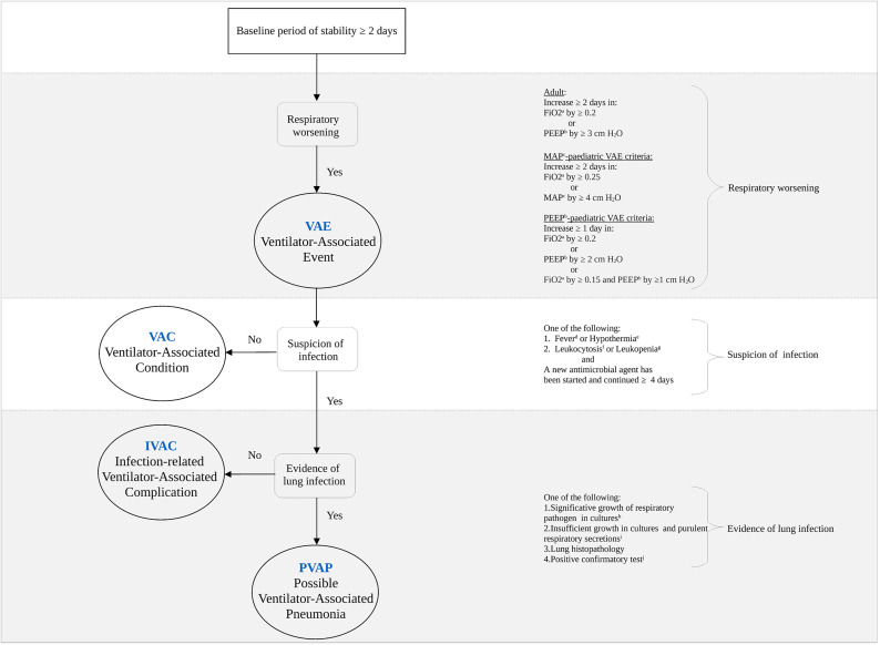 Figure 1: Hierarchical surveillance algorithm for Ventilator-Associated Events (VAE) established by the CDC NHSN, categorizing progressive respiratory deterioration into Ventilator-Associated Condition (VAC), Infection-related Ventilator-Associated Complication (IVAC), and Possible/Probable Ventilator-Associated Pneumonia (PVAP). Reproduced from Zilahi et al., Journal of Intensive Medicine (2022), DOI: 10.1016/j.jointm.2022.09.004 under CC BY-NC-ND 4.0.
1. Tier 1: Ventilator-Associated Condition (VAC)
A VAC represents an objective, non-specific signal of acute pulmonary deterioration regardless of underlying etiology (e.g., pulmonary edema, atelectasis, mucous plugging, pneumothorax, pulmonary embolism, or infection).
- Baseline Period: The patient must demonstrate at least $\ge 2$ consecutive calendar days of stable or decreasing daily minimum oxygenation requirements:
- Minimum daily Positive End-Expiratory Pressure ($\text{PEEP}_{\min}$), or
- Minimum daily Fraction of Inspired Oxygen ($\text{FiO}_{2,\min}$).
- Worsening Oxygenation Threshold: Following the baseline period, the patient experiences acute, sustained oxygenation worsening for $\ge 2$ consecutive calendar days, defined by either:
- An increase in daily minimum $\text{PEEP}$ of $\ge 3\text{ cmH}_2\text{O}$ over the daily minimum $\text{PEEP}$ of the baseline period: $$\Delta \text{PEEP}_{\min} \ge 3\text{ cmH}_2\text{O}$$
- An increase in daily minimum $\text{FiO}_2$ of $\ge 20\text{ points}$ ($\ge 0.20$) over the daily minimum $\text{FiO}_2$ of the baseline period: $$\Delta \text{FiO}_{2,\min} \ge 0.20\text{ (i.e., }+20\%\text{)}$$
- Exclusion Criteria: Days on mechanical ventilation prior to day 3 of mechanical ventilation cannot trigger a VAE (i.e., the earliest a VAC can occur is mechanical ventilation day 3, with days 1–2 serving as baseline).
2. Tier 2: Infection-related Ventilator-Associated Complication (IVAC)
An IVAC narrows the scope of VAC to episodes driven by systemic infection or severe inflammation that prompt new therapeutic antimicrobial administration.
- Underlying Requirement: Must strictly satisfy all criteria for VAC.
- Systemic Inflammatory Trigger: On or within $\pm 2$ calendar days of the onset of worsening oxygenation (the VAC window period), the patient exhibits at least one of the following:
- Abnormal Temperature: Core body temperature $< 36.0^\circ\text{C}$ ($< 96.8^\circ\text{F}$) or $> 38.0^\circ\text{C}$ ($> 100.4^\circ\text{F}$).
- Abnormal Leukocyte Count: White blood cell (WBC) count $\le 4{,}000/\mu\text{L}$ (leukopenia) or $\ge 12{,}000/\mu\text{L}$ (leukocytosis).
- Antimicrobial Requirement: A new qualifying antimicrobial agent (QAA) is initiated on or within $\pm 2$ calendar days of the VAC onset date and is continued for $\ge 4$ consecutive Qualifying Antimicrobial Days (QADs).
3. Tier 3: Possible and Probable Ventilator-Associated Pneumonia (VAP)
Tier 3 identifies respiratory tract-specific infection based on objective laboratory and microbiological markers of purulence and bacterial growth.
- Underlying Requirement: Must strictly satisfy all criteria for IVAC.
- Microbiological / Laboratory Verification (obtained within the VAE window period):
- Purulent Respiratory Secretions: Direct microscopic examination of respiratory secretions demonstrating:
- $\ge 25$ neutrophils (polymorphonuclear cells [PMNs]) per low-power field ($\text{LPF}, \times 100$), and
- $\le 10$ squamous epithelial cells per $\text{LPF}$ (indicating low oropharyngeal contamination).
- Quantitative or Semi-Quantitative Microbial Cultures: Positive growth of pathogenic microorganisms meeting standard diagnostic colony-forming unit (CFU) thresholds:
- Bronchoalveolar Lavage (BAL): $\ge 10^4\text{ CFU/mL}$ (or $\ge 1+$ / moderate/heavy semi-quantitative growth).
- Protected Specimen Brush (PSB) / Mini-BAL: $\ge 10^3\text{ CFU/mL}$.
- Endotracheal Aspirate (ETA): $\ge 10^5\text{ CFU/mL}$ (or $\ge 3+$ / heavy semi-quantitative growth).
- Alternative Diagnostic Criteria: Positive pleural fluid culture with organism matching respiratory tract isolate, histopathologic lung tissue evidence of pneumonia, or positive diagnostic tests for specific respiratory pathogens (e.g., Legionella urinary antigen, viral PCR for Influenza/RSV).
- Purulent Respiratory Secretions: Direct microscopic examination of respiratory secretions demonstrating:
Evidence-Based VAP Prevention Bundles (IHI, CDC, SHEA/IDSA)
Ventilator-Associated Pneumonia occurs primarily through the microaspiration of contaminated oropharyngeal secretions and gastric refluxates pooling above the endotracheal tube (ETT) cuff, coupled with biofilm formation on the luminal and extraluminal surfaces of the artificial airway. Implementing structured, multidisciplinary care bundles substantially reduces VAP incidence, ICU length of stay, and duration of mechanical ventilation.
                  ┌─────────────────────────────────────────────────────────┐
                  │          CORE VAP PREVENTION BUNDLE MEASURES            │
                  └─────────────────────────────────────────────────────────┘
                                               │
     ┌──────────────────────┬──────────────────┴──────────────────┬──────────────────────┐
     ▼                      ▼                                     ▼                      ▼
[ Positioning ]      [ Liberation ]                         [ Airway & Cuff ]      [ Circuit Integrity ]
• HOB 30°-45°        • Daily Sedation Interruption (DSI)    • Subglottic Suction   • No routine changes
• Lateral rotational • Spontaneous Breathing Trial (SBT)    • Cuff 20-30 cmH2O     • Humidifier / HME care
  positioning        • Early mobilization & rehab           • Non-chlorhexidine    • Closed inline suction
                                                              oral care
1. Head of Bed (HOB) Elevation ($30^\circ\text{--}45^\circ$)
- Mechanism: Semi-recumbent positioning leverages gravity to diminish passive gastroesophageal reflux, reduce retrograde pharyngeal colonization by enteric Gram-negative bacilli, and prevent macro- and microaspiration.
- Clinical Target: Maintain continuous head-of-bed elevation between $30^\circ$ and $45^\circ$ unless contraindicated by acute spinal trauma, hemodynamic instability, active prone positioning, or specific surgical procedures.
- Evidence Level: High (Standard of Care across IHI, SHEA, ATS/IDSA guidelines).
2. Daily Sedation Interruption (DSI) and Spontaneous Breathing Trials (SBT)
- Mechanism: Excessive, unmonitored continuous sedation suppresses protective airway reflexes, prolongs mechanical ventilation duration, promotes immobility-induced atelectasis, and drastically increases VAP risk.
- Protocolized Approach ("Wake Up and Breathe"):
- Spontaneous Awakening Trial (SAT): Daily temporary cessation of sedative and opioid infusions (titrated to light sedation, Richmond Agitation-Sedation Scale [RASS] target $-1$ to $0$).
- Spontaneous Breathing Trial (SBT): If SAT is passed without signs of severe agitation, tachypnea, or desaturation, proceed directly to an SBT (using T-piece, CPAP $5\text{ cmH}_2\text{O}$, or Pressure Support $\le 5\text{--}7\text{ cmH}_2\text{O}$ for $30\text{--}120\text{ minutes}$).
- Clinical Impact: Shortens mechanical ventilation time by an average of $2\text{--}4\text{ days}$, decreases ICU mortality, and expedites liberation.
3. Subglottic Secretion Drainage (SSD) Endotracheal Tubes
- Mechanism: Endotracheal intubation creates an anatomical dead space between the vocal cords and the inflated balloon cuff, where secretions continuously pool. SSD tubes feature an integrated dorsal suction lumen terminating just above the cuff ($0.5\text{--}1.0\text{ cm}$ cranial to the balloon), allowing continuous or intermittent evacuation of subglottic secretions.
- Operational Parameters:
- Continuous suction at low negative pressures ($-20\text{ to }-30\text{ mmHg}$) or intermittent automated suction at $-100\text{ to }-150\text{ mmHg}$ for $10\text{--}15\text{ seconds}$ every $1\text{--}2\text{ hours}$.
- Periodic clearance of the suction lumen with a small bolus of sterile air or saline if occlusion occurs.
- Clinical Impact: Meta-analyses show a $40\%\text{--}50\%$ relative risk reduction in VAP, particularly in patients intubated for $> 48\text{--}72\text{ hours}$.
4. Endotracheal Tube Cuff Pressure Optimization ($20\text{--}30\text{ cmH}_2\text{O}$)
- Physiological Imperative:
- Undercuffing ($P_{\text{cuff}} < 20\text{ cmH}_2\text{O}$): Insufficient seal allows pooling subglottic secretions, colonized saliva, and gastric contents to leak past the longitudinal microfolds of polyvinyl chloride (PVC) cuffs into the lower respiratory tract, triggering microaspiration.
- Overcuffing ($P_{\text{cuff}} > 30\text{ cmH}_2\text{O}$): Exceeds capillary perfusion pressure of the tracheal mucosal microvasculature ($\approx 25\text{--}30\text{ mmHg}$), inducing mucosal ischemia, ulceration, chondritis, cartilage necrosis, and eventual tracheomalacia or post-extubation tracheal stenosis.
- Monitoring Regimen: Measure and adjust cuff pressure at least every $6\text{--}8\text{ hours}$ using a calibrated handheld manometer, or deploy continuous pneumatic automated cuff controllers. Polyurethane (PU) cuffs with tapered geometry further minimize microchannel formation compared to standard thick PVC cuffs.
5. Oral Hygiene: Chlorhexidine vs. Toothbrushing
- Historical Practice: Routine application of $0.12\%$ or $0.2\%$ chlorhexidine gluconate (CHG) oral rinse was historically universal in VAP bundles.
- Contemporary Evidence & Guidelines:
- While CHG reduces bacterial burden and lower respiratory tract colonization, large randomized trials and meta-analyses revealed a concerning signal of increased all-cause mortality in non-cardiac surgical ICU patients exposed to routine chlorhexidine (likely due to chemical microaspiration, mucosal desquamation, and impaired local mucosal immunity).
- Current Recommendation (SHEA/IDSA 2022 Update): Routine chlorhexidine is no longer universally recommended for all medical ICU patients. Instead, comprehensive oral hygiene protocols emphasizing mechanical toothbrushing twice daily, regular suctioning of oral secretions, and moisturizing mucosal gels without routine antiseptic toxicity are prioritized.
6. Peptic Ulcer Disease (PUD) & Deep Vein Thrombosis (DVT) Prophylaxis
- PUD Prophylaxis:
- Indicated for high-risk patients (coagulopathy, mechanical ventilation anticipated $> 48\text{ hours}$, traumatic brain injury, thermal injury).
- Clinical Nuance: Histamine-2 receptor antagonists ($\text{H}_2\text{RAs}$) and Proton Pump Inhibitors (PPIs) elevate gastric pH, eliminating the natural acid barrier and facilitating gastric bacterial overgrowth (predominantly enteric Gram-negative bacilli). Sucralfate preserves gastric acidity and is associated with lower VAP rates than PPIs, though PPIs provide superior stress ulcer bleeding prophylaxis. Early enteral nutrition also provides mucosal cytoprotection.
- DVT Prophylaxis:
- Routine administration of low-molecular-weight heparin (LMWH) or unfractionated heparin (UFH) coupled with sequential compression devices (SCDs) prevents fatal pulmonary thromboembolism.
7. Ventilator Circuit Management
- Strict Avoidance of Routine Circuit Changes: Ventilator breathing circuits (tubing, Y-piece, exhalation valves) should NOT be changed on a scheduled, routine basis (e.g., every 7 days). Routine disconnection disrupts closed system integrity and aerosolizes colonized condensate directly into the patient's airway.
- Change Indications: Circuits should be changed only when visibly soiled, contaminated with blood/vomitus, or mechanically malfunctioning.
- Condensate Management: Closed-circuit in-line suction catheters should be used. Drainage traps must be placed at dependent loop positions, and heated wire humidifiers must be regulated to prevent pooled condensate from draining toward the patient during bed repositioning.
Mechanical and Structural Complications of Mechanical Ventilation
Beyond infectious risks, positive-pressure mechanical ventilation imposes profound biophysical and biomechanical stresses upon the respiratory system, thoracic vasculature, and airway anatomy.
                             POSITIVE-PRESSURE VENTILATION COMPLICATIONS
                                                  │
     ┌────────────────────────┬───────────────────┴───────────────────┬────────────────────────┐
     ▼                        ▼                                       ▼                        ▼
[ Barotrauma & Volutrauma ] [ Dynamic Hyperinflation ]     [ Airway Complications ]  [ Diaphragm Dysfunction ]
• Pneumothorax              • Auto-PEEP / Intrinsic PEEP   • Tracheal stenosis       • VIDD
• Pneumomediastinum         • Venous return obstruction    • Tracheomalacia          • Proteolysis & atrophy
• Subcutaneous emphysema    • Hemodynamic collapse         • Cuff pressure necrosis  • Delayed liberation
1. Barotrauma and Volutrauma
Barotrauma describes extra-alveolar air accumulation resulting from alveolar rupture under excessive airway pressures, transpulmonary pressures ($P_{\text{tp}}$), and end-inspiratory alveolar distension (volutrauma).
- Pathophysiological Cascade (Macklin Effect): Excessive alveolar wall tension causes alveolar epithelial-endothelial disruption at the alveolar-bronchovascular sheath junction. Escaping gas dissects centripetally along the bronchovascular sheaths into the mediastinum: $$\text{Alveolar Rupture} \longrightarrow \text{Pneumomediastinum} \longrightarrow \begin{cases} \text{Subcutaneous Emphysema (cervical/fascial planes)} \\ \text{Pneumothorax (rupture of mediastinal pleura)} \\ \text{Pneumoperitoneum / Pneumopericardium} \end{cases}$$
- Clinical Spectrum:
- Tension Pneumothorax: Sudden elevation in peak inspiratory pressure ($P_{\text{peak}}$) and plateau pressure ($P_{\text{plat}}$), abrupt arterial desaturation, absent unilateral breath sounds, tracheal deviation, and obstructive shock from right atrial collapse and impaired venous return.
- Pneumomediastinum & Subcutaneous Emphysema: Crepitus over the neck, chest wall, and axilla; Hamman's crunch on cardiac auscultation.
- Mitigation Strategy: Implement lung-protective ventilation:
- Low tidal volume ($V_t = 4\text{--}8\text{ mL/kg}$ Predicted Body Weight [PBW]).
- Limit plateau pressure: $P_{\text{plat}} \le 30\text{ cmH}_2\text{O}$.
- Minimize driving pressure: $\Delta P = P_{\text{plat}} - \text{PEEP} \le 14\text{--}15\text{ cmH}_2\text{O}$.
- Restrict total mechanical power: $\text{MP} < 17\text{--}20\text{ J/min}$.
2. Dynamic Hyperinflation, Auto-PEEP, and Hemodynamic Collapse
Dynamic hyperinflation occurs when the expiratory time ($T_e$) is insufficient to permit complete exhalation to the static functional residual capacity (FRC) prior to the initiation of the subsequent breath.
- Physiological Mechanisms:
- Severe airflow limitation (bronchospasm, airway inflammation, luminal secretions in COPD/Asthma).
- Short expiratory time (high respiratory rates, inverted I:E ratios).
- High minute ventilation demands.
- Pathophysiology of Intrinsic PEEP ($\text{PEEP}_i$): Alveolar gas remains trapped at positive pressure at end-expiration: $$\text{Total PEEP} = \text{Set PEEP} + \text{Auto-PEEP (\text{PEEP}_i)}$$
- Cardiovascular Collapse: The markedly elevated intrathoracic pressure exerts direct mechanical compression upon the superior and inferior vena cava, severely reducing right ventricular preload ($\downarrow \text{EDV}$). Simultaneously, increased pulmonary vascular resistance elevates right ventricular afterload, shifting the interventricular septum leftward (ventricular interdependence) and precipitating profound hypotension, electromechanical dissociation, and cardiac arrest.
- Bedside Recognition & Management:
- Measurement: Perform an end-expiratory occlusion hold maneuver on a passive patient to quantify $\text{Auto-PEEP}$. Observe failure of the expiratory flow waveform to return to zero before the next inhalation.
- Immediate Acute Rescue: Disconnect the patient from the ventilator circuit immediately to allow passive, uninterrupted exhalation.
- Ventilator Adjustments: Increase peak inspiratory flow rate ($70\text{--}90\text{ L/min}$ using a square or ramp waveform), reduce respiratory rate, decrease tidal volume, and optimize bronchodilator therapy to prolong $T_e$.
3. Airway and Tracheal Complications
Direct physical trauma from the endotracheal tube, stylets, and pneumatic cuff inflation can induce irreversible structural damage to the upper airway:
- Tracheal Ischemia and Necrosis: Sustained cuff pressures exceeding mucosal capillary pressure ($> 30\text{ cmH}_2\text{O}$) induce ischemic necrosis of the tracheal mucosa over the cartilaginous rings within $48\text{ hours}$.
- Tracheomalacia: Destruction of tracheal cartilaginous support causes dynamic airway collapse during expiration.
- Tracheal Stenosis: Fibroblastic proliferation and cicatricial scarring during the healing phase cause circumferential subglottic or tracheal narrowing, presenting weeks to months post-extubation with progressive stridor and dyspnea.
- Tracheoesophageal (TE) Fistula: Prolonged pressure necrosis between an overinflated ETT cuff and an indwelling rigid nasogastric tube erodes the posterior tracheal wall into the anterior esophagus, causing massive aspiration, recurrent pneumonia, and gastric distension with ventilator gas.
- Tracheoinnominate Artery Fistula (TIF): A catastrophic, life-threatening complication of low tracheostomy tube placement (below the 3rd–4th tracheal ring), where the cuff or distal tip erodes into the brachiocephalic (innominate) artery, causing massive hemoptysis.
4. Ventilator-Induced Diaphragmatic Dysfunction (VIDD)
Ventilator-Induced Diaphragmatic Dysfunction (VIDD) represents rapid, progressive loss of diaphragmatic force-generating capacity and muscle mass induced by prolonged mechanical unloading of the respiratory pump.
                      PROLONGED CONTROLLED MECHANICAL VENTILATION
                                           │
         ┌─────────────────────────────────┴─────────────────────────────────┐
         ▼                                                                   ▼
[ Myofibrillar Inactivity & Unloading ]                          [ Cellular Stress Cascades ]
• Absence of mechanical strain                                   • Mitochondrial ROS generation
• Downregulation of protein synthesis                            • Oxidative contractile protein damage
         │                                                                   │
         └─────────────────────────────────┬─────────────────────────────────┘
                                           ▼
                       [ Accelerated Proteolytic Activation ]
                       • Ubiquitin-Proteasome System (MuRF1, Atrogin-1)
                       • Autophagy-Lysosome Degradation
                       • Calpain & Caspase-3 Protease Cleavage
                                           │
                                           ▼
                       [ Diaphragm Atrophy & Force Deficit ]
                       • Rapid loss of cross-sectional area (up to 50% in 48h)
                       • Severe weaning failure & prolonged mechanical ventilation
- Cellular Mechanisms:
- Complete mechanical silencing (controlled mechanical ventilation with heavy sedation or neuromuscular blockade) halts sarcomeric protein synthesis and triggers hyperactivation of endogenous proteolytic pathways:
- The Ubiquitin-Proteasome System (upregulation of muscle-specific E3 ligases MuRF1 and Atrogin-1).
- The Autophagy-Lysosome Pathway.
- Calpains and Caspase-3 proteases activated by mitochondrial reactive oxygen species (ROS).
- Diaphragmatic cross-sectional muscle fiber area declines by as much as $50\%$ within the first $18\text{--}48\text{ hours}$ of full ventilator support—a rate substantially faster than that observed in peripheral skeletal muscles.
- Complete mechanical silencing (controlled mechanical ventilation with heavy sedation or neuromuscular blockade) halts sarcomeric protein synthesis and triggers hyperactivation of endogenous proteolytic pathways:
- Clinical Consequences:
- Failure of spontaneous breathing trials, difficult weaning, prolonged ICU dependency, and heightened mortality.
- Prevention & Preservation:
- Transition from fully controlled to assisted/spontaneous modes (e.g., Pressure Support Ventilation, Neurally Adjusted Ventilatory Assist [NAVA], Proportional Assist Ventilation [PAV+]) as early as clinically feasible.
- Maintain modest diaphragmatic contractile effort (targeting an inspiratory esophageal pressure swing $\Delta P_{\text{es}} = 3\text{--}8\text{ cmH}_2\text{O}$ or a diaphragm thickening fraction on ultrasound $\text{TF}_{\text{diaph}} = 15\%\text{--}30\%$). Avoid both diaphragmatic over-assistance (atrophy) and under-assistance (fatigue and load-induced injury).
Comprehensive Summary Table: Complications, Mechanisms, and Bundled Interventions
| Complication Category | Specific Pathophysiology | Key Diagnostic / Surveillance Criteria | Evidence-Based Prevention Bundle Element |
|---|---|---|---|
| Infectious (VAE Tier 1: VAC) | Alveolar derecruitment, atelectasis, fluid overload, pulmonary edema | $\ge 2$ days baseline stability followed by $\Delta \text{PEEP} \ge 3\text{ cmH}_2\text{O}$ or $\Delta \text{FiO}_2 \ge 0.20$ for $\ge 2$ days | Conservative fluid management, lung-protective ventilation, lung recruitment. |
| Infectious (VAE Tier 2: IVAC) | Systemic inflammatory response to hospital-acquired infection | Meets VAC criteria + Temp $<36^\circ\text{C}$ or $>38^\circ\text{C}$ / WBC $\le 4\text{k}$ or $\ge 12\text{k}$ + $\ge 4$ QADs of new antimicrobial | Targeted antibiotic stewardship, strict aseptic catheter management, diagnostic workup. |
| Infectious (VAE Tier 3: PVAP) | Bacterial colonization, biofilm formation, microaspiration of secretions | Meets IVAC + purulent sputum ($\ge 25\text{ PMNs}, \le 10\text{ SECs/LPF}$) OR quantitative culture ($\text{BAL} \ge 10^4$, $\text{ETA} \ge 10^5\text{ CFU/mL}$) | Subglottic suction ETTs, HOB $30^\circ\text{--}45^\circ$, mechanical toothbrushing, avoid routine circuit changes. |
| Barotrauma / Volutrauma | High transpulmonary pressure, alveolar overdistension, Macklin effect | $P_{\text{plat}} > 30\text{ cmH}_2\text{O}$, $\Delta P > 15\text{ cmH}_2\text{O}$, extra-alveolar air on chest radiograph / CT / ultrasound | Low tidal volume ($4\text{--}8\text{ mL/kg}$ PBW), limit $P_{\text{plat}} \le 30$, limit $\Delta P \le 14\text{ cmH}_2\text{O}$, mechanical power $< 17\text{ J/min}$. |
| Dynamic Hyperinflation | Incomplete exhalation, airway resistance, air trapping | $\text{Auto-PEEP} > 0\text{ cmH}_2\text{O}$, incomplete expiratory flow, hypotension, thoracic hyperresonance | Prolong $T_e$, optimize inspiratory flow ($70\text{--}90\text{ L/min}$), reduce respiratory rate, disconnect for acute arrest. |
| Airway Mucosal Necrosis / Stenosis | Capillary perfusion pressure exceeded by cuff ($> 30\text{ cmH}_2\text{O}$) | Measured cuff pressure $> 30\text{ cmH}_2\text{O}$, post-extubation stridor, subglottic narrowing | Maintain cuff pressure $20\text{--}30\text{ cmH}_2\text{O}$ via manometer every $6\text{--}8\text{ hours}$, polyurethane cuffs. |
| Diaphragm Dysfunction (VIDD) | Diaphragmatic unloading, ubiquitin-proteasome proteolysis, oxidative stress | Diaphragmatic ultrasound thickening fraction $< 15\%$, low maximal inspiratory pressure ($P_{I,\max}$) | Daily sedation interruption, early SAT/SBT, titration to assisted modes (PSV/NAVA/PAV+) maintaining modest effort. |
Clinical Practice Pearls
- Surveillance vs. Clinical Diagnosis: The CDC VAE criteria are strictly epidemiological surveillance tools designed for objective hospital benchmarking; they do not dictate individual bedside antimicrobial initiation. A patient may require targeted antimicrobial therapy for clinical VAP even if VAE criteria are not fully met, and conversely, a VAC may be triggered by cardiogenic pulmonary edema requiring diuresis rather than antibiotics.
- Cuff Pressure Pitfall: Never rely on subjective digital palpation of the pilot balloon to assess endotracheal cuff pressure. Palpation fails to detect both severe underinflation ($< 15\text{ cmH}_2\text{O}$) and hazardous overinflation ($> 40\text{ cmH}_2\text{O}$); always use an inline calibrated analog or digital manometer.
- Sudden Deterioration ("DOPE" Mnemonic): When an intubated patient experiences acute, unexplained respiratory deterioration or hemodynamic collapse, systematically assess:
- Dislodgement / Displacement of ETT (right mainstem or accidental extubation).
- Obstruction of ETT (mucous plug, blood clot, tube kinking, biting).
- Pneumothorax / Pulmonary embolism / Auto-PEEP.
- Equipment failure (ventilator malfunction, gas source disconnect, circuit leak).
References & Seminal Literature
- Magill SS, Klompas M, Balk R, et al. Developing a new, objective, predictable agent for surveillance of ventilator-associated events in intensive care units. Crit Care Med. 2013;41(11):2467-2475.
- Klompas M, Branson R, Eichenwald EC, et al. Strategies to prevent ventilator-associated pneumonia in acute care hospitals: 2014 Updates. Infect Control Hosp Epidemiol. 2014;35(8):915-936.
- Zilahi G, Artigas A, Martin-Loeches I. Ventilator-associated events: From surveillance to optimizing management. J Intensive Med. 2022;3(2):107-113. DOI: 10.1016/j.jointm.2022.09.004. (PMCID: PMC10391577).
- Klompas M, Li L, Kleinman K, et al. Associations Between Ventilator Bundle Components and Outcomes in Ventilated Patients. JAMA Intern Med. 2016;176(9):1277-1283.
- Vassilakopoulos T, Petrof BJ. Ventilator-induced diaphragmatic dysfunction. Am J Respir Crit Care Med. 2004;169(3):336-341.
- Papazian L, Klompas M, Luyt CE. Ventilator-associated pneumonia in adults: a narrative review. Intensive Care Med. 2020;46(5):888-906.