Positive airway pressure fundamentally couples the mechanics of the respiratory system with the hemodynamics of the cardiovascular circuit. In positive pressure ventilation (PPV), the heart and the intrathoracic great vessels reside within an engineered pressure chamber where fluctuations in airway, alveolar, and pleural pressures directly dictate cardiac preload, right and left ventricular afterload, myocardial transmural pressure gradients, and systemic organ perfusion.
Mastery of cardiopulmonary interactions is indispensable for the intensivist. Titration of positive end-expiratory pressure (PEEP) and mean airway pressure ($mPaw$) involves a delicate equilibrium: recruiting atelectatic lung units, optimizing functional residual capacity (FRC), and minimizing cyclic atelectotrauma against the hazards of alveolar overdistension, pulmonary vascular resistance (PVR) escalation, right ventricular (RV) failure, and systemic hypoperfusion.
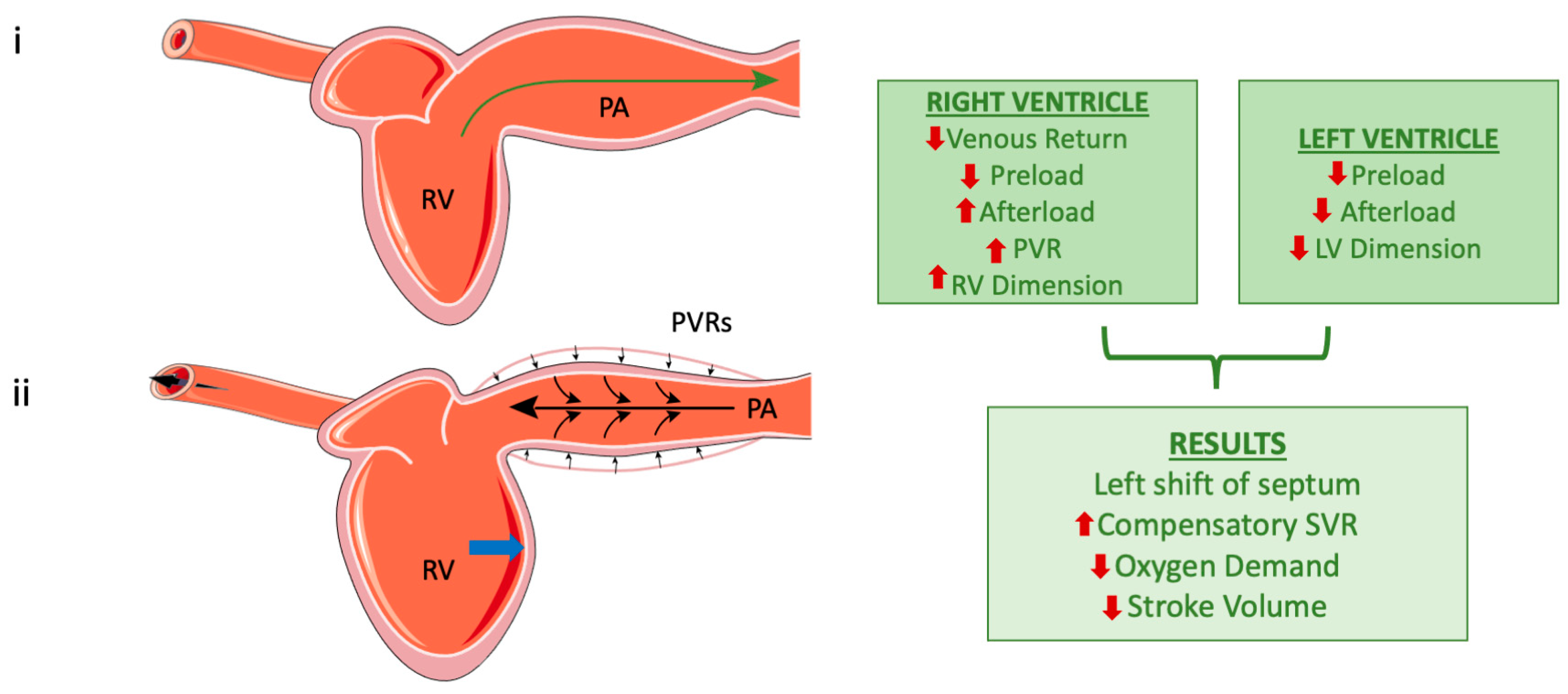 Figure 1: Pathophysiological cascade of heart-lung interactions under positive pressure ventilation. Top: Normal spontaneous breathing with physiological subatmospheric pleural pressure facilitating systemic venous return. Bottom: Positive pressure ventilation with elevated PEEP and mean airway pressure causes alveolar capillary compression, increases pulmonary vascular resistance (PVR), elevates RV afterload, and induces RV chamber enlargement. RV dilation shifts the interventricular septum toward the left ventricle (reversed Bernheim effect), impairing LV diastolic filling, reducing stroke volume, and depressing mean arterial pressure. Sourced from Zakynthinos et al., J Clin Med 2024;13(24):7788. DOI: 10.3390/jcm13247788 (Open Access / CC BY 4.0).
Positive End-Expiratory Pressure and Mean Airway Pressure ($mPaw$)
Mathematical Derivation and Determinants of $mPaw$
Mean airway pressure ($mPaw$ or $\bar{P}_{aw}$) represents the time-weighted average of airway pressure across an entire respiratory cycle ($T_{total} = T_i + T_e$). It serves as the primary physical correlate of mean alveolar distending volume and oxygenation in passive lungs.
For a rectangular (square) pressure waveform (typical of Pressure-Controlled Ventilation or Pressure Support):
$$mPaw = \frac{P_{peak} \times T_i + PEEP \times T_e}{T_{total}} = (P_{peak} - PEEP) \times \frac{T_i}{T_{total}} + PEEP$$Where:
- $P_{peak}$ is the peak inspiratory pressure ($\text{cmH}_2\text{O}$).
- $PEEP$ is the extrinsic positive end-expiratory pressure ($\text{cmH}_2\text{O}$).
- $T_i$ is the inspiratory time ($\text{seconds}$).
- $T_e$ is the expiratory time ($\text{seconds}$).
- $T_{total} = T_i + T_e = \frac{60}{RR}$ ($\text{seconds}$).
- $\frac{T_i}{T_{total}}$ is the inspiratory duty cycle.
In constant-flow Volume-Controlled Ventilation (where inspiratory pressure rises linearly with an inspiratory pause $T_{pause}$):
$$mPaw \approx PEEP + \frac{T_i}{T_{total}} \left( \frac{P_{peak} + P_{plat}}{2} - PEEP \right)$$Key physiological determinants that elevate $mPaw$:
- PEEP: Shifts the baseline pressure upward throughout both inspiration and expiration, exerting the strongest continuous effect on $mPaw$.
- Inspiratory Time / I:E Ratio: Prolonging $T_i$ (e.g., from 1:2 to 1:1 or inverse ratio ventilation 2:1) expands the fraction of the cycle spent at high inspiratory pressures.
- Inspiratory Driving Pressure ($\Delta P = P_{plat} - PEEP$) or Peak Inspiratory Pressure: Increases the amplitude of the pressure curve.
- Flow Waveform Profile: Decelerating ramp flow waveforms maintain higher average inspiratory pressures across $T_i$ than square flow waveforms, slightly elevating $mPaw$ for identical delivered tidal volumes.
Pressure (cmH2O)
  ^
  |          +-----------------+ (Pplat / Pinsp)
  |         /|                 |
  |        / |    Inspiration  |
  |       /  |     (Ti area)   |
  |      /   |                 \
  |     /    |                  \
  |----+     |                   +---------------------------+ (PEEP)
  |          |                   |        Expiration         |
  |          |                   |         (Te area)         |
  +----------+-------------------+---------------------------+--> Time
             <-------- Ti -------><------------ Te ----------->
             <---------------------- Ttotal ------------------>

             mPaw = [ Area(Ti) + Area(Te) ] / Ttotal
Impact of PEEP and $mPaw$ on Functional Residual Capacity (FRC)
In health, Functional Residual Capacity (FRC) represents the volume of gas remaining in the lungs at the end of a passive exhalation, where the inward elastic recoil of the lung balances the outward elastic recoil of the chest wall.
In acute respiratory failure (ARDS, acute lung injury, severe atelectasis, supine anesthesia), FRC is dramatically contracted due to:
- Surfactant deactivation and alveolar capillary leak flooding.
- Dependent compressive atelectasis from increased lung mass (edematous lung weight).
- Upward displacement of the relaxed hemidiaphragms by intra-abdominal contents.
When end-expiratory lung volume falls below the Closing Capacity (CC), dependent terminal bronchioles and alveoli collapse during every expiration, resulting in:
- Intrapulmonary Shunting ($V/Q = 0$): Mixed venous blood bypasses ventilated gas exchange units, leading to severe refractory hypoxemia.
- Atelectotrauma: Cyclic opening and collapse of unstable alveolar units generate colossal shear stresses at the interfaces between open and collapsed alveoli, tearing alveolar-capillary membranes and precipitating biotrauma.
Application of therapeutic PEEP restores and expands FRC above Closing Capacity. By maintaining a continuous positive distending pressure during expiration, PEEP recruits recruitable alveolar units, stabilizes open alveoli, splints small airways open, and converts non-ventilated shunt fractions ($V/Q = 0$) into functional exchange units ($V/Q > 0$).
Cardiopulmonary Hemodynamic Interactions Under Positive Pressure Ventilation
The thoracic cage behaves as an isobaric chamber surrounding the cardiac chambers, pulmonary vascular bed, and intrathoracic portions of the venae cavae and aorta. Applying positive pressure within the airway transmits pressure across the lung parenchyma into the pleural space ($P_{pl}$), pericardial space ($P_{peri}$), and right atrium ($P_{ra}$).
$$\Delta P_{pl} = \Delta P_{aw} \times \left( \frac{C_L}{C_L + C_{cw}} \right) = \Delta P_{aw} \times \left( \frac{E_{cw}}{E_{rs}} \right)$$In stiff ARDS lungs (low lung compliance $C_L$, high lung elastance $E_L$), less airway pressure is transmitted to the pleural space ($20\% - 30\%$). Conversely, in patients with high chest wall elastance $E_{cw}$ (morbid obesity, abdominal compartment syndrome, tight chest wall binding), up to $50\% - 80\%$ of airway pressure is transmitted into the pleural space, imposing extreme hemodynamic loads.
       +-------------------------------------------------------------+
       |             POSITIVE PRESSURE VENTILATION / PEEP            |
       +-------------------------------------------------------------+
                     |                                 |
                     v                                 v
       +---------------------------+     +---------------------------+
       |  INCREASED PLEURAL / RA   |     |     ALVEOLAR EXPANSION    |
       |       PRESSURE (Pra)      |     |     & HIGH LUNG VOLUMES   |
       +---------------------------+     +---------------------------+
                     |                                 |
         +-----------+-----------+                     v
         |                       |        +--------------------------+
         v                       v        |  COMPRESSION OF ALVEOLAR |
+------------------+   +------------------+   CAPILLARIES (WEST Z. 1)|
| DECREASED VENOUS |   | DECREASED LV     |   +--------------------------+
| RETURN GRADIENT  |   | TRANSMURAL       |                |
| (Pmsf - Pra)     |   | AFTERLOAD        |                v
+------------------+   +------------------+   +--------------------------+
         |                       |            |  INCREASED PULMONARY     |
         v                       v            |  VASCULAR RESISTANCE(PVR)|
+------------------+   +------------------+   +--------------------------+
| DECREASED RV     |   | FAVORS LV        |                |
| PRELOAD (OUTPUT) |   | SYSTOLIC EJECTION|                v
+------------------+   +------------------+   +--------------------------+
         |                                    |  INCREASED RV AFTERLOAD  |
         +-----------------+                  +--------------------------+
                           |                               |
                           v                               v
                 +---------------------------------------------------+
                 |      RV DILATION & D-SHAPED LV (SEPTAL SHIFT)     |
                 |             (REVERSED BERNHEIM EFFECT)            |
                 +---------------------------------------------------+
                                           |
                                           v
                 +---------------------------------------------------+
                 | DECREASED LV DIASTOLIC COMPLIANCE & PRELOAD FILL  |
                 +---------------------------------------------------+
                                           |
                                           v
                 +---------------------------------------------------+
                 |    PRECIPITOUS FALL IN STROKE VOLUME & MAP        |
                 |            (HEMODYNAMIC COLLAPSE)                 |
                 +---------------------------------------------------+
1. Right Ventricular Interactions
A. Venous Return Impedance (Reduced RV Preload)
Venous return ($VR$) to the right atrium is governed by Guyton's hydraulic relationship:
$$VR = \frac{P_{msf} - P_{ra}}{R_{vr}}$$Where:
- $P_{msf}$ is the Mean Systemic Filling Pressure (the driving pressure upstream in the compliant venous capacitance vessels, typically $7 - 10\text{ mmHg}$).
- $P_{ra}$ is the Right Atrial Pressure (the downstream back-pressure opposing venous inflow).
- $R_{vr}$ is the resistance to venous return.
During positive pressure inspiration and with increasing PEEP, the rise in intrathoracic pleural pressure directly elevates $P_{ra}$. As $P_{ra}$ approaches $P_{msf}$, the pressure gradient $(P_{msf} - P_{ra})$ narrows, decelerating blood flow from the superior and inferior vena cava into the right atrium. In hypovolemic patients with low unstressed venous volume ($P_{msf}$ reduced to $4 - 6\text{ mmHg}$), even moderate PEEP ($5 - 10\text{ cmH}_2\text{O}$) can completely obliterate the venous return gradient, precipitating immediate circulatory collapse.
B. Biphasic Behavior of Pulmonary Vascular Resistance (RV Afterload)
Pulmonary Vascular Resistance (PVR) exhibits a distinctive U-shaped relationship with lung volume:
$$PVR = \frac{MPAP - PAWP}{CO}$$The pulmonary vasculature comprises two distinct anatomical and functional compartments:
- Alveolar Microvessels: Fragile capillary networks embedded within alveolar septa. As lung volume expands toward Total Lung Capacity (TLC) under high PEEP or tidal overdistension, alveolar septa stretch and thin out, compressing alveolar capillaries and drastically increasing alveolar vascular resistance. In West Zone 1 conditions ($P_{alv} > P_{art} > P_{ven}$), alveolar capillaries completely collapse, creating non-perfused alveolar dead space ($V/Q \to \infty$).
- Extra-Alveolar Vessels: Larger pulmonary arteries and veins coursing through bronchovascular bundles. These vessels are distended by the radial outward traction of surrounding lung parenchyma as lung volume rises. At low lung volumes (atelectasis, near Residual Volume), loss of radial tethering and hypoxic pulmonary vasoconstriction (HPV) cause extra-alveolar vessel collapse, increasing extra-alveolar resistance.
Pulmonary Vascular
Resistance (PVR)
  ^
  |        \               /  <-- Combined Total PVR Curve
  |         \   Minimum   /
  |          \   at FRC  /
  |           \   ____  /
  |   Extra-   \_/    \/    Alveolar Capillary Compression
  |   Alveolar             (Overdistension / High PEEP)
  |   Collapse
  |   (Atelectasis)
  +------------------------------------------------------------->
     Residual Volume        FRC                     TLC
                            Lung Volume
Clinical Takeaway: PVR is minimized at normal FRC. Optimal PEEP titration lowers PVR by reversing atelectasis and relieving hypoxic pulmonary vasoconstriction. Conversely, excessive PEEP expands lungs into the hyperinflated right arm of the curve, driving up PVR and RV afterload.
C. RV Dilation and Interventricular Ventricular Interdependence (Reversed Bernheim Effect)
The right ventricle is a thin-walled, highly compliant crescent-shaped pump designed to work into a low-impedance pulmonary circulation. When acute elevations in PEEP or lung overdistension abruptly increase PVR:
- RV end-systolic volume increases, leading to acute RV dilation.
- The dilated RV exceeds the non-distensible pericardial constraint.
- The interventricular septum shifts and bulges leftward during diastole (flattening the septum and producing the classic echocardiographic "D-shaped" left ventricle in parasternal short-axis view).
- This reversed Bernheim effect mechanically restricts LV diastolic filling, reducing LV end-diastolic volume (LVEDV) and stroke volume independently of intrinsic LV myocardial performance.
2. Left Ventricular Interactions
A. LV Preload Alterations
LV preload is compromised through two distinct, synergistic mechanisms during PPV:
- Serial Transit Limitation: Reduction in RV stroke volume (caused by impaired venous return and elevated PVR) decreases pulmonary venous outflow into the left atrium after a 2- to 3-beat transit delay.
- Direct Diastolic Ventricular Interdependence: Leftward septal shifting impairs LV compliance, elevating LV end-diastolic pressure (LVEDP) despite a lower true LV volume.
B. LV Transmural Afterload Reduction (The Favorable Effect in Systolic Heart Failure)
Left ventricular afterload is physically determined by the systolic LV transmural pressure ($P_{tm,LV}$):
$$P_{tm,LV} = P_{intraventricular} - P_{pericardial/pleural} = P_{systolic,LV} - P_{pl}$$- During Spontaneous Breathing: Vigorous inspiratory efforts generate highly negative pleural pressures ($P_{pl} \approx -10\text{ to } -25\text{ cmH}_2\text{O}$). This makes $P_{tm,LV} = P_{LV} - (-20) = P_{LV} + 20\text{ mmHg}$, substantially augmenting the systolic wall stress that the LV must overcome to eject blood into the extrathoracic aorta.
- Under Positive Pressure Ventilation and PEEP: Positive pleural pressure ($P_{pl} > 0\text{ mmHg}$) acts as an external squeezing force on the ventricular wall, reducing $P_{tm,LV} = P_{LV} - (+10) = P_{LV} - 10\text{ mmHg}$.
Cardiogenic Shock & Acute Pulmonary Edema: In severe dilated cardiomyopathy with acute pulmonary edema, applying CPAP or PEEP ($8 - 12\text{ cmH}_2\text{O}$) simultaneously:
- Reduces LV afterload by boosting $P_{pl}$.
- Decreases pathological LV preload (moving down the flat top of the Starling curve).
- Clears alveolar edema, improves compliance, and aborts respiratory muscle exhaustion.
- Increases cardiac index and stroke volume without requiring inotropic escalation.
"Best PEEP" Titration Strategies: Bedside Physiology and Protocols
Selecting the optimal PEEP level requires balancing alveolar recruitment against hemodynamic impairment and overdistension. Several evidence-based paradigms are employed at the bedside:
                            PEEP TITRATION PARADIGMS
                                       |
    +-------------------+--------------+--------------+-------------------+
    |                   |                             |                   |
    v                   v                             v                   v
+---------------+ +--------------------+ +--------------------+ +-------------------+
|  MAXIMAL CRS  | |  DECREMENTAL PEEP  | |    STRESS INDEX    | |    TRANSPULMONARY |
|  & LOWEST DP  | |   TITRATION POST-RM| |   (CONSTANT FLOW)  | |   ESOPHAGEAL (Pes)|
+---------------+ +--------------------+ +--------------------+ +-------------------+
| Target:       | | Target:            | | Target:            | | Target:           |
| min (Pplat-   | | PEEP 2 cmH2O above | | Straight linear    | | End-expiratory    |
| PEEP) for     | | sudden drop in     | | Paw waveform       | | PL,exp >= 0 to    |
| fixed Vt      | | compliance / PaO2  | | (SI = 1.0)         | | +2 cmH2O          |
+---------------+ +--------------------+ +--------------------+ +-------------------+
1. Maximum Compliance and Lowest Driving Pressure ($\Delta P$)
Amato et al. (NEJM 2015) identified driving pressure ($\Delta P = P_{plat} - PEEP = \frac{V_T}{C_{rs}}$) as the mechanical ventilatory variable most strongly correlated with ARDS survival.
- Protocol: With tidal volume strictly fixed at $6\text{ mL/kg PBW}$, PEEP is titrated incrementally or decrementally in $2\text{ cmH}_2\text{O}$ steps.
- End-Point: The PEEP level yielding the lowest $\Delta P$ (and thus the highest $C_{rs}$) represents the point where alveolar recruitment exceeds alveolar overdistension.
- Safety Bounds: Maintain $P_{plat} \le 30\text{ cmH}_2\text{O}$ and $\Delta P \le 14 - 15\text{ cmH}_2\text{O}$.
2. Decremental PEEP Titration Following a Recruitment Maneuver
Decremental titration exploits the hysteresis loop of the respiratory system: the pressure required to maintain recruited alveoli open along the deflation limb is significantly lower than the opening pressure required along the inflation limb.
Volume (mL)
  ^                                  Deflation Limb (Open Alveoli)
  |                                 /==================+ (Point of Overdistension)
  |                                //                  /
  |                               //                  /
  |                              //                  /  Inflation Limb
  |           Optimal PEEP ---> //                  /  (Recruitment required)
  |           (Closing Point + 2) \                /
  |                             \  \              /
  |                              \  \            /
  |                               \  \__________/ <-- Lower Inflection Point
  |                                \____________/
  +------------------------------------------------------------->
  0                                                              Pressure (cmH2O)
- Protocol Execution:
- Perform a controlled recruitment maneuver (e.g., CPAP $40\text{ cmH}_2\text{O}$ for 40 seconds, or stepwise staircase recruitment up to $P_{peak} = 45\text{ cmH}_2\text{O} / PEEP = 25\text{ cmH}_2\text{O}$ in deeply sedated/paralyzed patients).
- Set initial PEEP at $20 - 24\text{ cmH}_2\text{O}$ with $V_T = 6\text{ mL/kg PBW}$.
- Reduce PEEP downward in decrements of $2\text{ cmH}_2\text{O}$ every $2 - 3\text{ minutes}$.
- Record $C_{stat}$, $P_{plat}$, and peripheral $SpO_2$ at each step.
- Identify the "collapse point" (derecruitment PEEP), defined as the PEEP level where $C_{stat}$ abruptly decreases by $> 5 - 10\%$ or $SpO_2$ drops.
- Re-recruit the lung briefly with a recruitment maneuver, then set the definitive clinical PEEP at $2\text{ cmH}_2\text{O}$ above the collapse point.
3. Stress Index Dynamic Assessment
In volume-controlled ventilation with a constant (square) inspiratory flow profile, the shape of the dynamic airway pressure-time curve reflects instantaneous compliance changes across the tidal inflation:
$$P_{aw}(t) = a \cdot t^b + c$$Where the exponent $b$ is the Stress Index (SI):
- $SI < 1.0$ (Downward concavity / progressive flattening): Compliance is improving during inflation. Alveoli are undergoing continuous tidal recruitment. Action: Increase PEEP.
- $SI = 1.0$ (Straight linear rise): Compliance remains constant throughout tidal inflation. Ventilation is operating within the linear, safe zone without recruitment or overdistension. Action: Ideal PEEP setting.
- $SI > 1.0$ (Upward concavity / progressive steepening): Compliance is worsening as lung volume expands. Alveoli have reached their elastic limit and are undergoing severe tidal overdistension. Action: Decrease PEEP or reduce $V_T$.
4. Esophageal Manometry and Transpulmonary Pressure ($P_L$)
In patients with altered chest wall elastance (severe obesity, ascites, elevated intra-abdominal pressure, pleural effusions), airway pressure ($P_{aw}$) misrepresents the true distending stress applied across the lung tissue. Transpulmonary pressure ($P_L$) isolates lung stress:
$$P_L = P_{aw} - P_{es}$$Where $P_{es}$ (esophageal balloon pressure) serves as a validated surrogate for dependent pleural pressure ($P_{pl}$).
- Direct End-Expiratory Target: $$P_{L,exp} = PEEP - P_{es,exp}$$ Titrate PEEP to ensure end-expiratory transpulmonary pressure is slightly positive ($P_{L,exp} = 0\text{ to } +2\text{ cmH}_2\text{O}$), ensuring dependent alveolar units do not collapse at end-expiration.
- End-Inspiratory Safety Threshold: $$P_{L,insp} = P_{plat} - P_{es,insp}$$ Maintain end-inspiratory transpulmonary pressure below safety limits ($P_{L,insp} \le 20 - 25\text{ cmH}_2\text{O}$) to prevent barotrauma and overdistension volutrauma.
Peri-Intubation Hemodynamic Collapse: Mechanisms and Resuscitation
Tracheal intubation in critically ill, hypoxemic patients is one of the highest-risk resuscitation procedures in the intensive care unit. Peri-intubation cardiac arrest or severe shock occurs in $10\% - 25\%$ of emergency intubations.
Mechanisms of Collapse
- Sudden Withdrawal of Endogenous Sympathetic Tone: Critically ill patients in respiratory distress survive through intense endogenous catecholamine surges maintaining peripheral vasomotor tone and heart rate. Induction agents (propofol, midazolam, even reduced doses of etomidate or ketamine) abruptly abolish sympathetic output, causing immediate venodilation and loss of systemic vascular resistance ($SVR$).
- Transition from Negative to Positive Intrathoracic Pressure: Spontaneous breathing generates negative intrathoracic suction that augments venous return. Positive pressure ventilation immediately raises $P_{ra}$, causing an acute drop in $(P_{msf} - P_{ra})$ and collapsing right ventricular filling.
- Acute RV Strain from Acidosis and Hypoxia: Peri-intubation hypoxemia and hypercapnic acidosis trigger acute pulmonary vasoconstriction, driving up RV afterload at the exact instant RV preload is depleted.
Preventive Pre-Load Resuscitation and Vasopressor Protocols
| Domain | High-Risk Presentation | Pre-Intubation Optimization Protocol | Intra- and Post-Intubation Protocol |
|---|---|---|---|
| Volume Status & Preload | Hypovolemic, septic shock, pre-existing tachycardia, dehydration | Rapid crystalloid infusion ($500 - 1000\text{ mL}$) or blood product resuscitation prior to induction. | Avoid high initial tidal volumes; keep $V_T = 6\text{ mL/kg PBW}$. |
| Vasoactive Support | Baseline MAP $< 65\text{ mmHg}$ or norepinephrine requirement | Start push-dose vasopressors (phenylephrine $50 - 200\text{ mcg}$ or epinephrine $10 - 20\text{ mcg}$) or up-titrate continuous norepinephrine infusion before giving sedatives. | Target post-intubation MAP $> 65 - 70\text{ mmHg}$ to sustain coronary perfusion. |
| Induction Strategy | Shock index ($\frac{HR}{SBP} > 1.0$) | Use hemodynamically favorable agents (ketamine $1 - 1.5\text{ mg/kg}$ or etomidate $0.15 - 0.2\text{ mg/kg}$). Completely avoid propofol. | Ensure rapid neuromuscular blockade (rocuronium $1.2\text{ mg/kg}$) to prevent violent coughing. |
| Initial PEEP Strategy | Refractory hypoxemia, low systemic compliance | Start with modest PEEP ($5 - 8\text{ cmH}_2\text{O}$) during the initial 5 minutes of stabilization. | Gradually up-titrate PEEP in $2\text{ cmH}_2\text{O}$ increments only after confirming arterial pressure stability. |
Comprehensive Clinical Summary & Key Formulas
Core Cardiopulmonary Mathematical Formulas
Mean Airway Pressure (Rectangular Waveform):
$$mPaw = \frac{P_{peak} \times T_i + PEEP \times T_e}{T_{total}}$$Driving Pressure:
$$\Delta P = P_{plat} - PEEP = \frac{V_T}{C_{stat}}$$Transpulmonary Pressure:
$$P_L = P_{aw} - P_{pl} \approx P_{aw} - P_{es}$$Venous Return Gradient:
$$VR \propto \frac{P_{msf} - P_{ra}}{R_{vr}}$$Left Ventricular Transmural Systolic Pressure (Afterload):
$$P_{tm,LV} = P_{LV,systolic} - P_{pl}$$
Clinical Decision Matrix: Hemodynamics vs. Oxygenation
| Clinical Scenario | Underlying Pathophysiology | PEEP / Ventilator Strategy | Hemodynamic Management |
|---|---|---|---|
| Severe ARDS with Normal RV Function | Alveolar collapse, low FRC, severe $V/Q$ mismatch | High PEEP ($12 - 18\text{ cmH}_2\text{O}$) titrated to lowest $\Delta P$; prone positioning $16+\text{ hrs/day}$. | Maintain euvolemia; monitor dynamic pulse pressure variation (PPV) or SVV. |
| Acute Cor Pulmonale / RV Failure in ARDS | High PVR, RV dilation, septal flattening, low RV output | Limit PEEP ($\le 10 - 12\text{ cmH}_2\text{O}$); strictly cap $P_{plat} \le 27 - 28\text{ cmH}_2\text{O}$ and $\Delta P < 14\text{ cmH}_2\text{O}$; avoid hypercapnic acidosis. | Inodilators (milrinone / dobutamine), inhaled nitric oxide (iNO $10 - 20\text{ ppm}$) or prostacyclin; avoid fluid overload. |
| Cardiogenic Pulmonary Edema / Systolic Heart Failure | Elevated LV afterload, high pulmonary venous congestion | Moderate-to-high PEEP ($8 - 14\text{ cmH}_2\text{O}$) to reduce LV transmural afterload and relieve preload. | Diuretics, vasodilators, afterload reduction; positive pressure is therapeutic. |
| Severe Hypovolemia / Hemorrhagic Shock | Depleted unstressed venous volume ($P_{msf} < 6\text{ mmHg}$) | Minimal PEEP ($5\text{ cmH}_2\text{O}$) until volume resuscitation established. | Rapid crystalloid / blood resuscitation before increasing airway pressures. |