Arterial blood gas (ABG) analysis is the foundational diagnostic tool in mechanical ventilation and critical care medicine. It provides real-time quantitative data on alveolar ventilation ($PaCO_2$), pulmonary gas exchange efficiency ($PaO_2$, $P/F$ ratio, $A\text{-}a$ gradient), and systemic acid-base homeostasis ($pH$, $[HCO_3^-]$, base excess, anion gap). Interpreting blood gases in the intensive care unit requires a structured, multi-step physiological approach that integrates blood sample type, patient physiology, ventilator mechanics, and metabolic status to guide lung-protective ventilator adjustments.
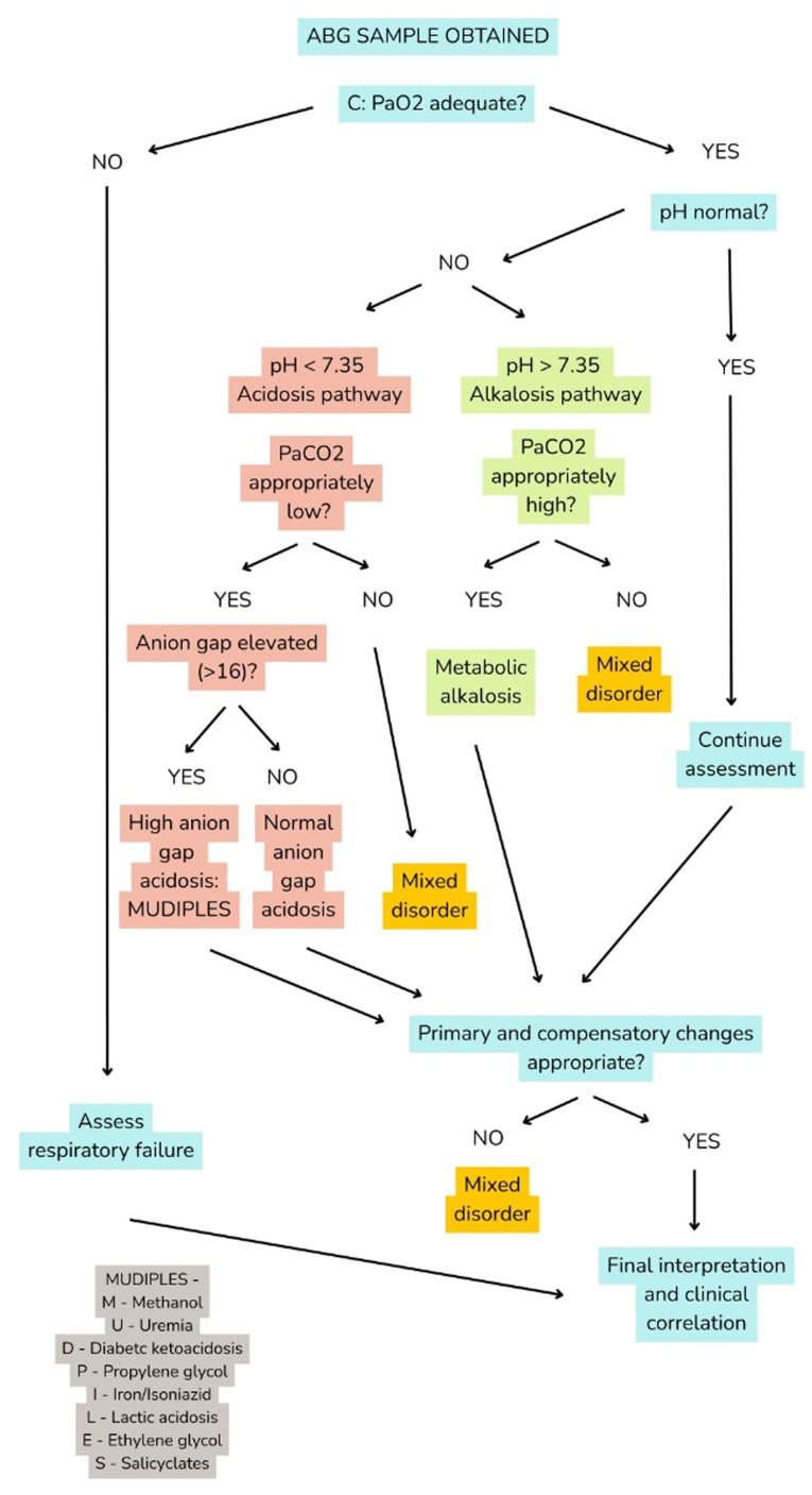 Figure 1: Systematic approach to arterial blood gas interpretation using the CLEAR framework — Check oxygenation, Look at pH, Evaluate compensation, Assess anion gap, Review for mixed disorders — integrating the oxygenation and acid–base steps into a single reproducible bedside sequence to identify simple and mixed disorders. (Source: Sanagustín MN, et al. Medicine International 2026. DOI:
10.3892/mi.2025.291
, PMCID: PMC12746054).
Systematic 6-Step ABG Interpretation Algorithm
A structured, sequential methodology prevents diagnostic errors in complex critically ill patients:
flowchart TD
    A[Step 1: Evaluate Arterial pH] -->|pH < 7.35| B1[Acidemia]
    A -->|pH > 7.45| B2[Alkalemia]
    A -->|pH 7.35-7.45| B3[Normal or Mixed/Fully Compensated]

    B1 --> C1[Step 2: Determine Primary Disorder]
    B2 --> C1
    B3 --> C1

    C1 -->|PaCO2 matches pH| D1[Primary Respiratory Disorder]
    C1 -->|HCO3 matches pH| D2[Primary Metabolic Disorder]

    D1 --> E[Step 3: Assess Compensation Rules]
    D2 --> E

    E --> F[Step 4: Calculate Anion Gap & Albumin Correction]
    F --> G[Step 5: Calculate Delta Gap / Delta Ratio]
    G --> H[Step 6: Assess Oxygenation & Gas Exchange Indices]
Step 1: Arterial pH Assessment
- Normal Range: $7.35 - 7.45$ (corresponding to $[H^+] \approx 35 - 45\text{ nmol/L}$, where $[H^+] = 24 \times \frac{PaCO_2}{[HCO_3^-]}$).
- Acidemia: $pH < 7.35$ ($[H^+] > 45\text{ nmol/L}$).
- Alkalemia: $pH > 7.45$ ($[H^+] < 35\text{ nmol/L}$).
- Clinical Pearl: A normal $pH$ does not rule out severe acid-base pathology; it may indicate a fully compensated disorder or two coexisting, opposing primary disorders (e.g., severe high-anion-gap metabolic acidosis combined with a primary respiratory alkalosis).
Step 2: Primary Disorder Identification
Compare the directional shift of $PaCO_2$ and $[HCO_3^-]$ relative to $pH$:
- Respiratory Acidosis: $pH < 7.35$ with elevated $PaCO_2 > 45\text{ mmHg}$.
- Respiratory Alkalosis: $pH > 7.45$ with decreased $PaCO_2 < 35\text{ mmHg}$.
- Metabolic Acidosis: $pH < 7.35$ with decreased $[HCO_3^-] < 22\text{ mEq/L}$.
- Metabolic Alkalosis: $pH > 7.45$ with elevated $[HCO_3^-] > 26\text{ mEq/L}$.
Step 3: Secondary Compensation Rules
The human body does not overcompensate; compensation never returns $pH$ completely past the baseline median ($7.40$). If observed compensation differs from expected values, a secondary mixed disorder is present.
| Primary Disorder | Compensatory Response | Quantitative Expected Compensation Formula | Timeframe for Full Compensation |
|---|---|---|---|
| Acute Respiratory Acidosis | Acute cellular buffering | $\Delta [HCO_3^-] = 1\text{ mEq/L}$ per $10\text{ mmHg} \uparrow PaCO_2$ Expected $[HCO_3^-] = 24 + \frac{PaCO_2 - 40}{10} \times 1$ | Minutes to 2 hours |
| Chronic Respiratory Acidosis | Renal $HCO_3^-$ retention | $\Delta [HCO_3^-] = 3.5 - 4.0\text{ mEq/L}$ per $10\text{ mmHg} \uparrow PaCO_2$ Expected $[HCO_3^-] = 24 + \frac{PaCO_2 - 40}{10} \times 3.5$ | $3 - 5\text{ days}$ (72–120 hours) |
| Acute Respiratory Alkalosis | Acute cellular buffering | $\Delta [HCO_3^-] = 2\text{ mEq/L}$ per $10\text{ mmHg} \downarrow PaCO_2$ Expected $[HCO_3^-] = 24 - \frac{40 - PaCO_2}{10} \times 2$ | Minutes to 2 hours |
| Chronic Respiratory Alkalosis | Renal $HCO_3^-$ excretion | $\Delta [HCO_3^-] = 4 - 5\text{ mEq/L}$ per $10\text{ mmHg} \downarrow PaCO_2$ Expected $[HCO_3^-] = 24 - \frac{40 - PaCO_2}{10} \times 5$ | $2 - 3\text{ days}$ (48–72 hours) |
| Metabolic Acidosis | Alveolar hyperventilation | Winter's Formula: Expected $PaCO_2 = (1.5 \times [HCO_3^-]) + 8 \pm 2$ (Alternative quick rule: Expected $PaCO_2 \approx \text{last 2 digits of } pH$) | $12 - 24\text{ hours}$ |
| Metabolic Alkalosis | Alveolar hypoventilation | Expected $PaCO_2 = 40 + [0.7 \times ([HCO_3^-] - 24)] \pm 5$ (Alternative: Expected $PaCO_2 = (0.7 \times [HCO_3^-]) + 20 \pm 5$) | Rapid (limited by hypoxemic ventilatory drive, rarely $PaCO_2 > 55-60$) |
Step 4: Serum Anion Gap & Albumin Correction
Calculate the serum anion gap ($AG$) in all cases of metabolic acidosis, even when $[HCO_3^-]$ appears normal, because hypoalbuminemia frequently masks significant unmeasured organic anions:
$$AG = [Na^+] - ([Cl^-] + [HCO_3^-])$$- Normal Anion Gap: $10 - 12\text{ mEq/L}$ (reference based on normal serum albumin of $4.0\text{ g/dL}$ or $40\text{ g/L}$).
- Albumin-Corrected Anion Gap: Serum albumin carries the majority of unmeasured negative charges. For every $1.0\text{ g/dL}$ drop in serum albumin below $4.0\text{ g/dL}$, the normal baseline $AG$ drops by $2.5\text{ mEq/L}$:
- Etiology Classification:
- High Anion Gap Metabolic Acidosis (HAGMA): $\text{MUDPILES / GOLDMARK}$ (Methanol, Uremia, Diabetic ketoacidosis, Propylene glycol, Isoniazid/Iron, L-lactic acidosis, D-lactic acidosis, Ethylene glycol, Salicylates, Pyroglutamic acid/Oxoprolin).
- Normal Anion Gap Metabolic Acidosis (NAGMA / Hyperchloremic): Renal tubular acidosis (RTA), severe diarrhea, enteric fistulas, normal saline ($0.9\%\text{ NaCl}$) resuscitation, acetazolamide.
Step 5: Delta Gap ($\Delta AG$) and Delta Ratio ($\Delta / \Delta$)
The Delta Ratio evaluates whether a HAGMA is isolated or coexists with a hidden NAGMA or metabolic alkalosis:
$$\text{Delta Ratio} = \frac{\Delta AG}{\Delta [HCO_3^-]} = \frac{AG_{\text{corrected}} - 12}{24 - [HCO_3^-]}$$| Delta Ratio Value | Physiological Interpretation | Common Clinical Scenario |
|---|---|---|
| $< 0.4$ | Pure NAGMA (Hyperchloremic acidosis) | Diarrhea, RTA, massive normal saline infusion |
| $0.4 - 0.8$ | Mixed HAGMA and NAGMA | DKA with normal saline resuscitation, sepsis with severe diarrhea |
| $0.8 - 2.0$ | Pure / Uncomplicated HAGMA | Typical DKA, pure lactic acidosis from shock |
| $> 2.0$ | HAGMA with concurrent Metabolic Alkalosis or Pre-existing Chronic Hypercapnia | DKA or lactic acidosis with severe vomiting/nasogastric suctioning; COPD patient developing sepsis |
Step 6: Systematic Oxygenation & Gas Exchange Assessment
Oxygenation must be evaluated relative to inspired oxygen fraction ($FiO_2$) and mean airway pressure ($mPaw$):
+------------------------------------------------------------------------------------+
|                         OXYGENATION ASSESSMENT MATRIX                              |
+------------------------------------+-----------------------------------------------+
| PARAMETER                          | CLINICAL FORMULA & THRESHOLDS                 |
+------------------------------------+-----------------------------------------------+
| PaO2 / FiO2 Ratio (P/F)            | PaO2 / FiO2 (mmHg)                            |
|                                    | - Normal: > 400-500                           |
|                                    | - Mild ARDS: 200 - 300 (PEEP >= 5)            |
|                                    | - Moderate ARDS: 100 - 200 (PEEP >= 5)        |
|                                    | - Severe ARDS: < 100 (PEEP >= 5)              |
+------------------------------------+-----------------------------------------------+
| Alveolar Gas Equation              | PAO2 = (Patm - PH2O) * FiO2 - (PaCO2 / R)     |
|                                    | - At sea level: PAO2 = (760 - 47)*FiO2 - (PaCO2 / 0.8) |
|                                    |               = 713 * FiO2 - (1.25 * PaCO2)   |
+------------------------------------+-----------------------------------------------+
| A-a Oxygen Gradient                | P(A-a)O2 = PAO2 - PaO2                        |
|                                    | - Normal young adult: < 10 - 15 mmHg          |
|                                    | - Age-adjusted normal: (Age / 4) + 4          |
|                                    | - Elevated: V/Q mismatch, Shunt, Diffusion defect |
|                                    | - Normal in Hypoxemia: Hypoventilation, High altitude |
+------------------------------------+-----------------------------------------------+
| Arterial-to-Alveolar Ratio (a/A)   | a/A = PaO2 / PAO2                             |
|                                    | - Normal: > 0.75 - 0.80                       |
|                                    | - Moderate impairment: 0.30 - 0.74            |
|                                    | - Severe shunting/ARDS: < 0.15                |
+------------------------------------+-----------------------------------------------+
| Oxygenation Index (OI)             | OI = (mPaw * FiO2 * 100) / PaO2               |
|                                    | - Normal: < 5                                 |
|                                    | - Severe hypoxemic failure: > 15 - 20         |
|                                    | - Inhaled NO / Proning threshold: > 20 - 25   |
|                                    | - ECMO evaluation threshold: > 35 - 40        |
+------------------------------------+-----------------------------------------------+
| Oxygen Saturation Index (OSI)      | OSI = (mPaw * FiO2 * 100) / SpO2              |
|                                    | - Non-invasive surrogate for OI               |
|                                    | - OSI > 6.5 corresponds to OI > 10 (ARDS)     |
+------------------------------------+-----------------------------------------------+
Arterial vs Central Venous ($ScvO_2$) vs Peripheral Venous vs Capillary Blood Gases
Selecting and interpreting blood gas modalities requires understanding their physiological gradients and clinical limits.
                  =======================================================
                  BLOOD GAS SAMPLE SITES & CIRCULATORY PHYSIOLOGY
                  =======================================================

                   [ Lungs (Arterialization) ]
                                |
                                v
                   [ Arterial Blood (ABG) ] ---------> (Reflects Gas Exchange & Ventilation)
                                |
                                v
                   [ Tissue Microcirculation ] ------> (Extracts O2, Adds CO2 & Lactate)
                                |
                                v
                   [ Venous Blood ]
                     |           |
                     v           v
                 (Central)   (Peripheral)
                  ScvO2       PVBG
                     |
                     v
             (Pulmonary Artery)
                   SvO2
Physiological Correlation Gradients
| Blood Gas Parameter | Arterial Blood Gas (ABG) | Central Venous Gas ($ScvO_2$ / CVBG) | Peripheral Venous Gas (PVBG) | Capillary Blood Gas (CBG) | Clinical Gradient ($\Delta$) & Significance |
|---|---|---|---|---|---|
| $pH$ | $7.35 - 7.45$ | $7.32 - 7.42$ | $7.31 - 7.41$ | $7.35 - 7.45$ | Venous $pH$ is $0.03 - 0.05$ lower than arterial. Excellent correlation ($r > 0.95$). |
| $PCO_2$ | $35 - 45\text{ mmHg}$ | $40 - 50\text{ mmHg}$ | $42 - 52\text{ mmHg}$ | $36 - 46\text{ mmHg}$ | Venous $PCO_2$ is $4 - 6\text{ mmHg}$ higher than arterial in stable hemodynamics. |
| $[HCO_3^-]$ | $22 - 26\text{ mEq/L}$ | $23 - 27\text{ mEq/L}$ | $23 - 28\text{ mEq/L}$ | $22 - 26\text{ mEq/L}$ | Venous $HCO_3^-$ is $1 - 2\text{ mEq/L}$ higher than arterial. Clinically interchangeable. |
| $PO_2$ | $80 - 100\text{ mmHg}$ | $35 - 45\text{ mmHg}$ | $30 - 40\text{ mmHg}$ | Variable ($45 - 75$) | No correlation. Venous $PO_2$ cannot assess oxygenation or calculate $P/F$ ratio. |
| $SO_2$ | $95 - 99\%$ | $70 - 75\%$ ($ScvO_2$) | Variable ($60 - 80\%$) | Reflects $SpO_2$ | $ScvO_2$ reflects balance between systemic oxygen delivery ($DO_2$) and consumption ($VO_2$). |
| Lactate | $0.5 - 1.5\text{ mmol/L}$ | $0.5 - 1.5\text{ mmol/L}$ | $0.5 - 2.0\text{ mmol/L}$ | $0.5 - 1.8\text{ mmol/L}$ | Venous lactate correlates well with arterial, but tourniquet stasis causes false elevations in PVBG. |
Venous-to-Arterial $PCO_2$ Difference ($\Delta PCO_2$ or "$\text{PCO}_2$ Gap")
The central venous-to-arterial carbon dioxide gradient is defined as:
$$\Delta PCO_2 = P_{cv}CO_2 - PaCO_2$$- Normal Value: $\le 6\text{ mmHg}$ ($0.8\text{ kPa}$).
- Pathophysiology: By the Fick principle applied to $CO_2$, $CO_2$ production ($\dot{V}CO_2$) equals cardiac output ($CO$) multiplied by the venoarterial difference in $CO_2$ content ($C_vCO_2 - C_aCO_2$): $$\Delta PCO_2 \propto \frac{\dot{V}CO_2}{CO}$$
- Clinical Significance in Shock Resuscitation:
- If $ScvO_2 \ge 70\%$ but $\Delta PCO_2 > 6\text{ mmHg}$, the patient suffers from ischemic / stagnant hypoxia (inadequate cardiac output and microvascular hypoperfusion despite normal macrovascular arterial saturation).
- An elevated $\Delta PCO_2 > 6\text{ mmHg}$ indicates persistent tissue hypoperfusion and predicts failure to clear lactate during septic and cardiogenic shock resuscitation.
Central Venous Oxygen Saturation ($ScvO_2$) vs Mixed Venous ($SvO_2$)
- $SvO_2$ (True Mixed Venous): Sampled from the distal port of a pulmonary artery catheter (PAC) in the pulmonary artery. It reflects complete mixing of blood from the superior vena cava (SVC), inferior vena cava (IVC), and coronary sinus. Normal: $65 - 70\%$.
- $ScvO_2$ (Central Venous): Sampled from a central venous catheter tip in the internal jugular or subclavian vein located in the distal SVC / cavoatrial junction. Normal: $70 - 75\%$ ($ScvO_2$ runs $3 - 5\%$ higher than $SvO_2$ due to low oxygen saturation in coronary sinus blood).
===================================================================================
                       CLINICAL INTERPRETATION OF ScvO2
===================================================================================
   ScvO2 < 65-70% (Hypoperfusion / Inadequate DO2)
   -------------------------------------------------
   - Decreased DO2:
       * Low Cardiac Output (Cardiogenic shock, Hypovolemia, Tamponade)
       * Severe Anemia (Hemoglobin < 7.0 g/dL)
       * Severe Arterial Hypoxemia (PaO2 < 55 mmHg, SpO2 < 88%)
   - Increased VO2:
       * Severe Shivering, Fever, Malignant Hyperthermia, Pain, Seizures

   ScvO2 > 80-85% (Microvascular Shunting / Impaired Extraction)
   --------------------------------------------------------------
   - Cytopathic Dysoxia & Mitochondrial Poisoning (Severe Sepsis, Cyanide Toxicity)
   - Microcirculatory Shunting (Septic Shock)
   - Markedly Decreased VO2 (Deep Hypothermia, Deep Sedation / Neuromuscular Blockade)
   - Severe Left-to-Right Intracardiac Shunts
===================================================================================
Capillary Blood Gas (CBG): Indications and Limitations
- Technique: Arterialization of the skin via localized heating ($40 - 42^\circ\text{C}$ for $5 - 10\text{ minutes}$) on the lateral heel (neonates) or earlobe/fingertip (pediatrics/adults), followed by puncture.
- Reliability:
- $pH$ and $PCO_2$ show high correlation with ABG ($r \approx 0.90 - 0.95$).
- $PO_2$ is unreliable and significantly underestimates true arterial $PaO_2$ due to tissue capillary oxygen extraction.
- Contraindications & Failure States:
- Shock, severe hypotension, and systemic vasoconstriction (vasopressor therapy).
- Edema or local tissue induration.
- Peripheral vascular disease or cold extremities.
- Critical titration of $FiO_2$ in severe ARDS (requires arterial line).
Ventilator Adjustments Based on Blood Gas Results
Arterial $PaCO_2$ is inversely proportional to alveolar ventilation ($\dot{V}_A$):
$$PaCO_2 = \frac{\dot{V}CO_2 \times 0.863}{\dot{V}_A} = \frac{\dot{V}CO_2 \times 0.863}{\dot{V}_E \times (1 - V_D/V_T)}$$Where $\dot{V}CO_2$ is metabolic $CO_2$ production, $\dot{V}_E$ is minute ventilation ($\text{Minute Ventilation} = RR \times V_T$), and $V_D/V_T$ is the physiological dead space fraction.
                      ==============================================
                      VENTILATOR PaCO2 CORRECTION DECISION PATHWAY
                      ==============================================

                                  Target PaCO2 Required
                                            |
                         +------------------+------------------+
                         |                                     |
                [ Change Rate (RR) ]                  [ Change Volume (VT) ]
                         |                                     |
               Preferred in:                         Preferred in:
               - Established lung-protective VT      - Severe airflow obstruction
                 (e.g., 6 mL/kg PBW in ARDS)           (Asthma/COPD: low RR to
               - Preserving driving pressure           prolong Te and reduce auto-PEEP)
                         |                                     |
                         v                                     v
                 New RR = (RR_curr * PaCO2_curr) / PaCO2_target
                 New VT = (VT_curr * PaCO2_curr) / PaCO2_target
Rapid Titration Formulas
1. Adjusting Respiratory Rate ($RR$)
When tidal volume is fixed at lung-protective limits ($6\text{ mL/kg}$ PBW in ARDS):
$$\text{New } RR = \frac{\text{Current } RR \times \text{Current } PaCO_2}{\text{Target } PaCO_2}$$Example: Patient on $RR = 18\text{ bpm}$, $PaCO_2 = 56\text{ mmHg}$. Target $PaCO_2 = 40\text{ mmHg}$.
$$\text{New } RR = \frac{18 \times 56}{40} = 25.2 \to \mathbf{25\text{ bpm}}$$2. Adjusting Tidal Volume ($V_T$)
When adjusting volume in non-ARDS conditions without violating plateau pressure ($P_{plat} \le 30\text{ cmH}_2\text{O}$) or driving pressure ($\Delta P \le 14\text{ cmH}_2\text{O}$):
$$\text{New } V_T = \frac{\text{Current } V_T \times \text{Current } PaCO_2}{\text{Target } PaCO_2}$$3. Adjusting Total Minute Ventilation ($\dot{V}_E$)
$$\text{New } \dot{V}_E = \frac{\text{Current } \dot{V}_E \times \text{Current } PaCO_2}{\text{Target } PaCO_2}$$Dead Space Ventilation ($V_D/V_T$) Management
The Enghoff modification of the classic Bohr equation quantifies total physiological dead space:
$$\frac{V_D}{V_T} = \frac{PaCO_2 - P\bar{E}CO_2}{PaCO_2}$$Where $P\bar{E}CO_2$ is the mixed expired carbon dioxide partial pressure measured via volumetric capnography.
- Normal Values:
- Healthy spontaneous breathing: $V_D/V_T = 0.25 - 0.35$ ($25 - 35\%$).
- Intubated, mechanically ventilated ICU patient: $V_D/V_T = 0.40 - 0.55$.
- Severe ARDS / Pulmonary embolism: $V_D/V_T > 0.65 - 0.75$ ($65 - 75\%$).
- Prognostic Value: In ARDS, an elevated $V_D/V_T$ is a stronger predictor of mortality than the $P/F$ ratio alone, reflecting extensive pulmonary microvascular thrombosis, diffuse alveolar destruction, and high mechanical overdistension.
- Strategies to Reduce Instrumental and Alveolar Dead Space:
- Remove unnecessary tubing connectors, swivel adaptors, and dead-space flex tubes.
- Switch from heat-and-moisture exchangers (HME filters, adding $50 - 100\text{ mL}$ instrumental dead space) to active heated humidification in hypercapnic respiratory failure.
- Optimize PEEP: Decreasing excessive PEEP reduces West Zone 1 alveolar capillary compression; increasing suboptimal PEEP recruits atelectatic alveoli.
- Prone positioning: Improves ventilation-perfusion matching and reduces physiological dead space.
Oxygenation Titration & Hemoglobin Dissociation Dynamics
     100 |---------------------------------------------...--- SpO2 ~ 97-100%
         |                                   ...''''
         |                             ..''''
      80 |                       ...'''  <--- P50 ~ 26.8 mmHg (50% Saturation)
         |                   ..''
  Hb SO2 |               ..''             LEFT SHIFT (Increased Affinity):
   (%)   |             .'                 - pH ^ (Alkalosis), PCO2 v, Temp v, 2,3-DPG v
      50 |----------..'                   - HbF, COHb, MetHb
         |        .'
         |      .'                        RIGHT SHIFT (Decreased Affinity / Bohr Effect):
         |    .'                          - pH v (Acidosis), PCO2 ^, Temp ^, 2,3-DPG ^
      20 |  .'                            - Facilitates peripheral tissue O2 unloading
         |.'
       0 +-------------------------------------------------------
         0       20       40       60       80      100     120
                               PaO2 (mmHg)
The Oxyhemoglobin Dissociation Curve & $P_{50}$
- $P_{50}$ Definition: The partial pressure of oxygen at which $50\%$ of hemoglobin binding sites are saturated with oxygen. Normal human adult blood at $37^\circ\text{C}$ and $pH = 7.40$ has a $P_{50} = 26.8\text{ mmHg}$ ($3.57\text{ kPa}$).
- Bohr Effect (Right Shift): Decreases hemoglobin's oxygen affinity, shifting the curve to the right and increasing $P_{50}$. This enhances oxygen unloading at tissue capillaries where $CO_2$ and $[H^+]$ are elevated.
- Triggers: Acidemia ($\downarrow pH$), Hypercapnia ($\uparrow PaCO_2$), Hyperthermia ($\uparrow \text{Temperature}$), and elevated $2,3\text{-Diphosphoglycerate (2,3-DPG)}$.
- Left Shift: Increases hemoglobin's oxygen affinity, shifting the curve to the left and lowering $P_{50}$. While this enhances pulmonary capillary oxygen uptake, it impairs offloading to hypoxic tissues.
- Triggers: Alkalemia ($\uparrow pH$), Hypocapnia ($\downarrow PaCO_2$), Hypothermia, depleted $2,3\text{-DPG}$ (massive transfusion of banked red blood cells), carboxyhemoglobin ($COHb$), and methemoglobin ($MetHb$).
Target Oxygenation Thresholds & Avoiding Hyperoxia Toxicity
- General ICU Targets: $PaO_2 = 60 - 80\text{ mmHg}$ ($8.0 - 10.7\text{ kPa}$), corresponding to $SpO_2 = 92 - 96\%$.
- ARDS Targets: $PaO_2 = 55 - 80\text{ mmHg}$ ($7.3 - 10.7\text{ kPa}$), corresponding to $SpO_2 = 88 - 95\%$.
- Traumatic Brain Injury / Post-Cardiac Arrest: $PaO_2 = 80 - 100\text{ mmHg}$ ($SpO_2 \ge 95\%$), avoiding hypoxemia ($PaO_2 < 60$) while strictly preventing severe hyperoxia ($PaO_2 > 200 - 300\text{ mmHg}$).
- Mechanisms of Hyperoxia Harm:
- Reactive oxygen species (ROS) generation leading to lipid peroxidation and alveolar-capillary damage.
- Absorption atelectasis: High $FiO_2$ washes out inert nitrogen ($N_2$), leading to alveolar collapse behind occluded small airways.
- Coronary and cerebral vasoconstriction reducing microvascular organ blood flow.
PEEP vs $FiO_2$ Titration Protocols (ARDSNet)
Titrate PEEP and $FiO_2$ in tandem to maintain oxygenation targets ($PaO_2\ 55-80\text{ mmHg}$ or $SpO_2\ 88-95\%$):
+---------------------------------------------------------------------------------------------------------------+
|                                    ARDSNet PEEP - FiO2 TITRATION PROTOCOLS                                    |
+---------------------------------------------------------------------------------------------------------------+
| Lower PEEP / Higher FiO2 Strategy (Standard ARDS):                                                            |
| FiO2: 0.30 | 0.40 | 0.40 | 0.50 | 0.50 | 0.60 | 0.70 | 0.70 | 0.80 | 0.90 | 0.90 | 1.00                    |
| PEEP:  5   |  5   |  8   |  8   |  10  |  10  |  10  |  12  |  14  |  14  |  16  | 18-24                   |
+---------------------------------------------------------------------------------------------------------------+
| Higher PEEP / Lower FiO2 Strategy (Moderate-to-Severe ARDS with High Recruitable Fraction):                  |
| FiO2: 0.30 | 0.30 | 0.30 | 0.30 | 0.30 | 0.40 | 0.40 | 0.50 | 0.50 | 0.60 | 0.70 | 0.80 | 0.90 | 1.00   |
| PEEP:  5   |  8   |  10  |  12  |  14  |  14  |  16  |  16  |  18  |  20  |  20  |  22  |  22  | 22-24  |
+---------------------------------------------------------------------------------------------------------------+
Permissive Hypercapnia in Mechanical Ventilation
Permissive hypercapnia is a deliberate clinical strategy in which alveolar hypoventilation is tolerated to prioritize lung-protective ventilation and avoid ventilator-induced lung injury (VILI).
   [ Low Tidal Volume Ventilation (4-6 mL/kg) & Pressure Limits (Pplat <= 30) ]
                                    |
                                    v
                       [ Alveolar Hypoventilation ]
                                    |
                                    v
     ===============================================================
                     PERMISSIVE HYPERCAPNIA STATE
     ===============================================================
     Benefits:
     - Prevents Volutrauma, Barotrauma, and Atelectrauma
     - Reduces Driving Pressure (Delta P <= 14 cmH2O)
     - Attenuates Systemic Pro-inflammatory Cytokine Release (Biotrauma)

     Tolerated Parameters:
     - PaCO2: 55 - 80+ mmHg
     - Arterial pH: >= 7.20 (>= 7.15 in refractory ARDS)
     ===============================================================
Primary Clinical Indications
- Acute Respiratory Distress Syndrome (ARDS): Maintaining $V_T \le 4 - 6\text{ mL/kg}$ predicted body weight (PBW) to prevent alveolar overdistension and cyclic opening/collapse.
- Status Asthmaticus & Severe COPD Exacerbation: Decreasing respiratory rate ($RR \le 10 - 14\text{ bpm}$) and minute ventilation to prolong expiratory time ($T_E$), promote complete lung emptying, and minimize dynamic hyperinflation and intrinsic PEEP ($\text{auto-PEEP}$).
Physiological Contraindications to Permissive Hypercapnia
+------------------------------------------------------------------------------------+
|                ABSOLUTE & RELATIVE CONTRAINDICATIONS TO HYPERCAPNIA                |
+------------------------------------+-----------------------------------------------+
| CLINICAL CONDITION                 | UNDERLYING PATHOPHYSIOLOGICAL MECHANISM       |
+------------------------------------+-----------------------------------------------+
| Acute Intracranial Hypertension    | PaCO2 is a potent cerebral vasodilator.       |
| Traumatic Brain Injury (TBI)       | Increased PaCO2 -> increased cerebral blood   |
| Acute Ischemic/Hemorrhagic Stroke  | flow (CBF) -> rapid, catastrophic increase in |
| Cerebral Edema                     | intracranial pressure (ICP) & brain herniation.|
+------------------------------------+-----------------------------------------------+
| Severe Right Ventricular Failure   | Hypercapnic acidosis causes marked precapillary|
| Acute Cor Pulmonale                | pulmonary arterial vasoconstriction (HPV),    |
| Severe Pulmonary Hypertension      | increasing RV afterload -> acute RV dilation,  |
|                                    | tricuspid regurgitation, and cardiogenic shock.|
+------------------------------------+-----------------------------------------------+
| Severe Hemodynamic Instability     | Acidemia (pH < 7.15) decreases myocardial     |
| Refractory Shock                   | beta-adrenergic receptor sensitivity and      |
| Malignant Cardiac Arrhythmias      | lowers the threshold for ventricular tachycardias.|
+------------------------------------+-----------------------------------------------+
| Severe Pre-existing Metabolic      | Inability to buffer respiratory acidosis;     |
| Acidosis (e.g., pH < 7.10)         | severe combined acidosis causes lethal        |
|                                    | cellular enzymatic failure.                   |
+------------------------------------+-----------------------------------------------+
Summary & Bedside Clinical Checklist
+------------------------------------------------------------------------------------+
|                         BEDSIDE ABG INTERPRETATION CHECKLIST                       |
+----+-------------------------------------------------------------------------------+
| [ ]| 1. Verify Sample Source: Is this Arterial (ABG), Central Venous (ScvO2),      |
|    |    Peripheral Venous (PVBG), or Capillary (CBG)? (Do not assess PO2 on venous)|
+----+-------------------------------------------------------------------------------+
| [ ]| 2. Check pH Status: Acidemia (< 7.35) vs Alkalemia (> 7.45) vs Normal.        |
+----+-------------------------------------------------------------------------------+
| [ ]| 3. Identify Primary Driver: Match directional changes in PaCO2 and HCO3-.     |
+----+-------------------------------------------------------------------------------+
| [ ]| 4. Test Secondary Compensation: Apply Winter's formula (metabolic acidosis)   |
|    |    or acute/chronic respiratory compensation ratios.                          |
+----+-------------------------------------------------------------------------------+
| [ ]| 5. Calculate Anion Gap: Always adjust for serum albumin:                     |
|    |    AG_corr = AG + 2.5 * (4.0 - Albumin). Calculate Delta Ratio if AG elevated. |
+----+-------------------------------------------------------------------------------+
| [ ]| 6. Quantify Oxygenation: Calculate P/F ratio, A-a gradient, and Oxygenation   |
|    |    Index (OI). Titrate PEEP/FiO2 per ARDSNet tables.                          |
+----+-------------------------------------------------------------------------------+
| [ ]| 7. Ventilator Adjustments: Use New RR = (Current RR * Current PaCO2) / Target |
|    |    PaCO2. Respect lung-protective limits (Pplat <= 30, Delta P <= 14).        |
+----+-------------------------------------------------------------------------------+
| [ ]| 8. Check Contraindications: Ensure no ICP elevation, severe RV failure, or   |
|    |    myocardial ischemia before permitting hypercapnia.                         |
+----+-------------------------------------------------------------------------------+