Proportional assist modes represent a fundamental paradigm shift in mechanical ventilation: transitioning from conventional preset targets (fixed tidal volume or fixed airway pressure) to closed-loop assistance that varies continuously in proportion to the patient's instantaneous respiratory drive and muscle effort. In conventional Pressure Support Ventilation (PSV), the ventilator delivers a constant, square pressure block regardless of whether the patient's demand fluctuates within or between breaths. This uncoupling between patient neural demand and pneumatic delivery frequently causes severe patient-ventilator dyssynchrony, over-assistance, respiratory muscle unloading with subsequent ventilator-induced diaphragmatic dysfunction (VIDD), or under-assistance and eccentric muscle fatigue.
Neurally Adjusted Ventilatory Assist (NAVA) and Proportional Assist Ventilation with Load-Adjusted Speed of Response (PAV+) restore physiological neuro-ventilatory coupling by dynamically matching ventilatory assistance to instantaneous patient demand on a millisecond-by-millisecond basis.
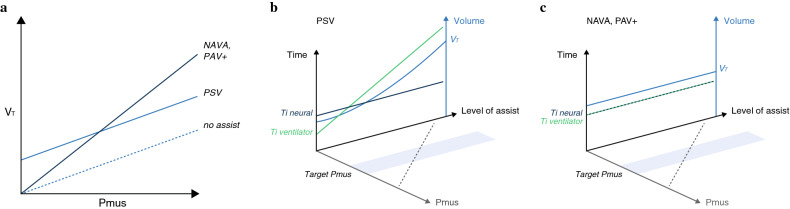 Figure: Physiological comparison of patient-ventilator interaction and neuro-ventilatory coupling in Pressure Support Ventilation (PSV) versus proportional modes (NAVA and PAV+). In PSV, a fixed level of assist generates an inflexible pressure target where tidal volume ($V_T$) is disproportionately high at low patient efforts ($P_{mus}$), driving over-assistance, hyperventilation, and central apnea; conversely, during high neural demand, assistance remains fixed, risking exhaustion. In NAVA and PAV+, ventilatory support scales linearly with instantaneous patient effort ($P_{mus}$ or $Edi$), preserving natural breath-to-breath tidal volume variability, maintaining intrinsic feedback loops (Hering-Breuer reflex), and aligning neural inspiratory time ($T_{i,\text{neural}}$) with mechanical ventilator inflation time ($T_{i,\text{ventilator}}$).
Neuro-Ventilatory Coupling & The Proportional Assist Paradigm
Under spontaneous physiological conditions, the respiratory control center in the brainstem adjusts phrenic nerve firing rate and diaphragmatic muscle activation in response to afferent inputs from central/peripheral chemoreceptors, lung stretch receptors (Hering-Breuer reflex), and chest wall mechanoreceptors.
Brainstem Respiratory Centers
       │
       ▼ (Phrenic Nerve Action Potentials)
Diaphragmatic Depolarization (Edi: μV) ───► [NAVA Sensing Point]
       │
       ▼ (Diaphragm Contraction / Pmus)
Pleural & Alveolar Pressure Deflection (ΔPpl / ΔPalv)
       │
       ▼ (Equation of Motion: Flow & Volume)
Pneumatic Airway Flow (V') & Volume (V) ──► [PAV+ Sensing Point]
Limitations of Conventional Pressure Support (PSV)
In PSV, the clinician sets a fixed inspiratory pressure ($PS$), a pneumatic trigger sensitivity (flow or pressure), and an expiratory cycling threshold (percentage of peak inspiratory flow, $E_{\text{sens}}$):
- Fixed Assistance vs. Dynamic Demand: If patient effort doubles within a breath, the ventilator delivers the exact same airway pressure, causing subjective air hunger and under-assistance dyssynchrony. If the patient relaxes or falls asleep, the fixed pressure delivery over-assists, driving $V_T$ above neural requirements, suppressing $P_a\text{CO}_2$ below the apneic threshold, and causing central apneas and ineffective trigger efforts.
- Pneumatic Trigger & Cycling Delays: Triggering requires flow or pressure drop generated at the airway opening, which is delayed by intrinsic PEEP ($PEEP_i$), circuit compliance, and airway resistance. Cycling off relies on a fixed percentage drop in peak flow, which fails in obstructive lung disease (delayed cycling and breath stacking) or air leaks (cycling failure).
The Proportional Solution
Proportional modes link machine output to real-time patient physiological signals:
- NAVA captures the signal at the neural depolarization level (the root of the cascade), using diaphragmatic electromyography ($Edi$).
- PAV+ captures the signal at the musculoskeletal mechanics level, using instantaneous flow and volume to solve the Equation of Motion every 5 milliseconds.
Neurally Adjusted Ventilatory Assist (NAVA)
NAVA (Getinge/Maquet Servo-i, Servo-u, Servo-n) utilizes the electrical activity of the crural diaphragm ($Edi$, measured in microvolts, $\mu\text{V}$) to control both the timing and the magnitude of airway pressure delivered by the mechanical ventilator.
       ┌────────────────────────────────────────────────────────┐
       │   Brainstem Respiratory Centers (Phrenic Firing)      │
       └──────────────────────────┬─────────────────────────────┘
                                  │
                                  ▼
       ┌────────────────────────────────────────────────────────┐
       │ Diaphragm Depolarization: 8-Electrode Edi Catheter (μV)│
       └──────────────────────────┬─────────────────────────────┘
                                  │
                                  ▼
       ┌────────────────────────────────────────────────────────┐
       │ Microprocessor Filtering & Real-Time Processing (16 kHz)│
       └──────────────────────────┬─────────────────────────────┘
                                  │
                                  ▼
 ┌────────────────────────────────────────────────────────────────────┐
 │ Dynamic Pressure Output: Paw(t) = NAVA Level × [Edi(t) - Edi_min]  │
 ├────────────────────────────────────────────────────────────────────┤
 │ • Neural Trigger: ΔEdi ≥ 0.5 μV above baseline                     │
 │ • Continuous Titration: Paw updates every 2 milliseconds           │
 │ • Neural Cycle-Off: Edi drops to 70% of Edi_peak (30% decay)       │
 └────────────────────────────────────────────────────────────────────┘
The $Edi$ Signal & Transesophageal Catheter Array
The electrical activity of the diaphragm reflects spatial and temporal recruitment of diaphragmatic motor units.
- Catheter Architecture: A specialized, medical-grade polyurethane feeding tube embedded with an array of 9 miniature ring electrodes (creating 8 sequential bipolar electrode pairs) positioned transesophageally at the level of the crural diaphragm.
- Signal Filtering & Spatial Processing:
- The raw signal is sampled at high frequency (16 kHz).
- Advanced digital signal processing subtracts cardiac electrocardiographic signals (QRS complexes and P-waves) using automated cross-talk cancellation algorithms.
- Baseline esophageal noise, motion artifacts, and peristaltic waves are eliminated with bandpass filters.
- A real-time spatial filter continuously scans across all 8 electrode pairs, identifying the single bipolar pair spanning the electrical center of the crural diaphragm. As the diaphragm shifts during lung inflation or changes in patient posture, the algorithm dynamically switches channels to maintain an invariant, authentic $Edi$ amplitude.
Catheter Placement & Verification Protocol
Accurate anatomical placement is mandatory for reliable NAVA operation:
- Catheter Size Selection: Sized according to patient height (adults) or crown-rump/heel length (neonates/pediatrics), ranging from 6 Fr to 16 Fr.
- Insertion Depth Estimation: Calculated using standardized anthropometric distance: $$\text{Insertion Depth (cm)} = \frac{\text{Nose-to-Ear-Lobe Distance} + \text{Xiphoid Distance}}{1.0} \times \text{correction factor}$$
- Bedside Oscilloscope Positioning Tool:
- The ventilator displays 4 simultaneous raw bipolar lead waveforms alongside a dedicated $Edi$ positioning graphic.
- Correct Placement Criteria:
- Retrocardiac P-waves and large QRS complexes are prominent on the top (proximal) leads.
- Electrocardiographic complexes progressively diminish in amplitude from top to bottom leads.
- No P-waves are visible on the bottom (distal) leads.
- The central channels demonstrate robust, symmetric, phasic diaphragmatic electromyographic bursts during inspiration.
- Blue highlighted bars on the screen indicate that the center of the electrode array aligns with the diaphragm.
NAVA Delivery Equation
During inspiration, the ventilator delivers pressure according to the continuous formula:
$$P_{aw}(t) = \text{NAVA Level} \times [Edi(t) - Edi_{\min}] + PEEP$$Where:
- $P_{aw}(t)$ is the instantaneous airway pressure (cmH$_2$O).
- $\text{NAVA Level}$ is the clinician-set proportionality factor ($\text{cmH}_2\text{O}/\mu\text{V}$).
- $Edi(t)$ is the instantaneous diaphragmatic electromyography amplitude ($\mu\text{V}$).
- $Edi_{\min}$ is the tonic baseline electrical activity at end-expiration ($\mu\text{V}$).
- $PEEP$ is the extrinsic positive end-expiratory pressure ($\text{cmH}_2\text{O}$).
Neural Triggering and Neural Cycling
NAVA decouples triggering and cycling from pneumatic delays:
- Neural Trigger: The breath is initiated when $Edi$ rises by $0.5\ \mu\text{V}$ above the resting electrical baseline ($Edi_{\min}$), regardless of flow or airway pressure. Trigger delay is reduced from 100–300 ms (pneumatic) to $< 20\text{ ms}$ (neural). Pneumatic trigger backup is active simultaneously.
- Neural Cycling: The inspiratory phase terminates immediately when the $Edi$ signal drops to $70\%$ of its peak value (i.e., when $Edi$ decays by $30\%$ from $Edi_{\text{peak}}$). This synchronizes mechanical expiration precisely with the onset of neural diaphragmatic relaxation and post-inspiratory braking activity.
- Pneumatic Safety Overrides: If $Edi$ remains elevated abnormally (e.g., severe hiccups or artifact), cycling occurs if inspiratory time exceeds $T_{i,\max}$ or if airway pressure exceeds $P_{\text{insp,max}}$.
Non-Invasive NAVA (NIV-NAVA)
In conventional non-invasive ventilation (NIV), massive and variable leaks around masks or prongs impair pneumatic triggering and cycling, leading to severe dyssynchrony and high NIV failure rates.
- In NIV-NAVA, because the trigger, pressure contour, and cycle-off mechanisms are governed entirely by electrical signals from the phrenic-diaphragmatic axis, cuff leaks and mask leaks have zero effect on trigger delay or cycling accuracy.
- Neonatal & Pediatric Applications: NIV-NAVA is widely utilized in extremely low birth weight (ELBW) premature neonates with Respiratory Distress Syndrome (RDS) and bronchopulmonary dysplasia (BPD). It markedly reduces peak inspiratory pressures, prevents intubation, accelerates weaning from invasive ventilation, and reduces extubation failure rates compared to non-invasive CPAP or BiPAP.
Proportional Assist Ventilation with Load-Adjusted Speed of Response (PAV+)
PAV+ (Medtronic Puritan Bennett 840/980) delivers ventilatory assistance in proportion to instantaneous patient effort ($P_{mus}$), calculated continuously from the Equation of Motion of the Respiratory System.
$$\begin{aligned} P_{\text{total}}(t) &= P_{mus}(t) + P_{vent}(t) \\ &= [V'(t) \times R_{aw}] + [V(t) \times E_{rs}] + PEEP_{\text{total}} \end{aligned}$$Where $P_{vent}(t) = P_{aw}(t) - PEEP$.
                 [Patient Effort: Pmus(t)]
                           │
                           ▼
 ┌────────────────────────────────────────────────────────┐
 │ Airway Opening: Flow V'(t) & Delivered Volume V(t)     │
 ├────────────────────────────────────────────────────────┤
 │ Auto-Calculated Every 5 ms:                            │
 │ • Resistive Work: Flow × Raw                           │
 │ • Elastic Work: Volume × Ers                           │
 └──────────────────────────┬─────────────────────────────┘
                            │
                            ▼
 ┌────────────────────────────────────────────────────────┐
 │ Random Micro-Pauses (300 ms every 4-10 breaths)        │
 │ • Measures Static End-Inspiratory Plateau (Pplat)      │
 │ • Recalculates Raw, Ers, and Crs dynamically           │
 └──────────────────────────┬─────────────────────────────┘
                            │
                            ▼
 ┌────────────────────────────────────────────────────────┐
 │ Ventilator Support Delivery:                           │
 │ Paw(t) = % Assist × [(V' × Raw) + (V × Ers)] + PEEP    │
 └────────────────────────────────────────────────────────┘
Real-Time Mechanics & Micro-Pause Maneuvers
To deliver proportional pressure without requiring an esophageal catheter, the ventilator must accurately know the patient's respiratory system resistance ($R_{aw}$) and elastance ($E_{rs} = 1 / C_{rs}$).
- Automatic Semi-Continuous Micro-Pauses:
- Every 4 to 10 breaths (randomized to prevent patient anticipation and respiratory reflex perturbation), the ventilator seamlessly executes a 300-millisecond end-inspiratory pause.
- During this micro-pause, airway flow ceases ($V' = 0$), allowing the system to measure static plateau pressure ($P_{\text{plat}}$).
- The ventilator computes instantaneous compliance ($C_{rs} = \Delta V / [P_{\text{plat}} - PEEP]$) and instantaneous airway resistance ($R_{aw} = [P_{\text{peak}} - P_{\text{plat}}] / V'_{\text{peak}}$).
- The newly updated $R_{aw}$ and $E_{rs}$ values are applied to subsequent breaths for proportional pressure computation.
Setting Percentage Support (% Assist)
The clinician selects the % Support (percentage of total resistive and elastic work assumed by the ventilator):
$$P_{aw}(t) = \left(\frac{\% \text{ Assist}}{100}\right) \times \left[V'(t) \cdot R_{aw} + V(t) \cdot E_{rs}\right] + PEEP$$- Patient Contribution: The patient's respiratory muscles generate the remaining fraction ($100 - \% \text{ Assist}$): $$P_{mus}(t) = \left(1 - \frac{\% \text{ Assist}}{100}\right) \times \left[V'(t) \cdot R_{aw} + V(t) \cdot E_{rs}\right]$$
- Standard Clinical Range: Set between 50% and 70%.
- 70% Assist: Provides substantial unloading for acute respiratory recovery.
- 50% Assist: Normal physiological workload partition for conditioning and weaning.
- > 80% Assist (Runaway Risk): Prohibited or capped by safety algorithms because over-amplification of flow/volume signals can cause self-perpetuating, non-terminating positive feedback pressure escalation ("runaway condition").
Monitoring Patient Work of Breathing ($WOB_p$)
PAV+ calculates and displays real-time work of breathing metrics on every breath cycle:
- Patient Work of Breathing ($WOB_p$): Expressed in Joules per liter (J/L).
- Target Physiological Range: $0.3 \text{ to } 0.7\ \text{J/L}$.
- Over-Assistance / Muscle Atrophy: $WOB_p < 0.2\ \text{J/L}$.
- Under-Assistance / Muscle Fatigue: $WOB_p > 1.0\ \text{J/L}$.
- Total Work of Breathing ($WOB_{\text{tot}}$): Partitioned into patient work ($WOB_p$) and ventilator work ($WOB_{vent}$).
- Rapid Shallow Breathing Index (RSBI): Continuously measured and updated online.
Head-to-Head Physiological Comparison
| Feature / Domain | Pressure Support Ventilation (PSV) | Neurally Adjusted Ventilatory Assist (NAVA) | Proportional Assist Ventilation (PAV+) |
|---|---|---|---|
| Control Signal | Airway pressure target ($PS$) | Diaphragmatic electromyography ($Edi$) | Instantaneous flow ($V'$) and volume ($V$) |
| Trigger Mechanism | Pneumatic (Flow: $1\text{--}3\text{ L/min}$ or $\Delta P: 0.5\text{--}1.5\text{ cmH}_2\text{O}$) | Neural: $\Delta Edi \ge 0.5\ \mu\text{V}$ (pneumatic backup) | Pneumatic (Flow trigger) |
| Trigger Delay | $100 - 300\text{ ms}$ (long, sensitive to $PEEP_i$) | $< 20\text{ ms}$ (near-instantaneous) | $100 - 200\text{ ms}$ |
| Inspiratory Pressure Contour | Fixed square rectangular block | Continuous proportionality to $Edi(t)$ | Continuous proportionality to $P_{mus}(t)$ |
| Cycling Mechanism | Pneumatic peak flow decay ($E_{\text{sens}}\ 25\text{--}50\%$) | Neural decay: $70\%$ of $Edi_{\text{peak}}$ ($30\%$ drop) | Pneumatic flow decay threshold |
| Adaptation to Leaks | Highly vulnerable (autotriggering, delayed cycling) | Completely immune to leaks (NIV-NAVA) | Moderately vulnerable (leaks distort $R_{aw}/C_{rs}$) |
| Invasive Hardware | Standard ETT/Tracheostomy | Dedicated 8-electrode $Edi$ Catheter | Standard ETT/Tracheostomy (No extra catheter) |
| Respiratory Mechanics Measurement | Requires manual inspiratory/expiratory holds | Evaluated via Neuroventilatory Efficiency ($NVE$) | Automatic continuous micro-pauses |
| Over-Assistance Protection | None (drives hyperventilation and central apnea) | Intrinsically self-limiting (Hering-Breuer feedback) | Self-limiting within $50\text{--}70\%$ assist envelope |
| Risk of Runaway Pressure | None (fixed pressure limit) | None (limited by neural drive feedback) | Present if assist $>80\%$ (managed via safety limits) |
Bedside Titration & Clinical Protocols
               [Initiate Proportional Assist Mode]
                               │
               ┌───────────────┴───────────────┐
               ▼                               ▼
       [NAVA Protocol]                  [PAV+ Protocol]
               │                               │
    Place & Verify Edi Catheter         Verify Stable Mechanics
    (Positioning Tool Criteria)         (No Massive Circuit Leak)
               │                               │
    Set NAVA Level: 0.5 - 1.5 cmH2O/μV  Set % Assist: 50% - 70%
    Set Backup PC/VC Mode               Set Backup PC/VC Mode
               │                               │
               ▼                               ▼
    Target Edi_peak: 5 - 15 μV          Target WOBp: 0.3 - 0.7 J/L
    Target Vt: 6 - 8 mL/kg PBW          Target Vt: 6 - 8 mL/kg PBW
               │                               │
      ┌────────┴────────┐             ┌────────┴────────┐
      ▼                 ▼             ▼                 ▼
Edi < 5 μV        Edi > 15 μV   WOBp < 0.3 J/L    WOBp > 0.7 J/L
(Over-Assisted)   (Fatigue)     (Over-Assisted)   (Fatigue)
      │                 │             │                 │
Reduce NAVA Level Increase NAVA  Reduce % Assist   Increase % Assist
by 0.2 - 0.5      Level by 0.2    by 5% - 10%       by 5% - 10%
NAVA Titration Protocol
- Initial NAVA Level Setting:
- Start at $0.8 \text{ to } 1.0\ \text{cmH}_2\text{O}/\mu\text{V}$ (or match peak inspiratory pressure from preceding PSV: $\text{NAVA Level} = [P_{\text{peak,PSV}} - PEEP] / Edi_{\text{peak}}$).
- Titration Target:
- Optimal $Edi_{\text{peak}}$ Range: $5 \text{ to } 15\ \mu\text{V}$.
- Tonic $Edi_{\min}$: $< 3\ \mu\text{V}$ (elevated baseline $> 5\ \mu\text{V}$ indicates intrinsic PEEP, active exhalation, or diaphragmatic hypertonicity).
- Target Tidal Volume: $6 \text{ to } 8\ \text{mL/kg}$ predicted body weight (PBW).
- Titration Adjustments:
- If $Edi_{\text{peak}} < 5\ \mu\text{V}$ (over-assistance, excessive unloading): Decrease NAVA Level by $0.2 - 0.5\ \text{cmH}_2\text{O}/\mu\text{V}$.
- If $Edi_{\text{peak}} > 15\ \mu\text{V}$ (under-assistance, tachypnea, accessory muscle use): Increase NAVA Level by $0.2 - 0.5\ \text{cmH}_2\text{O}/\mu\text{V}$.
- Neuroventilatory Efficiency ($NVE$):
$$NVE = \frac{\text{Delivered Tidal Volume } (V_T)}{Edi_{\text{peak}}}$$
- Measures how effectively diaphragmatic neural electrical depolarization translates into mechanical tidal volume (expressed in $\text{mL}/\mu\text{V}$). A decline in $NVE$ indicates worsening lung elastance, high resistance, or diaphragmatic neuromuscular weakness.
PAV+ Titration Protocol
- Initial Setting:
- Select $60\% \text{ Assist}$ (or $70\%$ in severe obstructive/restrictive loading; $50\%$ in weaning).
- Monitoring & Target Parameters:
- Patient Work of Breathing ($WOB_p$): Maintain within $0.3 \text{ to } 0.7\ \text{J/L}$.
- Target $V_T$: $6 \text{ to } 8\ \text{mL/kg}$ PBW.
- Titration Adjustments:
- If $WOB_p < 0.3\ \text{J/L}$ (over-assisted, risk of VIDD): Wean % Assist in increments of $5\% \text{ to } 10\%$.
- If $WOB_p > 0.7\ \text{J/L}$ (under-assisted, excessive loading): Increase % Assist by $5\% \text{ to } 10\%$ (max $75\text{--}80\%$).
- If $WOB_p$ remains high despite $80\%$ assist, assess for severe dynamic hyperinflation, mucous plugging, or transition to controlled ventilation.
Troubleshooting & Clinical Red Flags
Loss of $Edi$ Signal & Backup Apnea Switching (NAVA):
- Etiology: Neuromuscular blockade, severe central respiratory depression (high-dose opioids/propofol), catastrophic catheter dislodgement into stomach or pharynx, or acute phrenic nerve transection.
- Ventilator Response: If no $Edi$ signal is detected within the preset apnea time (default: 5–10 s), the ventilator automatically falls back into conventional pressure-controlled (PC) or volume-controlled (VC) backup ventilation.
- Action: Re-enter the $Edi$ catheter positioning tool. Advance or withdraw the catheter in 1 cm increments until retrocardiac P-waves appear on proximal leads and the blue center bar aligns.
Severe Air Leaks Distorting Mechanics (PAV+):
- PAV+ relies on accurate micro-pause measurements of flow and volume. In the presence of bronchopleural fistulas, uncuffed tracheostomies, or uncuffed pediatric endotracheal tubes with $> 20\%$ leaks, calculated $R_{aw}$ and $C_{rs}$ become erroneous. Under-calculated elastance can lead to profound under-assistance or inappropriate pressure delivery.
- Action: Switch patient to NAVA (which is insensitive to pneumatic leaks) or conventional PSV.
Diaphragmatic Myotrauma Prevention: Maintain patient effort within the physiological "goldilocks" zone ($Edi_{\text{peak}} = 5\text{--}15\ \mu\text{V}$, $WOB_p = 0.3\text{--}0.7\ \text{J/L}$). Avoid total muscular silencing ($Edi < 3\ \mu\text{V}$, $WOB_p < 0.15\ \text{J/L}$) which triggers rapid myofibrillar proteolysis and diaphragmatic atrophy within 18–24 hours.
Key Clinical Takeaways
- True Physiological Closed-Loop Control: NAVA and PAV+ deliver proportional pressure assist tailored to instantaneous patient effort, eliminating the neuro-ventilatory uncoupling inherent to fixed-pressure modes like PSV.
- Elimination of Dyssynchrony: NAVA virtually eradicates trigger delay, ineffective triggering, and premature/delayed cycling by triggering and cycling off the electrical activity of the diaphragm ($Edi$).
- Leak Compensation: NIV-NAVA maintains flawless trigger and cycling synchrony even in the face of massive pneumatic mask or prong leaks, making it the premier mode for non-invasive support in neonates and complex pediatric patients.
- Automated Mechanics & Work Monitoring: PAV+ provides continuous bedside measurement of respiratory compliance ($C_{rs}$), resistance ($R_{aw}$), and patient work of breathing ($WOB_p$), allowing precise titration within the physiological safety window ($0.3\text{--}0.7\ \text{J/L}$).
- Diaphragm-Protective Ventilation: Both modes protect against ventilator-induced diaphragmatic dysfunction (VIDD) from over-assistance while preventing muscle exhaustion and eccentric load injury from under-assistance.