Dual-controlled and adaptive closed-loop ventilation modes combine the volume-guaranteeing security of conventional volume-targeted ventilation with the decelerating flow kinetics and peak airway pressure attenuation of pressure-controlled ventilation. By measuring patient mechanics (respiratory system compliance $C_{rs}$, airway resistance $R_{aw}$, and expiratory time constant $\tau_{RC}$) on a breath-by-breath basis, modern microprocessors dynamically adjust inspiratory drive to achieve target tidal volumes ($V_T$) or optimize the total work of breathing ($WOB$).
While these modes offer substantial clinical benefits in passive or smoothly breathing patients, their automated feedback algorithms introduce unique vulnerabilities—most notably the "PRVC trap" of paradoxical under-assistance during periods of high patient drive and agitation.
Dual-Controlled Mode Taxonomy & Proprietary Nomenclature
Closed-loop dual-targeting modes can be classified according to the timing of control feedback (within-a-breath vs. breath-to-breath) and patient breathing state (mandatory continuous ventilation vs. spontaneous support):
Dual Control Breath-to-Breath (Mandatory Breaths): Pressure-regulated, volume-targeted mandatory ventilation. The ventilator delivers a pressure-controlled breath with a decelerating flow pattern and alters the target pressure level for subsequent breaths based on measured delivered volume.
- PRVC (Pressure-Regulated Volume Control – Getinge / Maquet Servo-i / Servo-u)
- VC+ (Volume Control Plus – Medtronic / Puritan Bennett 840/980)
- AutoFlow (Dräger Evita Infinity / V500 / V800)
- Adaptive Pressure Control (APC) (Philips Respironics V680)
- Volume Targeted Pressure Control (VTPC) (Hamilton Medical)
Dual Control Breath-to-Breath (Spontaneous Breaths): Pressure support with a guaranteed tidal volume. Every breath is patient-triggered and flow-cycled, but the applied pressure support level automatically adjusts step-wise between breaths to maintain target $V_T$.
- Volume Support (VS) (Getinge / Maquet)
- Variable Pressure Support (VPS) (Medtronic / PB 840/980)
Adaptive closed-loop / Optimal Work of Breathing Modes: Advanced multivariate closed-loop modes that govern both pressure delivery and mandatory rate based on mathematical cost-function optimization (e.g., minimal work of breathing, minimal force).
- Adaptive Support Ventilation (ASV) (Hamilton Medical G5/C6/T1)
- INTELLiVENT-ASV (Hamilton Medical automated oxygenation and ventilation closed-loop)
========================================================================================
                     DUAL-CONTROLLED & ADAPTIVE VENTILATION TAXONOMY
========================================================================================
                                     |
       +-----------------------------+-----------------------------+
       |                                                           |
MANDATORY BREATHS                                           SPONTANEOUS BREATHS
[PRVC / VC+ / AutoFlow]                                     [Volume Support (VS)]
- Trigger: Time / Patient                                   - Trigger: Patient Only
- Limit: Pressure (Autotitrated)                            - Limit: Pressure (Autotitrated)
- Cycle: Time (preset T_i)                                  - Cycle: Flow (% peak flow)
- Target: Preset Tidal Volume (V_T)                         - Target: Preset Tidal Volume (V_T)
       |                                                           |
       +-----------------------------+-----------------------------+
                                     |
                         ADAPTIVE CLOSED-LOOP (ASV)
                         - Target: %MinVol (Target \dot{V}_E)
                         - Algorithm: Otis Equation of Minimal WOB
                         - Dynamically Titrates: Pinsp, RR, I:E Ratio
========================================================================================
Pressure-Regulated Volume Control (PRVC)
The Feedback Control Architecture
PRVC functions as a time-triggered (or patient-triggered), pressure-limited, time-cycled mode with a volume-targeting closed-loop algorithm. The ventilator aims to deliver the operator-set tidal volume ($V_{T,\text{target}}$) at the absolute lowest inspiratory pressure possible.
PRVC CLOSED-LOOP ARCHITECTURE
----------------------------------------------------------------------------------------
       [ Clinician Set: Target V_T, T_i, RR, PEEP, P_max ]
                                |
                                v
               [ Initial Test Breath: VC @ 5 cmH2O ]
                                |
                                v
                [ Measure Dynamic Compliance (C_rs) ]
                                |
                                v
             [ Calculate Required P_insp = V_T,target / C_rs ]
                                |
                                v
               [ Deliver Pressure-Controlled Breath ]
              (Decelerating Flow, Constant P_insp)
                                |
                                v
                   [ Measure Delivered Volume (V_T) ]
                                |
                 +--------------+--------------+
                 |                             |
          Delivered V_T < Target        Delivered V_T > Target
                 |                             |
                 v                             v
           Increase P_insp               Decrease P_insp
         (Max +3 cmH2O/breath)         (Max -3 cmH2O/breath)
                 |                             |
                 +--------------+--------------+
                                |
                     [ Check Upper Limit ]
                   (P_insp <= P_max - 5 cmH2O)
----------------------------------------------------------------------------------------
Breath-by-Breath Operational Algorithm
- The Test Breath Initialization:
- When PRVC is initiated, the ventilator delivers an unassisted test breath—typically a volume-controlled breath with a constant square flow (or a low-pressure test breath of $5\text{ cmH}_2\text{O}$ above PEEP) to measure baseline respiratory system compliance ($C_{rs}$): $$C_{rs} = \frac{V_{T,\text{delivered}}}{P_{\text{plat}} - \text{PEEP}}$$
- Target Pressure Calculation:
- The microprocessor calculates the inspiratory pressure ($P_{\text{insp}}$) required for the subsequent breath: $$P_{\text{target}} = \frac{V_{T,\text{target}}}{C_{rs}} + \text{PEEP}$$
- Step-Wise Pressure Adjustment Rate Limits:
- To prevent hemodynamic instability or rapid pressure swings from single-breath artifacts (e.g., patient swallow, transient tube kink), the control loop imposes strict step limits: $$\Delta P_{\text{insp}} \le \pm 3.0\text{ cmH}_2\text{O per breath}$$
- Most platforms restrict incremental changes to between $1.0\text{ and }3.0\text{ cmH}_2\text{O}$ per respiratory cycle until $V_{T,\text{delivered}} = V_{T,\text{target}}$.
- Decelerating Flow Profile:
- Once $P_{\text{insp}}$ is set, gas is delivered identically to Pressure-Controlled Continuous Mandatory Ventilation (PC-CMV). Flow immediately peaks to establish the target driving pressure, then decays exponentially as alveolar pressure ($P_{\text{alv}}$) approaches set airway pressure ($P_{aw}$): $$\dot{V}(t) = \frac{P_{\text{insp}} - P_{\text{alv}}(0)}{R_{aw}} \cdot e^{-\frac{t}{R_{aw} \cdot C_{rs}}}$$
- Safety Pressure Ceilings:
- The ventilator enforces an absolute upper pressure boundary. In most ventilators, the working pressure limit is capped at $5\text{ cmH}_2\text{O}$ below the operator-set high airway pressure alarm limit ($P_{\text{max}}$): $$P_{\text{insp, allowed}} = P_{\text{max}} - 5\text{ cmH}_2\text{O}$$
- If the target volume cannot be achieved within this ceiling due to severely deteriorating compliance, the ventilator flags a "Volume Not Delivered" or "Regulation Pressure Limit Reached" alert while delivering whatever volume is generated at that pressure ceiling.
Clinical Waveform Morphology in PRVC & Adaptive Modes
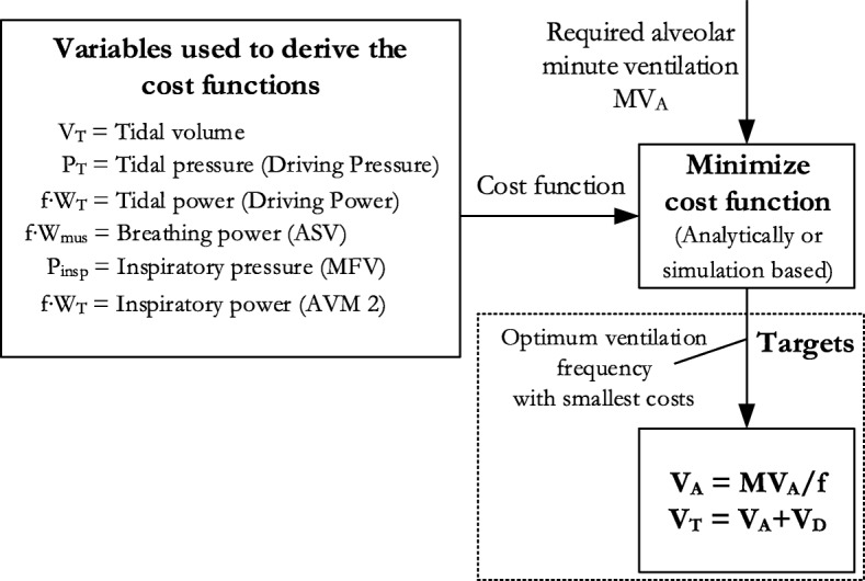 Figure 1: Mathematical cost-function optimization framework for closed-loop adaptive ventilation modes (ASV/AVM). Depicts the derivation of optimal respiratory frequency ($f_{opt}$) and target tidal volume ($V_T$) by minimizing mathematical cost functions—including the Otis work-rate formulation and Mead minimal force principle—to ensure target alveolar minute ventilation ($\dot{V}_A$) across heterogeneous respiratory mechanics (obstructive, restrictive, and normal profiles). Source: Brunner JX et al. Intensive Care Medicine Experimental 2018; 6:31. DOI: 10.1186/s40635-018-0195-6 [CC BY 4.0].
PRVC WAVEFORM SCALAR: RESPONSE TO ACUTE COMPLIANCE REDUCTION
========================================================================================
Breath #         (1) Baseline          (2) Acute Stiffening    (3) Titrating Up (+3)   (4) Target Restored
----------------------------------------------------------------------------------------
Pressure  P_max | - - - - - - - - - - - - - - - - - - - - - - - - - - - - - - - - - - -
(cmH2O)         |
                |                     +-------+               +---------+             +-----------+
                |     +-----+         |       |               |         |             |           |
                |     |     | (20)    |  (20) |               |   (23)  |             |    (26)   |
          PEEP  |-----+-----+---------+-------+---------------+---------+-------------+-----------+
                +-----------------------------------------------------------------------
Flow      Insp  | |\                  |\                      |\                      |\
(L/min)       0 |-\-\------------------\-\---------------------\-\---------------------\-\-----
                |    \                    \                       \                       \
          Exp   |     \--                  \--                     \--                     \--
                +-----------------------------------------------------------------------
Volume    V_T   |      /\  (450 mL)            (320 mL)                (390 mL)            /\  (450 mL)
(mL)            |     /  \                    /\                      /\                  /  \
              0 +----+----+------------------+--+--------------------+--+----------------+----+--
Mechanics:       Normal C_rs (30)     C_rs drops to 16        Loop increments P       Equilibrium at 26
========================================================================================
Clinical Advantages of PRVC / AutoFlow
- Automatic Pressure Minimization:
- Under passive conditions, PRVC guarantees that the selected tidal volume is delivered using the minimum driving pressure possible, reducing the risk of volutrauma and barotrauma during dynamic improvements in lung compliance (e.g., following surfactant, recruitment, or diuresis).
- Decelerating Flow Benefits:
- Compared to square-wave VC-CMV, the decelerating flow pattern reduces peak inspiratory pressures ($P_{\text{peak}}$), improves alveolar gas distribution across lung units with heterogeneous time constants, and shortens dead space fraction ($V_D/V_T$).
- Automatic Compensation for Postural / Bronchomotor Fluctuations:
- Minor shifts in chest wall compliance (turning, patient repositioning) or mild bronchospasm are automatically compensated for by incremental pressure upward titration without clinician intervention.
Critical Pitfalls & The "PRVC Trap"
Despite its computational elegance, PRVC possesses significant mechanical and physiological vulnerabilities when applied in actively breathing, dyssynchronous, or unstable patients.
========================================================================================
                              THE "PRVC PARADOXICAL TRAP"
========================================================================================

   PATIENT AGITATION / SEVERE AIR HUNGER
                     |
                     v
   Vigorous Patient Inspiratory Effort ($P_{mus} \uparrow\uparrow$, e.g., $-15\text{ cmH}_2\text{O}$)
                     |
                     v
   High Instantaneous Flow Demand + Augmented Transpulmonary Driving Pressure
                     |
                     v
   Delivered Tidal Volume Surpasses Target ($V_{T,\text{delivered}} \gg V_{T,\text{target}}$)
                     |
                     v
   PRVC Closed Loop Interprets Breath as "Excess Compliance / Overventilation"
                     |
                     v
   Ventilator Systematically REDUCES Inspiratory Driving Pressure ($P_{\text{insp}} \downarrow\downarrow$)
   (Down to minimum PEEP $+ 0\text{ to }2\text{ cmH}_2\text{O}$)
                     |
                     v
   Patient Receives Near-Zero Ventilator Work Assistance During Extreme Air Hunger
                     |
                     v
   Catastrophic Work of Breathing Exhaustion, Patient-Ventilator Asynchrony, & Diaphragm Injury
========================================================================================
Detailed Pathomechanisms of Failure
- The "PRVC Trap" (Demand-Support Mismatch):
- When a patient experiences severe metabolic acidosis, sepsis, or neurological agitation, their endogenous neuromuscular drive ($P_{mus}$) surges. The patient sucks gas vigorously from the circuit.
- Because $V_T = C_{rs} \cdot (P_{\text{vent}} + P_{mus})$, the vigorous $P_{mus}$ generates an oversized tidal volume (e.g., $650\text{ mL}$ when target is $420\text{ mL}$).
- The PRVC algorithm reacts by reducing machine pressure support in successive steps ($\Delta P = -3\text{ cmH}_2\text{O}$ per breath).
- In severe cases, the machine drops applied pressure down to baseline (PEEP), effectively converting the mode into an unassisted spontaneous breath through a restricted circuit while the patient is in acute respiratory failure.
- Coughing & Transient Artifact De-escalation:
- A vigorous cough generates high circuit pressures ($> 40\text{ cmH}_2\text{O}$) that trigger the $P_{\text{max}}$ ceiling, prematurely cycling the breath. The delivered volume is truncated ($< 150\text{ mL}$).
- The algorithm senses hypoventilation and inappropriately drives up the target pressure for the subsequent breath, potentially over-pressurizing the airway immediately after the cough clears.
- Variable Endotracheal Tube Resistance Artifacts:
- Partial ETT occlusion (secretions, biting) sharply elevates resistive load. In PRVC, the ventilator cannot differentiate resistive pressure drop from loss of alveolar compliance, driving up airway pressure to achieve volume and risking alveolar overdistension if secretions clear suddenly.
- Fixed Inspiratory Time vs. Flow Cycling:
- PRVC remains a time-cycled mandatory mode. If the patient's neural inspiratory time ($T_{i,\text{neural}}$) is shorter or longer than the machine set $T_i$, severe cycle dyssynchrony (premature or delayed cycling) occurs.
Volume Support (VS)
Volume Support is the spontaneous counterpart to PRVC. It is a dual-controlled mode where every breath is patient-triggered and flow-cycled (identical to Pressure Support Ventilation), but the ventilator continuously modulates the pressure support level ($\Delta P_{\text{supp}}$) to maintain a preset target tidal volume ($V_{T,\text{target}}$).
========================================================================================
                        PRVC vs. VOLUME SUPPORT (VS) COMPARISON
========================================================================================
Variable / Feature          PRVC (VC+ / AutoFlow)          Volume Support (VS)
----------------------------------------------------------------------------------------
Trigger                     Time or Patient                Patient Only (Spontaneous)
Target Variable             Preset Tidal Volume (V_T)      Preset Tidal Volume (V_T)
Control Variable            Pressure (Step-adjusted)       Pressure (Step-adjusted)
Cycle Variable              Time (Set T_i)                 Flow (% Peak Expiratory Trigger)
Backup Rate                 Mandatory Backup Rate          Apnea Ventilation Protocol Only
Patient Effort Increase     Drops P_insp (Underassist)     Drops P_supp (Auto-weans)
Patient Apnea               Delivers Time-Triggered Breaths Shifts to Apnea Backup Mode
========================================================================================
Auto-Weaning Mechanics and Hazards in VS
- Auto-Weaning Potential: In recovering, stable post-operative patients, as endogenous diaphragm strength returns and compliance improves, VS automatically steps down pressure support ($\Delta P_{\text{supp}} \to 0\text{ cmH}_2\text{O}$), seamlessly weaning the patient.
- Hypopnea / Apnea Hazard: If the patient's respiratory drive drops (e.g., re-narcotization from remifentanil/hydromorphone), respiratory rate drops. Because VS only titrates individual breath volume and does not control rate, the patient can become severely hypercapnic despite normal single-breath tidal volumes until apnea backup time expires.
Adaptive Support Ventilation (ASV)
Adaptive Support Ventilation (ASV, Hamilton Medical) is an advanced, multivariate closed-loop mode that automatically selects the target tidal volume ($V_T$), respiratory rate ($RR$), inspiratory time ($T_i$), and inspiratory pressure ($P_{\text{insp}}$) for both mandatory and spontaneous breaths.
Rather than targeting a clinician-set tidal volume, the clinician programs:
- Patient Ideal Body Weight (IBW / PBW) (which dictates baseline minute ventilation: typically $100\text{ mL/min/kg}$).
- Target Minute Ventilation Percentage (%MinVol): Adjustable from $25\%\text{ to }350\%$ (where $100\% = 0.1\text{ L/min/kg}$).
- PEEP and $\text{FiO}_2$.
- High Pressure Alarm Limit ($P_{\text{max}}$).
========================================================================================
                           ASV CLOSED-LOOP CONTROL ALGORITHM
========================================================================================
  [ Inputs: Height, Gender -> PBW -> Baseline MV ]  <-- [ Clinician sets %MinVol ]
                                |
                                v
               [ Target Minute Ventilation: \dot{V}_E ]
                                |
                                v
     [ Continuous Measurement of Expiratory Time Constant: \tau_RC = R_aw * C_rs ]
                                |
                                v
             [ OTIS EQUATION MINIMAL WORK-RATE OPTIMIZATION ]
             f_opt = \frac{-1 + \sqrt{1 + 4 \pi^2 \tau_RC (\dot{V}_A / V_D)}}{2 \pi^2 \tau_RC}
                                |
                                v
           [ Optimal Target Coordinates: (f_target, V_T,target) ]
                                |
                                v
                [ Apply Safety Ventilation Window ]
          (P_max limit, V_T,max limit, V_T,min limit, Auto-PEEP boundary)
                                |
                                v
         [ Deliver Breaths via Adaptive Pressure Regulation ]
========================================================================================
Mathematical Foundations: Otis and Mead Formulations
The core physiological premise of ASV is based on the classical work of Arthur Otis (1950) and Jere Mead (1960), which demonstrated that mammalian respiratory control naturally adopts a breathing frequency ($f$) and tidal volume ($V_T$) that minimizes either the total mechanical work rate of breathing ($\dot{W}$) or the average force developed by the respiratory muscles.
1. Total Work Rate of Breathing Equation
Total mechanical work rate ($\dot{W}$) to move air against elastic and resistive forces is:
$$\dot{W} = \dot{W}_{\text{elastic}} + \dot{W}_{\text{resistive}}$$$$\dot{W} = \frac{1}{2 C_{rs}} \cdot f \cdot V_T^2 + \frac{\pi^2}{4} \cdot R_{aw} \cdot f^2 \cdot V_T^2$$Substituting alveolar minute ventilation $\dot{V}_A = f \cdot (V_T - V_D)$, where $V_D$ is anatomical dead space:
$$V_T = \frac{\dot{V}_A}{f} + V_D$$$$\dot{W}(f) = \frac{1}{2 C_{rs}} \cdot f \cdot \left(\frac{\dot{V}_A}{f} + V_D\right)^2 + \frac{\pi^2}{4} \cdot R_{aw} \cdot f^2 \cdot \left(\frac{\dot{V}_A}{f} + V_D\right)^2$$2. The Otis Formula for Optimal Frequency ($f_{\text{opt}}$)
To find the frequency that minimizes work rate, the derivative $\frac{d\dot{W}}{df}$ is set to zero, yielding the Otis Equation:
$$f_{\text{opt}} = \frac{-1 + \sqrt{1 + 4 \pi^2 \cdot \tau_{RC} \cdot \left(\frac{\dot{V}_A}{V_D}\right)}}{2 \pi^2 \cdot \tau_{RC}}$$Where:
- $\tau_{RC} = R_{aw} \cdot C_{rs}$ is the expiratory time constant of the respiratory system (measured continuously from the decline of the expiratory flow scalar).
- $V_D$ is anatomical dead space (estimated by ASV as $2.2\text{ mL/kg}$ of predicted body weight).
- $\dot{V}_A$ is the required target alveolar minute ventilation ($\dot{V}_E - f \cdot V_D$).
3. The Mead Formula for Minimal Average Muscle Force
Mead showed that if the respiratory controller minimizes average muscle pressure/force ($P_{\text{mus}}$) rather than total work:
$$f_{\text{opt, Mead}} = \frac{1}{2 \pi} \cdot \sqrt{\frac{\dot{V}_A}{V_D \cdot \tau_{RC}}}$$Both equations converge on identical physiological adaptations across differing mechanics:
| Respiratory Mechanics | Expiratory Time Constant ($\tau_{RC}$) | ASV Target Strategy | Clinical Rationale |
|---|---|---|---|
| Normal Lungs | Normal ($0.5 - 0.8\text{ s}$) | Moderate $RR$ ($12-16$), Moderate $V_T$ ($6-8\text{ mL/kg}$) | Standard physiological efficiency. |
| Restrictive / ARDS | Very Short ($\tau_{RC} < 0.5\text{ s}$, low $C_{rs}$) | High $RR$ ($22-35$), Low $V_T$ ($4-6\text{ mL/kg}$) | Avoids high elastic work; prevents alveolar overdistension. |
| Obstructive / COPD / Asthma | Very Long ($\tau_{RC} > 1.2\text{ s}$, high $R_{aw}$) | Low $RR$ ($8-12$), High $V_T$ ($8-10\text{ mL/kg}$) | Prolongs expiratory time ($T_e$); prevents dynamic hyperinflation & auto-PEEP. |
The ASV Safety Ventilation Window
To prevent the Otis equation from selecting extreme, unphysiological parameters (e.g., $RR = 60$ or $V_T = 1800\text{ mL}$), the microprocessor plots the target $(f, V_T)$ coordinate within a strict Safety Window bounded by four physiological safety corners:
========================================================================================
                             ASV SAFETY VENTILATION WINDOW
========================================================================================
V_T (mL)
  ^
  |        +-----------------------------------+  <-- High V_T Limit (Volutrauma / 1.5 * V_T,max)
  |        |                                   |      (Based on P_max ceiling & Compliance)
  |        |      [OBSTRUCTIVE]                |
  |        |        (Low f, Large V_T)         |
  |        |                 \                 |
  |        |                  * TARGET (Normal)|
  |        |                   \               |
  |        |                    [RESTRICTIVE]  |
  |        |                      (High f,     |
  |        |                       Low V_T)    |
  |        +-----------------------------------+  <-- Low V_T Limit (Dead space / V_T > 2 * V_D)
  |        |                                   |
  +--------+-----------------------------------+--------> Respiratory Frequency (f, bpm)
          Min f Limit                         Max f Limit
          (Auto-PEEP Boundary /               (Inadequate Expiratory Time /
           T_e < 2 * \tau_RC)                  T_i,min < 0.5 s)
========================================================================================
- Upper Volume Limit: Derived from clinician-set $P_{\text{max}} - 10\text{ cmH}_2\text{O}$ to prevent alveolar overdistension and barotrauma.
- Lower Volume Limit: Restricted to at least $2 \times V_D$ (approx. $> 4.4\text{ mL/kg}$) to prevent dead-space non-alveolar ventilation.
- Upper Frequency Limit: Capped to ensure minimum expiratory time $T_e \ge 2 \times \tau_{RC}$, preventing severe dynamic gas trapping.
- Lower Frequency Limit: Capped to maintain minimum minute ventilation and avoid severe hypoventilation during prolonged apnea.
Initial Settings & Bedside Titration Protocols
PRVC / AutoFlow Initial Bedside Setup Protocol
| Parameter | Initial Setting | Clinical Titration Goal / Bedside Safety Rules |
|---|---|---|
| Mode Selection | PRVC / VC+ / AutoFlow | Ensure patient is either deeply sedated/passive or has smooth, non-vigorous trigger efforts. |
| Target Tidal Volume ($V_T$) | $6.0\text{ mL/kg PBW}$ | Range $4.0-8.0\text{ mL/kg PBW}$. Set $6\text{ mL/kg}$ in ARDS; $7-8\text{ mL/kg}$ in normal mechanics. |
| Inspiratory Time ($T_i$) | $0.8 - 1.0\text{ s}$ | Adjust $T_i$ to ensure inspiratory flow decelerates to near zero before cycling without premature cut-off. |
| Backup Rate ($RR$) | $12 - 18\text{ breaths/min}$ | Titrate to meet minute ventilation target and maintain $PaCO_2\text{ }35-45\text{ mmHg}$ (or permissive hypercapnia). |
| High Pressure Limit ($P_{\text{max}}$) | $35\text{ cmH}_2\text{O}$ | Set strictly. Remember working pressure ceiling is $P_{\text{max}} - 5\text{ cmH}_2\text{O}$ ($30\text{ cmH}_2\text{O}$). |
| PEEP | $5 - 12\text{ cmH}_2\text{O}$ | Titrate according to ARDSNet PEEP/$\text{FiO}_2$ tables or decremental PEEP titration trials. |
| Trigger Sensitivity | Flow: $1.5-2.0\text{ L/min}$ | Minimize trigger delay without inducing autotriggering. |
ASV Initial Bedside Titration Protocol
========================================================================================
                             ASV CLINICAL TITRATION ALGORITHM
========================================================================================
1. INPUT PATIENT DEMOGRAPHICS:
   - Biological Sex & Measured Standing Height (cm) -> Computes Predicted Body Weight (PBW).
   - Baseline Minute Ventilation Calculation:
     $$\dot{V}_{E,\text{norm}} = 0.10\text{ L/min/kg (Adults)} \quad [0.20\text{ L/min/kg in Pediatrics}]$$

2. SET INITIAL %MINVOL (MINUTE VENTILATION PERCENTAGE):
   - Normal Lungs / Post-Op: Set 100%
   - ARDS / Sepsis / Severe Acidosis: Set 120% - 150% (compensate for high $V_D/V_T$ and metabolic rate)
   - Severe COPD / Asthma: Set 70% - 90% (lower target $RR$, prolong $T_e$, prevent auto-PEEP)

3. BEDSIDE BLOOD GAS & WORKLOAD ADJUSTMENTS:
   - If $PaCO_2 > 45\text{ mmHg}$ and $pH < 7.35$ (Respiratory Acidosis):
     --> Increase %MinVol in increments of 10% - 20%.
   - If $PaCO_2 < 35\text{ mmHg}$ and $pH > 7.45$ (Respiratory Alkalosis):
     --> Decrease %MinVol in increments of 10% - 20%.
   - If Patient exhibits excessive spontaneous breathing work ($P_{0.1} > 3.5\text{ cmH}_2\text{O}$ or accessory muscle use):
     --> Increase %MinVol to offload respiratory muscle pump.

4. WEANING & EXTUBATION READINESS:
   - When %MinVol is reduced to 70% - 100% and patient maintains spontaneous breathing ($f_{spont} > 0$),
     ASV acts effectively as automated Pressure Support.
   - If target $P_{\text{insp}} \le 8\text{ cmH}_2\text{O}$ above PEEP and $\text{FiO}_2 \le 0.40$ with stable gas exchange:
     --> Perform formal Spontaneous Breathing Trial (SBT) / Extubation Assessment.
========================================================================================
Bedside Troubleshooting & Red Flags
Paradoxical Under-Assistance in Agitated Patients ("PRVC Trap"): If an actively breathing patient exhibits tachypnea, suprasternal retractions, and high airway pressure alarms during PRVC, inspect the delivered $P_{\text{insp}}$. If $P_{\text{insp}}$ has dropped to near PEEP while patient is visibly struggling, immediately switch to pure Volume Control (VC-CMV) or Pressure Support (PSV). Do not attempt to treat the agitation by increasing sedation alone without correcting the mode mismatch.
Severe Airway Leaks (Bronchopleural Fistula or Deflated Cuff): In both PRVC and Volume Support, a large circuit or tracheal leak causes measured expiratory volume to be severely underestimated. The ventilator falsely assumes poor compliance and relentlessly steps up inspiratory pressure to the maximum alarm ceiling ($P_{\text{max}} - 5\text{ cmH}_2\text{O}$), creating dangerous focal peak pressures. In the presence of significant unsealed leaks, transition to conventional Pressure-Controlled Ventilation (PC-CMV).
ASV in Rapidly Shifting Mechanics (e.g., Bronchospasm Post-Suctioning): ASV calculates $\tau_{RC}$ on a running exponential average over multiple breaths. If severe acute bronchospasm occurs instantaneously, the algorithm requires several breaths to recalculate the prolonged $\tau_{RC}$ and reduce target frequency. Monitor for transient dynamic hyperinflation and auto-PEEP during sudden acute resistance surges.
Comparative Matrix: Conventional vs. Dual-Controlled Modes
| Ventilatory Feature | VC-CMV (Square Flow) | PC-CMV (Decelerating) | PRVC / AutoFlow | ASV (Hamilton) |
|---|---|---|---|---|
| Control Variable | Flow / Volume | Pressure | Pressure | Pressure & Rate |
| Tidal Volume Stability | Guaranteed absolute | Highly variable | Target guaranteed (breath-by-breath) | Target guaranteed within Otis window |
| Peak Airway Pressure | High (resistive spike) | Minimized | Minimized | Minimized |
| Flow Pattern | Constant / Square | Decelerating exponential | Decelerating exponential | Decelerating exponential |
| Response to High $P_{mus}$ | Fixed volume, high $WOB$ | Increases delivered $V_T$ | Drops support pressure (Pitfall) | Adjusts support to match target work |
| Spontaneous Breath Handling | Fixed flow assist | Full pressure assist | Fixed $T_i$ mandatory assist | Seamless transition mandatory $\leftrightarrow$ spontaneous |
| Primary Clinical Domain | Neurotrauma, passive ARDS | Severe ARDS, barotrauma risk | General post-op ICU, stable sedation | Post-cardiac surgery, automated weaning |
Key Takeaways & Summary
- Dual-controlled mandatory modes (PRVC / VC+ / AutoFlow) deliver pressure-regulated breaths with decelerating flow while dynamically titrating applied driving pressure in step-wise increments ($\le \pm 3\text{ cmH}_2\text{O}$/breath) to achieve a clinician-set target tidal volume.
- The PRVC Trap occurs during intense patient respiratory drive ($P_{mus}$): the high muscular effort augments tidal volume, causing the algorithm to falsely interpret lung mechanics as over-compliant and progressively withdraw machine pressure support, leaving the dyspneic patient unassisted.
- Volume Support (VS) applies PRVC principles to purely spontaneous, flow-cycled breaths, functioning as an auto-weaning pressure support mode that requires robust apnea backup vigilance.
- Adaptive Support Ventilation (ASV) optimizes minute ventilation using the Otis equation of minimal work of breathing and the Mead minimal force principle, dynamically determining the optimal $(f, V_T)$ coordinate based on the measured expiratory time constant ($\tau_{RC} = R_{aw} \cdot C_{rs}$).
- ASV lung protection automatically selects rapid, low-volume breathing patterns in restrictive pathologies ($\tau_{RC} < 0.5\text{ s}$) and slow, deep breathing patterns in obstructive pathologies ($\tau_{RC} > 1.2\text{ s}$) to prevent dynamic hyperinflation while protecting against volutrauma.