The management of critically ill patients receiving mechanical ventilation requires an intimate, quantitative understanding of respiratory mechanics. Rather than viewing mechanical ventilation as merely a gas-exchange modality, the intensivist must conceptualize the ventilator and respiratory system as an integrated mechanical circuit governed by physical laws. The bedrock of this physiological framework is the Equation of Motion of the Respiratory System, which characterizes how driving pressures overcome elastic recoil, frictional resistance, and positive end-expiratory pressure to achieve alveolar ventilation.
Understanding the components of this equation allows the clinician to partition total airway pressures into resistive and elastic work, measure lung and chest wall mechanics independently, detect dynamic hyperinflation, optimize patient-ventilator synchrony, and prevent ventilator-induced lung injury (VILI).
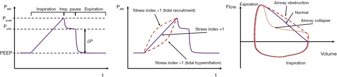 Figure 1: The single-compartment model of the respiratory system and partitioning of driving pressure during constant-flow volume-controlled ventilation. Total pressure ($P_{aw}$) balances resistive pressure drops ($\Delta P_{res} = R_{aw} \cdot \dot{V}$) across conducting airways and elastic recoil pressure ($\Delta P_{el} = V_T / C_{rs} = E_{rs} \cdot V_T$) of the respiratory system above PEEP. Measured via an end-inspiratory pause, $P_{peak}$ reflects combined resistive and elastic loads, whereas $P_{plat}$ isolates static elastic recoil. Sourced from Ball et al., Ann Transl Med 2018;6(19):379. DOI: 10.21037/atm.2018.09.50 (Open Access / CC BY-NC-ND 4.0).
The Single-Compartment Model and The Equation of Motion
The classic single-compartment linear mathematical model treats the respiratory system as a single distensible balloon (representing the combined elastance of lung parenchyma and chest wall) connected to a single rigid conducting pipe (representing the resistance of endotracheal tube, central airways, and peripheral bronchioles).
Under conditions of laminar flow and linear elasticity, the differential equation governing instantaneous pressure balance across the system at any given moment is:
$$P_{vent}(t) + P_{mus}(t) = E_{rs} \cdot V(t) + R_{aw} \cdot \dot{V}(t) + \text{PEEP}_{tot}$$Where:
- $P_{vent}(t)$: Pressure applied at the airway opening by the mechanical ventilator ($P_{ao}$).
- $P_{mus}(t)$: Pressure generated by the patient's inspiratory muscles (predominantly diaphragm and external intercostals), represented as a negative pleural pressure deflection during spontaneous breathing effort ($P_{mus} = 0$ in a fully passive, paralyzed patient).
- $E_{rs}$: Total elastance of the respiratory system ($\text{cmH}_2\text{O/L}$ or $\text{cmH}_2\text{O/mL}$), representing the inverse of compliance ($E_{rs} = 1 / C_{rs}$).
- $V(t)$: Instantaneous lung volume above functional residual capacity (FRC) or tidal volume ($V_T$) at time $t$.
- $R_{aw}$: Airway resistance ($\text{cmH}_2\text{O} \cdot \text{s/L}$), reflecting frictional opposition to gas flow through ventilator circuits, endotracheal tube, and tracheobronchial tree.
- $\dot{V}(t)$: Instantaneous inspiratory or expiratory gas flow rate ($\text{L/s}$ or $\text{L/min}$).
- $\text{PEEP}_{tot}$: Total Positive End-Expiratory Pressure, which represents the sum of applied extrinsic PEEP ($\text{PEEP}_e$) and intrinsic or auto-PEEP ($\text{PEEP}_i$).
Inertance: Why It Is Clinically Omitted
A complete fluid mechanics description includes an inertial pressure component:
$$P_{inertial} = I_{rs} \cdot \ddot{V}(t) = I_{rs} \cdot \frac{d^2V}{dt^2}$$where $I_{rs}$ represents the inertance (mass acceleration of gas and chest tissues). Under standard clinical mechanical ventilation frequencies ($f = 10\text{--}35\text{ breaths/min}$), inertial forces account for less than $0.5\text{ cmH}_2\text{O}$ of total pressure and are negligible. Inertance becomes physiologically significant only during High-Frequency Oscillatory Ventilation (HFOV) or High-Frequency Jet Ventilation (HFJV) at frequencies exceeding $3\text{--}15\text{ Hz}$ ($180\text{--}900\text{ breaths/min}$).
Elastic vs. Resistive Work of Breathing and Compliance Mechanics
Total Work of Breathing (WOB) performed per breath is the integral of distending pressure with respect to volume:
$$WOB = \int P(t) \, dV = \int_{0}^{V_T} \left( E_{rs} \cdot V + R_{aw} \cdot \dot{V} \right) dV$$This work is partitioned into two distinct physical entities:
- Elastic Work: Energy expended to overcome elastic recoil forces of lung elastin/collagen fibers, alveolar surface tension, and the thoracic wall cage. In a purely elastic structure, this potential energy is stored during inspiration and drives passive exhalation.
- Resistive (Frictional) Work: Energy dissipated entirely as thermal energy to overcome friction between gas molecules (viscosity) and airway walls, as well as tissue viscance.
       Airway Pressure (Paw)
         ^
  Ppeak  |-------------------/|
         |                  / |  <- Resistive Work: Raw * Flow
  Pplat  |.........../|    /  |
         |          / |   /   |  <- Elastic Work: 1/2 * Ers * (Vt)^2
         |         /  |  /    |     (Driving Pressure * Vt / 2)
  PEEP   |________/__ |_/_____|
         0                    Vt    Volume (V)
Static vs. Dynamic Compliance
Compliance ($C$) defines the volume change per unit change in distending pressure ($C = \Delta V / \Delta P$). In clinical practice, two separate compliance values are calculated:
1. Static Respiratory System Compliance ($C_{stat}$ or $C_{rs}$)
Measured under zero-flow static conditions at end-inspiration via an inspiratory pause maneuver:
$$C_{stat} = \frac{V_T}{P_{plat} - \text{PEEP}_{tot}} = \frac{V_T}{\Delta P}$$- Normal values: $50\text{--}100\text{ mL/cmH}_2\text{O}$ in healthy non-intubated individuals; $50\text{--}70\text{ mL/cmH}_2\text{O}$ in healthy anesthetized/intubated patients.
- Pathology: In moderate-to-severe ARDS, $C_{stat}$ characteristically falls to $< 30\text{ mL/cmH}_2\text{O}$ (frequently $15\text{--}25\text{ mL/cmH}_2\text{O}$) due to alveolar flooding, collapse, and functional lung volume reduction ("baby lung").
- Physical Meaning: $C_{stat}$ reflects the true elastic properties of the lung parenchyma and chest wall without confounding resistive pressures.
2. Dynamic Compliance ($C_{dyn}$)
Calculated using the peak inspiratory pressure during active flow:
$$C_{dyn} = \frac{V_T}{P_{peak} - \text{PEEP}_{tot}}$$- Clinical Utility: $C_{dyn}$ is always lower than $C_{stat}$ because $P_{peak}$ includes the resistive pressure drop. It reflects a composite of both elastic and resistive impediments.
- Diagnostic Discordance:
- If $C_{dyn}$ drops while $C_{stat}$ remains normal, the pathology is purely resistive (e.g., bronchospasm, mucus plugging, bitten endotracheal tube).
- If both $C_{dyn}$ and $C_{stat}$ drop in parallel, the underlying defect is elastic (e.g., ARDS progression, tension pneumothorax, massive pleural effusion, acute abdominal compartment syndrome).
3. Partitioning Respiratory Elastance ($E_{rs} = E_L + E_{cw}$)
Total respiratory elastance ($E_{rs}$) is the linear sum of lung elastance ($E_L$) and chest wall elastance ($E_{cw}$):
$$E_{rs} = E_L + E_{cw} \implies \frac{1}{C_{rs}} = \frac{1}{C_L} + \frac{1}{C_{cw}}$$Under normal conditions, chest wall compliance ($C_{cw} \approx 150\text{--}200\text{ mL/cmH}_2\text{O}$) and lung compliance ($C_L \approx 150\text{--}200\text{ mL/cmH}_2\text{O}$) combine to give a total $C_{rs} \approx 75\text{--}100\text{ mL/cmH}_2\text{O}$. In morbid obesity, massive ascites, chest wall burns, or high intra-abdominal pressure (IAP), decreased $C_{cw}$ elevates $P_{plat}$ without necessarily exposing alveolar units to excessive transpulmonary stress.
Airway Resistance ($R_{aw}$) and Rohrer's Non-Linearity
Airway resistance represents the pressure drop along the tracheobronchial tree per unit flow:
$$R_{aw} = \frac{\Delta P_{resistive}}{\dot{V}} = \frac{P_{peak} - P_{plat}}{\dot{V}}$$Important: The gas flow rate $\dot{V}$ in the denominator must be converted to $\text{L/s}$ (e.g., $60\text{ L/min} = 1.0\text{ L/s}$; $45\text{ L/min} = 0.75\text{ L/s}$).
Normal Values
- Healthy non-intubated adult: $1.0\text{--}2.5\text{ cmH}_2\text{O}\cdot\text{s/L}$.
- Intubated adult (ETT 7.5–8.5 mm ID): $5.0\text{--}10.0\text{ cmH}_2\text{O}\cdot\text{s/L}$.
- Severe bronchospasm / Status Asthmaticus / COPD: $15.0\text{--}35.0+\text{ cmH}_2\text{O}\cdot\text{s/L}$.
Laminar vs. Turbulent Flow: Rohrer's Equation
While the single-compartment model assumes a constant linear resistance (Poiseuille's law: $\Delta P = \frac{8\eta l}{\pi r^4} \cdot \dot{V}$), flow in large airways and through endotracheal tubes is transitional or turbulent. Rohrer's equation accounts for this non-linearity:
$$\Delta P = K_1 \cdot \dot{V} + K_2 \cdot \dot{V}^2$$- $K_1 \cdot \dot{V}$ represents laminar viscous dissipation.
- $K_2 \cdot \dot{V}^2$ represents turbulent convective acceleration and eddy formation (predominant inside the ETT and upper airway).
- Clinical Implication: Doubling inspiratory flow from $30\text{ L/min}$ to $60\text{ L/min}$ more than doubles the resistive pressure drop across the endotracheal tube due to the velocity-squared relationship.
The Respiratory Time Constant ($\tau$) and Expiratory Mechanics
The respiratory time constant ($\tau$, tau) describes the rate at which a viscoelastic lung compartment fills or empties when exposed to a step change in pressure. It is defined as the mathematical product of resistance and compliance:
$$\tau = R_{aw} \times C_{rs}$$Dimensionally:
$$\tau = \left(\frac{\text{cmH}_2\text{O}}{\text{L/s}}\right) \times \left(\frac{\text{L}}{\text{cmH}_2\text{O}}\right) = \text{seconds (s)}$$During passive exhalation, the instantaneous volume remaining in the lung $V(t)$ decays exponentially as a function of time:
$$V(t) = V_T \cdot e^{-t / \tau}$$Expiratory Volume Dissipation (% of Vt):
t = 1 tau: [=======> ] 63.2% exhaled (36.8% volume remains)
t = 2 tau: [==================> ] 86.5% exhaled (13.5% volume remains)
t = 3 tau: [=======================> ] 95.0% exhaled ( 5.0% volume remains)
t = 4 tau: [===========================> ] 98.2% exhaled ( 1.8% volume remains)
t = 5 tau: [=============================> ] 99.3% exhaled ( 0.7% volume remains)
| Time Constants ($\tau$) | Cumulative Volume Exchanged / Exhaled | Volume Remaining in Compartment | Clinical State |
|---|---|---|---|
| $1 \tau$ | $63.2\%$ | $36.8\%$ | Inadequate exhalation; massive air trapping |
| $2 \tau$ | $86.5\%$ | $13.5\%$ | Significant breath stacking |
| $3 \tau$ | $95.0\%$ | $5.0\%$ | Minimum acceptable expiratory time ($T_e$) |
| $4 \tau$ | $98.2\%$ | $1.8\%$ | Near-complete lung emptying |
| $5 \tau$ | $99.3\%$ | $0.7\%$ | Full physiological equilibration |
Clinical Pathophysiology: ARDS vs. COPD/Asthma
1. ARDS (Low $\tau$ System: "Fast Alveoli")
- Mechanics: Very low compliance ($C_{rs} \approx 20\text{ mL/cmH}_2\text{O} = 0.020\text{ L/cmH}_2\text{O}$), near-normal airway resistance ($R_{aw} \approx 8\text{ cmH}_2\text{O}\cdot\text{s/L}$).
- Time Constant: $\tau = 8 \times 0.020 = 0.16\text{ seconds}$.
- Full Expiration ($4\text{--}5 \tau$): Requires only $0.64\text{--}0.80\text{ seconds}$.
- Implications: Alveolar units fill and empty rapidly. The danger in ARDS is not gas trapping, but end-expiratory alveolar derecruitment and cyclic atelectrauma due to rapid collapse when expiratory time is prolonged without sufficient PEEP. High respiratory rates ($f = 28\text{--}35\text{ bpm}$) are well-tolerated without intrinsic PEEP generation.
2. COPD and Status Asthmaticus (High $\tau$ System: "Slow Alveoli")
- Mechanics: Extremely high airway resistance ($R_{aw} \approx 25\text{ cmH}_2\text{O}\cdot\text{s/L}$) and normal-to-increased compliance ($C_{rs} \approx 80\text{ mL/cmH}_2\text{O} = 0.080\text{ L/cmH}_2\text{O}$ in emphysema).
- Time Constant: $\tau = 25 \times 0.080 = 2.0\text{ seconds}$.
- Full Expiration ($4\text{--}5 \tau$): Requires $8.0\text{--}10.0\text{ seconds}$!
- Implications: If the ventilator's set expiratory time ($T_e$) is only $2.0\text{ seconds}$ ($1 \tau$), the patient will exhale only $63\%$ of the delivered tidal volume before the next breath triggers, resulting in severe breath-stacking, dynamic hyperinflation, occult auto-PEEP, high intrathoracic pressures, impaired venous return, and cardiovascular collapse.
- Management: Lower respiratory rate ($f = 10\text{--}14\text{ bpm}$), maximize $T_e$, and deliver high inspiratory flows ($70\text{--}80\text{ L/min}$) with square waveforms to shorten $T_i$.
3. Lung Heterogeneity and Pendelluft
In diseased lungs, adjacent alveolar units possess vastly different time constants ($\tau_{fast}$ vs $\tau_{slow}$). During both inspiration and post-inspiratory pause, gas redistributes from fast units to slow units without net external airflow—a phenomenon termed pendelluft. In spontaneously breathing ARDS patients, vigorous diaphragmatic contractions create localized negative pleural pressures that drive pendelluft from non-dependent to dependent lung regions, generating localized occult overdistension.
Stress Index and Mechanical Power
The Stress Index ($SI$)
The Stress Index evaluates the linearity of the pressure-time curve ($P_{aw}(t)$) during constant-flow (square wave) Volume-Controlled Ventilation (VCV) in passive patients. When inspiratory flow ($\dot{V}$) is held strictly constant, the resistive pressure drop ($R_{aw} \cdot \dot{V}$) is constant. Consequently, any curvature in $P_{aw}(t)$ reflects changes in instantaneous respiratory system compliance as lung volume expands.
The curve is modeled by the power equation:
$$P_{aw}(t) = a \cdot t^b + c$$where the exponent $b$ is the Stress Index.
Paw
 ^       SI > 1 (Upward Concavity) -> Overdistension / Decreasing Compliance
 |          __--'
 |        /
 |       /  SI = 1 (Linear) -> Constant Compliance / Safe Tidal Ventilation
 |      /
 |     / __-- SI < 1 (Downward Concavity) -> Tidal Recruitment / Increasing Compliance
 |    /'
 +-------------------------> Time (Inspiration with Constant Flow)
- $SI < 1.0$ (Downward Concavity / Decelerating slope):
- Pathophysiology: Dynamic compliance is increasing during lung inflation. Alveolar units that were collapsed at end-expiration are being progressively recruited throughout the breath.
- Clinical Action: PEEP is set below the lower inflection point. Increase PEEP to prevent cyclic end-expiratory collapse and atelectrauma.
- $SI = 1.0$ ($0.95\text{--}1.05$, Constant linear slope):
- Pathophysiology: Constant compliance throughout the tidal volume delivery. Ventilation is occurring along the linear, safe compliant zone of the respiratory pressure-volume curve.
- Clinical Action: Maintain current PEEP and tidal volume settings.
- $SI > 1.0$ (Upward Concavity / Accelerating slope):
- Pathophysiology: Dynamic compliance is decreasing as lung volume increases. Alveolar units have reached their maximum elastic limit and are undergoing progressive hyperinflation and overdistension (volutrauma/barotrauma).
- Clinical Action: Alveolar units are on the flat upper portion of the P-V curve. Decrease tidal volume ($V_T$) or decrease PEEP.
Mechanical Power ($\text{MP}$)
Mechanical power represents the amount of mechanical energy transmitted from the ventilator into the respiratory system per unit time, expressed in Joules per minute ($\text{J/min}$ or $\text{Watts}$). Introduced by Luciano Gattinoni and colleagues, mechanical power unifies all mechanical variables ($V_T$, $\Delta P$, flow, PEEP, and respiratory rate) into a single overarching biophysical metric of VILI risk.
1. Gattinoni Reference Equation (for Volume Control):
$$\text{MP}_{\text{VCV}} = 0.098 \cdot \text{RR} \cdot V_T \cdot \left[ P_{peak} - \frac{1}{2} \cdot \Delta P_{driving} \right]$$where $0.098$ is the conversion factor from $\text{L} \cdot \text{cmH}_2\text{O}$ to Joules ($\text{J}$), $\text{RR}$ is in breaths/min, and $V_T$ is in Liters.
2. Simplified Bedside Surrogates:
- Driving Pressure-Based Equation: $$\text{MP}_{\text{VCV}} = 0.098 \cdot \text{RR} \cdot V_T \cdot \left[ P_{peak} - \frac{P_{plat} - \text{PEEP}}{2} \right]$$
- Giosa / Chiumello Simplified Estimation: $$\text{MP} \approx 0.098 \cdot V_T \cdot \text{RR} \cdot \left( P_{peak} + \text{PEEP} + \frac{F}{6} \right)$$
- Pressure-Controlled Ventilation (PCV): $$\text{MP}_{\text{PCV}} = 0.098 \cdot \text{RR} \cdot V_T \cdot \left( \Delta P_{insp} + \text{PEEP} \right)$$
Clinical Thresholds
- Safe Range: $< 12\text{--}17\text{ J/min}$.
- High Risk of VILI: $> 22\text{--}27\text{ J/min}$ is strongly and independently associated with increased 28-day ICU mortality in ARDS patients.
- Normalized Mechanical Power: Power indexed to respiratory compliance ($\text{MP} / C_{rs}$) or indexed to estimated well-aerated lung weight ($\text{MP} / \text{kg lung tissue}$) provides even higher prognostic accuracy.
Bedside Diagnostic Maneuvers and Transpulmonary Pressure
Accurate bedside calculation of respiratory mechanics requires standardized pause maneuvers under conditions of zero flow ($\dot{V} = 0$).
Pressure (cmH2O)
  ^
35|        Ppeak
  |        /\
30|       /  \    Inspiratory Hold (V' = 0)
25|      /    \_______ Pplat (End-Inspiratory Pause)
  |     /             |
15|    /              \
  |   /                \                  Expiratory Hold (V' = 0)
 5|--/                  \_________________-------- PEEPi / PEEPtot
  +-------------------------------------------------------------> Time
Flow (L/min)
  ^
60|  |---------|
  |  |         |
 0|--|---------|______|___________________--------________________
  |                   | \               /
-60|                   |  \_____________/ (Expiratory Flow)
1. End-Inspiratory Hold ($P_{plat}$ Measurement)
- Technique: In a passive patient on VCV with constant flow, an end-inspiratory pause of $0.5\text{--}2.0\text{ seconds}$ is applied. Flow drops to zero ($\dot{V} = 0$).
- Physiological Effect: By eliminating $\Delta P_{resistive} = R_{aw} \cdot \dot{V} = 0$, airway opening pressure equilibrates with alveolar pressure.
- Calculations:
- Driving Pressure ($\Delta P$): $$\Delta P = P_{plat} - \text{PEEP}_{tot}$$ LUNG SAFE / Amato Trial threshold: Keep $\Delta P \le 14\text{--}15\text{ cmH}_2\text{O}$.
- Static Compliance ($C_{rs}$): $$C_{rs} = \frac{V_T}{P_{plat} - \text{PEEP}_{tot}}$$
- Airway Resistance ($R_{aw}$): $$R_{aw} = \frac{P_{peak} - P_{plat}}{\dot{V}}$$
2. End-Expiratory Hold ($\text{PEEP}_i$ / Auto-PEEP Measurement)
- Technique: An end-expiratory occlusion of $2.0\text{--}3.0\text{ seconds}$ is triggered immediately before the next inspiratory cycle begins.
- Physiological Effect: Flow ceases throughout the entire circuit. Trapped alveolar gas equilibrates across patent airways to the proximal pressure transducer.
- Calculations:
- Total PEEP: Measured plateau during expiratory hold.
- Intrinsic PEEP ($\text{PEEP}_i$, Auto-PEEP): $$\text{PEEP}_i = \text{PEEP}_{tot} - \text{PEEP}_{extrinsic}$$
- Significance: If dynamic airway collapse occurs (e.g., severe emphysema), $\text{PEEP}_i$ measured at the airway opening may underestimate true alveolar trapped gas pressure (closed-circuit uncommunicative gas units).
3. Esophageal Balloon Manometry and Transpulmonary Pressure ($P_{tp}$)
Transpulmonary pressure ($P_{tp}$) is the true distending pressure acting across the alveolar wall, defined as the difference between airway pressure ($P_{aw}$) and pleural pressure ($P_{pl}$):
$$P_{tp} = P_{aw} - P_{pl}$$Because direct pleural space catheterization carries high complication risks, esophageal pressure ($P_{es}$) measured via an air-filled balloon catheter positioned in the lower third of the esophagus serves as an established surrogate for local $P_{pl}$.
Clinical Calibration & Placement
- Placement: Advance catheter to stomach ($45\text{--}50\text{ cm}$ from nares), inflate recommended calibration volume ($0.5\text{--}1.0\text{ mL}$ of air), withdraw until cardiac oscillations appear on the pressure tracing (approx. $35\text{--}40\text{ cm}$ in adults).
- Baydur Occlusion Test (Dynamic Calibration):
- Perform an end-expiratory airway occlusion while the patient makes a spontaneous inspiratory effort.
- Because lung volume cannot change ($\Delta V = 0$), the change in airway pressure must exactly mirror the change in pleural pressure: $$\frac{\Delta P_{es}}{\Delta P_{aw}} = 1.0 \quad (\text{acceptable range: } 0.8\text{--}1.2)$$
- If the ratio is $< 0.8$, the balloon is underfilled or located in the stomach; if $> 1.2$, the balloon is overinflated or positioned too high (tracheal compression).
Transpulmonary Pressure Calculations & Targets
Method A: Direct End-Expiratory Transpulmonary Pressure (PEEP Titration)
$$P_{tp,exp} = \text{PEEP} - P_{es,exp}$$- Target: In severe ARDS, maintain end-expiratory transpulmonary pressure positive ($P_{tp,exp} \ge 0\text{ to }+2\text{ cmH}_2\text{O}$) to prevent end-expiratory atelectasis and cyclical derecruitment.
- Application: In severe chest wall stiffness or morbid obesity ($BMI > 40\text{ kg/m}^2$), $P_{es,exp}$ may reach $+15\text{--}20\text{ cmH}_2\text{O}$. Setting an applied PEEP of $10\text{ cmH}_2\text{O}$ yields a negative $P_{tp,exp} = 10 - 18 = -8\text{ cmH}_2\text{O}$ (alveolar collapse). Applied PEEP must be raised to $18\text{--}20\text{ cmH}_2\text{O}$ to achieve a neutral-to-positive transpulmonary pressure.
Method B: Elastance-Derived Transpulmonary Pressure (End-Inspiratory Stress Assessment)
Due to gravitational vertical pleural pressure gradients, the elastance-derived method estimates stress in the non-dependent, open lung zones:
$$P_{tp,insp} = P_{plat} \times \left( \frac{E_L}{E_{rs}} \right) = P_{plat} \times \left( \frac{\Delta P_{tp}}{\Delta P_{aw}} \right)$$- Target: Limit end-inspiratory transpulmonary pressure to prevent barotrauma/volutrauma: $$P_{tp,insp} \le 20\text{--}25\text{ cmH}_2\text{O}$$
Bedside Mechanical Titration Algorithms
                             [ High Peak Airway Pressure Alarm (Ppeak > 35 cmH2O) ]
                                                       |
                                            Perform Inspiratory Hold
                                                       |
                                    +------------------+------------------+
                                    |                                     |
                       Pplat Normal (<= 25 cmH2O)             Pplat Elevated (> 30 cmH2O)
                    [ Increased Resistive Gradient ]       [ Decreased Respiratory Compliance ]
                                    |                                     |
                    +---------------+---------------+     +---------------+---------------+
                    |               |               |     |               |               |
               Airway Secretions   ETT Kinked/    Severe  ARDS / Edema  Pneumothorax   Abdominal
               / Mucus Plug        Bitten        Spasm   Atelectasis   (Asymmetric    Hypertension
                    |               |               |     |             Breath Sounds) (Elevated IAP)
               [ Suction ]     [ Bite Block/   [ Broncho- [ Lung Prot.  [ Tube         [ Paracentesis /
                                Reposition ]   dilators ]  Ventilation/  Thoracostomy ] Decompression]
                                                           PEEP Titr. ]
1. The High Peak Airway Pressure ($P_{peak}$) Algorithm
When a ventilator generates a high peak pressure alarm, perform an immediate End-Inspiratory Pause:
- Calculate Resistive Pressure Drop: $\Delta P_{res} = P_{peak} - P_{plat}$
- If $\Delta P_{res} > 10\text{--}15\text{ cmH}_2\text{O}$ (Elevated Resistance, Normal $P_{plat}$):
- Etiologies: Endotracheal tube obstruction, biting, kinking, tracheobronchial mucus plugging, severe bronchospasm, water condensation in tubing.
- Management: Pass suction catheter; insert bite block; auscultate for expiratory wheezing; administer inhaled bronchodilators; check inline HME/filter resistance.
- If $\Delta P_{res} > 10\text{--}15\text{ cmH}_2\text{O}$ (Elevated Resistance, Normal $P_{plat}$):
- Calculate Elastic Driving Pressure: $\Delta P = P_{plat} - \text{PEEP}_{tot}$
- If both $P_{peak}$ and $P_{plat}$ are elevated ($\Delta P > 15\text{ cmH}_2\text{O}$, Low $C_{stat}$):
- Etiologies: Worsening pulmonary edema, progressive ARDS consolidation, tension pneumothorax, mainstem bronchus intubation, massive atelectasis, severe dynamic hyperinflation ($\text{PEEP}_i$), chest wall rigidity (e.g., fentanyl overdose), acute intra-abdominal hypertension.
- Management: Check bilateral breath sounds immediately; inspect tracheal tube depth; assess for tension pneumothorax (decompress if suspected); measure auto-PEEP with expiratory pause; measure bladder pressure; perform chest radiography or lung ultrasound.
- If both $P_{peak}$ and $P_{plat}$ are elevated ($\Delta P > 15\text{ cmH}_2\text{O}$, Low $C_{stat}$):
2. Comprehensive Bedside Monitoring Reference Values
| Physiological Parameter | Formula / Derivation | Normal Healthy Range | ARDS Targets / Thresholds | Obstructive (COPD/Asthma) Targets |
|---|---|---|---|---|
| Peak Airway Pressure ($P_{peak}$) | $E_{rs} \cdot V_T + R_{aw} \cdot \dot{V} + \text{PEEP}$ | $15\text{--}20\text{ cmH}_2\text{O}$ | $\le 35\text{ cmH}_2\text{O}$ | Frequently high ($> 40\text{ cmH}_2\text{O}$) due to $R_{aw}$ |
| Plateau Pressure ($P_{plat}$) | $E_{rs} \cdot V_T + \text{PEEP}_{tot}$ | $10\text{--}15\text{ cmH}_2\text{O}$ | $\le 28\text{--}30\text{ cmH}_2\text{O}$ | $\le 25\text{--}30\text{ cmH}_2\text{O}$ (Crucial safety limit) |
| Driving Pressure ($\Delta P$) | $P_{plat} - \text{PEEP}_{tot} = \frac{V_T}{C_{rs}}$ | $5\text{--}10\text{ cmH}_2\text{O}$ | $\le 14\text{--}15\text{ cmH}_2\text{O}$ | Keep minimized ($\le 14\text{ cmH}_2\text{O}$) |
| Static Compliance ($C_{stat}$) | $\frac{V_T}{P_{plat} - \text{PEEP}_{tot}}$ | $50\text{--}100\text{ mL/cmH}_2\text{O}$ | Marked drop ($15\text{--}30\text{ mL/cmH}_2\text{O}$) | Normal or elevated ($50\text{--}90\text{ mL/cmH}_2\text{O}$) |
| Airway Resistance ($R_{aw}$) | $\frac{P_{peak} - P_{plat}}{\dot{V}}$ | $5\text{--}10\text{ cmH}_2\text{O}\cdot\text{s/L}$ (ETT) | $5\text{--}12\text{ cmH}_2\text{O}\cdot\text{s/L}$ | Severe elevation ($15\text{--}35+\text{ cmH}_2\text{O}\cdot\text{s/L}$) |
| Time Constant ($\tau$) | $R_{aw} \times C_{rs}$ | $0.4\text{--}0.6\text{ seconds}$ | Short ($0.15\text{--}0.30\text{ s}$) | Prolonged ($1.5\text{--}3.0+\text{ s}$) |
| Stress Index ($SI$) | Shape of $P_{aw}(t)$ under constant flow | $1.0$ | Titrate PEEP until $SI = 0.95\text{--}1.05$ | N/A (Flow limited exhalation) |
| Mechanical Power ($\text{MP}$) | $0.098 \cdot \text{RR} \cdot V_T \cdot [P_{peak} - \frac{\Delta P}{2}]$ | $4\text{--}8\text{ J/min}$ | $< 17\text{ J/min}$ (Hazard $> 22\text{ J/min}$) | Minimize via low RR |
| End-Exp. Transpulmonary ($P_{tp,exp}$) | $\text{PEEP} - P_{es,exp}$ | $0\text{ to }+2\text{ cmH}_2\text{O}$ | $0\text{ to }+2\text{ cmH}_2\text{O}$ (Prevents collapse) | Non-negative |
| End-Insp. Transpulmonary ($P_{tp,insp}$) | $P_{plat} - P_{es,insp}$ (or elastance-based) | $< 10\text{ cmH}_2\text{O}$ | $\le 20\text{--}25\text{ cmH}_2\text{O}$ | $\le 20\text{ cmH}_2\text{O}$ |
Critical Bedside Pitfalls and Clinical Traps
Trap 1: Misinterpreting $P_{peak}$ as Alveolar Distending Pressure in Obstructive Disease In severe asthma or COPD exacerbations, $P_{peak}$ can easily reach $45\text{--}60\text{ cmH}_2\text{O}$ due to massive airway resistance ($\Delta P_{res} = R_{aw} \cdot \dot{V}$). Clinicians who inappropriately slash tidal volumes based on $P_{peak}$ alone cause profound hypercapnia and acidosis. Always obtain a $P_{plat}$ during an inspiratory pause. If $P_{plat}$ is low ($\le 25\text{ cmH}_2\text{O}$), the alveoli are completely protected from barotrauma/volutrauma despite alarming peak circuit pressures.
Trap 2: Performing Pause Maneuvers in Actively Breathing Patients Inspiratory and expiratory pause maneuvers are valid only when the patient is completely passive. Patient inspiratory effort ($P_{mus}$) during an inspiratory pause causes a negative deflection in $P_{plat}$, falsely overestimating compliance and underestimating driving pressure. Conversely, active abdominal contraction during an expiratory pause falsely inflates the measured auto-PEEP. If precise mechanics are required, ensure adequate sedation, transient neuromuscular blockade, or analyze esophageal pressure waveforms.
Trap 3: Overlooking Auto-PEEP When Setting Applied PEEP in ARDS vs. COPD In ARDS, dynamic collapse occurs without intrinsic PEEP generation; extrinsic PEEP is required to restore FRC. In COPD, flow limitation causes auto-PEEP ($\text{PEEP}_i$). In a passively ventilated COPD patient, adding extrinsic PEEP is additive and will increase total alveolar pressure and worsen hemodynamic compromise. In a spontaneously triggering COPD patient with flow limitation, setting extrinsic PEEP to $\approx 70\text{--}80\%$ of $\text{PEEP}_i$ counterbalances the threshold load required to trigger the ventilator without increasing total alveolar pressure ("waterfall effect").
Trap 4: Ignoring the Chest Wall Component in Obese and Surgical ICU Patients In patients with morbid obesity, tense ascites, open laparotomies with packings, or circumferential thoracic burns, an elevated $P_{plat}$ of $32\text{--}35\text{ cmH}_2\text{O}$ may reflect decreased chest wall compliance ($C_{cw}$) rather than stiff lungs. Calculating transpulmonary pressure ($P_{tp} = P_{aw} - P_{es}$) prevents the premature and hazardous withdrawal of necessary tidal volumes and distending pressures in patients whose lungs are not experiencing true excessive mechanical stress.
Clinical Summary & Takeaways
- The Equation of Motion ($P_{vent} + P_{mus} = E_{rs} \cdot V + R_{aw} \cdot \dot{V} + \text{PEEP}_{tot}$) governs every single mechanical breath and provides the physical foundation for interpreting ventilator graphics and pathology.
- $P_{peak}$ vs. $P_{plat}$ Partitioning: $P_{peak}$ reflects resistive + elastic loads; $P_{plat}$ (measured at zero flow) isolates the true elastic recoil load of the respiratory system.
- Driving Pressure ($\Delta P = P_{plat} - \text{PEEP}_{tot} = V_T / C_{rs}$) is the single strongest physiological predictor of mortality in ARDS; maintain $\Delta P \le 14\text{ cmH}_2\text{O}$.
- Time Constants ($\tau = R \times C$) dictate alveolar filling and emptying kinetics. ARDS lungs ("fast units", $\tau \approx 0.2\text{ s}$) require PEEP to prevent rapid collapse. Obstructive lungs ("slow units", $\tau \ge 2.0\text{ s}$) require prolonged expiratory times ($4\text{--}5 \tau \approx 8\text{--}10\text{ s}$) to prevent dynamic hyperinflation and cardiovascular collapse.
- Stress Index ($SI$) and Mechanical Power ($\text{MP}$) quantify dynamic compliance shifts and total energy transfer ($< 17\text{ J/min}$ target) to eliminate atelectrauma and overdistension.
- Esophageal Manometry ($P_{es}$) separates lung from chest wall mechanics, enabling direct calculation of transpulmonary distending pressure ($P_{tp} = P_{aw} - P_{es}$) to optimize PEEP and avoid VILI in complex thoracic/abdominal pathology.