Every chapter before this one explained a mechanism; this final chapter tests whether it can be recognised the moment the monitor alarms and the blood pressure falls. The ventilator screen is a continuous experiment in respiratory physiology: pressure, flow and volume against time, and their loops, hold almost everything needed to name an acute deterioration before an arterial gas, a radiograph or an echocardiogram is available.
The discipline is not a memory of shapes but a sequence: which scalar changed, in what direction, and what that change excludes. A rising peak with a normal plateau is a resistive problem in the tube or airways; a rising plateau at a fixed tidal volume is an elastic problem in the lung or chest wall; flow that never reaches baseline is trapped gas; pressure and volume that vanish together are a circuit problem.
Each vignette ends in a single question, and the answer gives the diagnosis, the governing number, the action and the parent chapter for the derivation. The gallery is for rapid pattern recognition, and the closing table maps every case to its abnormality, principle and home chapter.
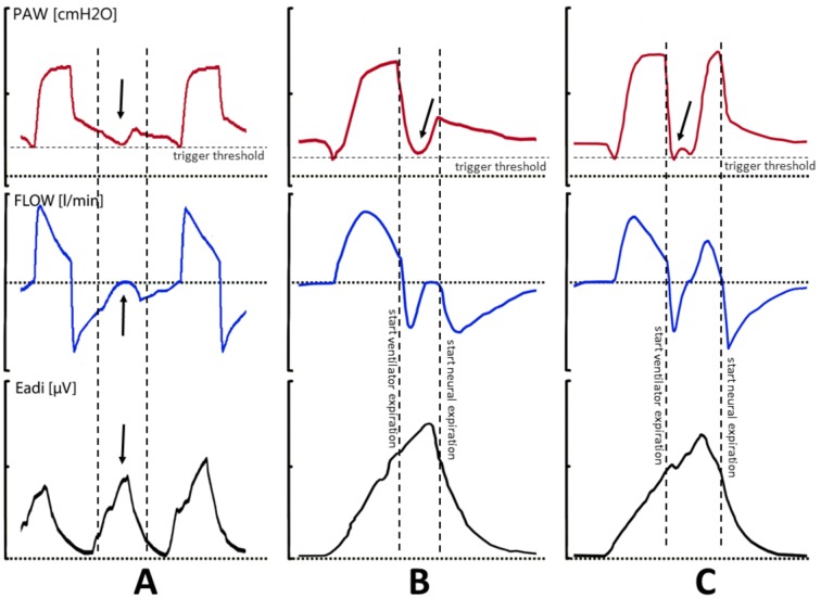 Figure 1: Three asynchronies every intensivist must recognise on the flow–time scalar — ineffective effort, premature cycling, and double triggering. (Source: Bulleri E, Fusi C, Bambi S, Pisani L. Acta Biomed 2018. DOI: 10.23750/abm.v89i7-S.7737).
The pressure–time curve reports the elastic and resistive loads; the flow–time curve reports the division of the cycle into inspiratory and expiratory time and is the earliest sign of gas trapping; the volume–time curve confirms what entered and left the lung.
flowchart TD
    A["Alarm on the ventilator"] --> B{"Which scalar changed?"}
    B -->|"Pressure rising"| C["End-inspiratory hold: read plateau and driving pressure"]
    B -->|"Expiratory flow never reaches baseline"| D["Suspect dynamic hyperinflation with intrinsic PEEP"]
    B -->|"Pressure and volume lost together"| E["Suspect circuit disconnection or tube obstruction"]
    B -->|"Extra, missing or stacked breaths"| F["Assess patient-ventilator asynchrony"]
    C --> G{"Compliance falling at a fixed tidal volume?"}
    G -->|"Yes"| H["Worsening ARDS, pneumothorax or abdominal distension"]
    G -->|"No"| I["Resistive problem: tube, secretions or bronchospasm"]
    D --> J["Disconnect and decompress, then lengthen expiratory time"]
    F --> K["Adjust sedation, rate, inspiratory time or mode"]
| Scalar | Normal appearance | First question when it changes |
|---|---|---|
| Pressure–time | Square plateau in volume control | Does the plateau move with the peak? |
| Flow–time | Expiratory flow reaches zero before the next breath | Is there an end-expiratory pause? |
| Volume–time | Returns to the starting volume each cycle | Is exhaled volume less than delivered volume? |
| Pressure–volume loop | Straight inspiratory limb | Is the limb concave toward the volume axis? |
| Flow–volume loop | Smooth, symmetric limbs | Are there notches on the expiratory limb? |
Case 1: The Rising Plateau at a Fixed Tidal Volume
A 48-year-old man with influenza ARDS is ventilated in volume control at a tidal volume of 6 mL/kg predicted body weight (400 mL), PEEP 14 cmH₂O and rate 22/min. Over four hours the peak pressure rises from 34 to 42 cmH₂O and the plateau pressure from 24 to 33 cmH₂O, while the delivered and exhaled tidal volumes are unchanged.
Question: What does the parallel rise in peak and plateau pressure mean, and what is the immediate action?
Driving pressure is $\Delta P = P_{plat} - PEEP_{tot} = 19\ \text{cmH}_2\text{O}$, up from $10$. With tidal volume and PEEP fixed, the only explanation is falling compliance, $C_{rs} = V_T/(P_{plat} - PEEP_{tot})$, now about half its earlier value. A rising driving pressure at a fixed tidal volume is the strongest mechanical signal of progressive lung injury. Confirm the hold, exclude a new pneumothorax, atelectasis and abdominal distension, reduce tidal volume toward $4$–$5\ \text{mL}/\text{kg}$, and reassess PEEP decrementally. If $\Delta P$ stays high with a $P_aO_2/F_iO_2$ below about $80$, refer for extracorporeal support. (Parent chapters 01 and 03.)
Attributing a rising peak pressure to secretions while ignoring a rising plateau. Secretions raise the peak alone; a rising plateau means elastic injury.
Case 2: The Obstructed Patient Who Crashes
A 71-year-old woman with a severe COPD exacerbation is intubated and ventilated in pressure control at rate 16/min, inspiratory time 1.0 s and PEEP 5 cmH₂O. Her blood pressure falls from 118/70 to 72/38 mmHg. The expiratory flow limb never touches the baseline, and an end-expiratory hold shows a total PEEP of 13 cmH₂O.
Question: What is the diagnosis, and what is the immediate manoeuvre?
Dynamic hyperinflation with intrinsic PEEP, here $PEEP_i = 13 - 5 = 8\ \text{cmH}_2\text{O}$. The trapped volume raises intrathoracic pressure, impedes venous return and lowers cardiac output. The immediate manoeuvre is mechanical: disconnect the endotracheal tube, allow passive deflation and compress the chest bilaterally, then reassess blood pressure. Rebuild the settings around expiratory time — rate 8–10/min, shorter inspiratory time, I:E of at least 1:3, low extrinsic PEEP — and deepen sedation, adding neuromuscular blockade if spontaneous effort defeats the strategy. (Parent chapter 05.)
Reaching for fluids or vasopressors first, or raising the rate. Both increase trapping and deepen the collapse.
Case 3: The Concave Pressure–Time Curve
An ARDS patient is ventilated in volume control with a constant inspiratory flow. The pressure–time curve bows upward throughout inspiration, and the ventilator's stress-index software reports 1.35.
Question: How is this read, and what change follows?
A concave, upward-bowing pressure–time curve gives a stress index above $1.1$: tidal overdistension, because each increment of volume requires more pressure as compliance falls during inflation. Reduce PEEP in steps of $2$–$3\ \text{cmH}_2\text{O}$ and re-measure; a curve that straightens toward $1.0$ confirms overdistension. The converse applies: a convex curve with a stress index below $0.9$ implies tidal recruitment and may tolerate more PEEP. (Parent chapter 03.)
Chasing hypoxaemia with more PEEP without checking the stress index, or measuring it during active inspiratory effort. The index is valid only in a passive patient on constant flow.
Case 4: Failing the Spontaneous Breathing Trial
A 62-year-old man is on day 6 of ventilation after septic shock. After 30 minutes of a spontaneous breathing trial on pressure support 8 cmH₂O over PEEP 5 cmH₂O, he is tachypnoeic at 38/min with a tidal volume of 210 mL.
Question: What is the rapid shallow breathing index, and what does it mean?
$\text{RSBI} = f / V_T = 38 / 0.21 \approx 181\ \text{breaths}/\text{min}/\text{L}$, far above the threshold of $105$. The rapid, shallow pattern reflects an unfavourable balance between respiratory load and capacity — resistance, reduced compliance, dynamic hyperinflation or cardiac dysfunction. Do not extubate; treat reversible factors (fluid overload, secretions, pain, delirium, ischaemia) and re-test later. (Parent chapter 13.)
Treating a single number as decisive. The index is a supportive predictor; a "pass" still requires an adequate cough, manageable secretions and a protected airway.
Case 5: The Double-Triggered Breath
A 40-year-old man with ARDS is ventilated in volume control at 420 mL and 18/min. Twice a minute two breaths appear back-to-back with no exhalation between them; the stacked tidal volume reaches about 800 mL and the second plateau pressure is 36 cmH₂O.
Question: What is this asynchrony, what is the risk, and what changes are made?
Double triggering, or breath stacking: most often reverse triggering, in which a diaphragmatic contraction is entrained to the mandatory breath in a sedated patient, or a short inspiratory time that leaves the patient to trigger again. The stacked volume causes regional overdistension and volutrauma and threatens barotrauma and haemodynamic compromise. Assess for reverse triggering, deepen sedation, lengthen inspiratory time or increase flow so the demand is met, and consider raising the set rate toward the intrinsic rhythm; refractory cases may need neuromuscular blockade. (Parent chapters 01 and 13, with adaptive modes in chapter 17.)
Lowering the set tidal volume to make the average look acceptable while continuing to accept stacked breaths. The injurious breath is the second one.
Case 6: The Ineffective Effort
A spontaneously breathing patient with COPD appears to breathe fast, but the ventilator reports a low total rate. The flow–time trace shows small negative deflections not followed by a pressure drop or a delivered breath. Measured auto-PEEP is 10 cmH₂O.
Question: What is the mechanism, and what is the role of extrinsic PEEP?
These are ineffective triggering efforts. Intrinsic PEEP is a threshold load: before flow can begin, the diaphragm must lower alveolar pressure below the airway opening pressure and then overcome the trigger sensitivity. Extrinsic PEEP at roughly $60$–$80\%$ of the measured auto-PEEP — here about $6$–$8\ \text{cmH}_2\text{O}$ — raises end-expiratory airway pressure toward alveolar pressure, reducing the threshold load without raising total PEEP while it stays below the intrinsic level. Titrate to comfort and recheck total PEEP. (Parent chapter 05.)
Assuming that added PEEP always worsens hyperinflation. In the spontaneously breathing, flow-limited patient it is usually therapeutic; in the passive patient it can raise end-expiratory lung volume, and the rate must instead be reduced.
Case 7: The Sawtooth
An intubated patient develops a flow–volume loop whose expiratory limb carries regular, small tooth-like notches, with minor oscillation of the inspiratory flow as well. Gas exchange is unchanged.
Question: What is the diagnosis, and what is the first action?
The sawtooth reflects a mobile or unstable airway — mucus strands dropping across the lumen, or oscillation of the airway wall — and is closely associated with retained secretions. It is not bronchospasm, although the two coexist. Suction, review humidification and position, and re-examine the loop; resolution confirms the cause. (Parent chapter 14, with airway mechanics in chapter 05.)
Escalating bronchodilators for a pattern caused by secretions, or misreading the sawtooth as auto-PEEP. The notches sit on the flow tracing, not the end-expiratory baseline.
Case 8: Silence on the Pressure Channel
The ventilator alarms for low minute volume. The pressure waveform is flat at zero, exhaled tidal volume has fallen to almost nothing, and the saturation drifts down while the patient still makes some chest movement.
Question: What is the diagnosis, and what is the first action?
A circuit disconnection or large leak — including endotracheal tube dislodgement or cuff failure. Trace the circuit from the Y-piece to the tube and check the cuff. Reconnect and manually ventilate with a self-inflating bag on 100% oxygen while completing the diagnosis; a displaced tracheostomy tube is a lost airway. (Parent chapters 10 and 14.)
Assuming the ventilator has failed and cycling the power. In a patient losing saturation, the airway, not the machine, is the priority.
Case 9: The Asthma Patient with Measured Auto-PEEP
A 26-year-old man with status asthmaticus is ventilated at rate 22/min, tidal volume 450 mL and PEEP 5 cmH₂O. Peak pressures are high and the blood pressure is 95/50 mmHg, and an end-expiratory hold reports a total PEEP of 15 cmH₂O.
Question: What is the correct ventilator response?
Deliberate controlled hypoventilation. Reduce the rate to $6$–$10$/min, shorten inspiratory time with high flow to an I:E of at least 1:4, keep tidal volume at 6–8 mL/kg, and accept hypercapnia to a pH floor of about 7.20–7.25. Deepen sedation and add neuromuscular blockade if spontaneous effort shortens expiratory time, and if the blood pressure falls, disconnect and decompress first. Recheck total PEEP after every adjustment. (Parent chapter 06.)
Pursuing normocapnia by raising the rate, which shortens expiratory time, worsens trapping and can precipitate pulseless electrical activity.
Case 10: Lung Rest on Veno-Venous ECMO
A patient with severe ARDS is established on veno-venous extracorporeal membrane oxygenation at 4 L/min with sweep gas titrated to carbon dioxide. The ventilator, in pressure control, delivers a driving pressure of 15 cmH₂O over PEEP 15 cmH₂O at rate 14/min, giving a tidal volume near 7 mL/kg predicted body weight.
Question: What should change now that the membrane is supporting gas exchange?
Reduce the ventilator to lung-rest parameters: tidal volume $1$–$3\ \text{mL}/\text{kg}$ predicted body weight, plateau pressure at or below $20$–$25\ \text{cmH}_2\text{O}$, driving pressure at or below $10\ \text{cmH}_2\text{O}$ (many centres target $\leq 8$), rate $4$–$10$/min with apnoea accepted, PEEP $10$–$15\ \text{cmH}_2\text{O}$, inspiratory time $1.5$–$2.0$ s, and $F_iO_2$ $0.21$–$0.40$. Mechanical power is collapsed while the membrane manages gas exchange. Watch for atelectasis at very low volumes and adjust sweep and blood flow, not the ventilator, for carbon dioxide. (Parent chapter 15.)
Continuing conventional protective ventilation because the numbers "look acceptable". The point of extracorporeal support is to unload the native lung, not to leave it at injurious stress.
Spotter Gallery
Spotter A: The blood gas that does not fit
pH 7.26, $P_aCO_2$ 70 mmHg, bicarbonate 31 mmol/L and $P_aO_2$ 58 mmHg on $F_iO_2$ 0.35 in a known COPD patient.
Acute-on-chronic hypercapnic respiratory failure: the raised bicarbonate indicates chronic compensation, so the fall in pH is smaller than the carbon dioxide rise alone would produce. Start controlled oxygen and non-invasive ventilation, and target the patient's chronic baseline rather than a normal $P_aCO_2$. (Parent chapters 05 and 07.)
Spotter B: The vanishing end-tidal trace
The end-tidal carbon dioxide plateau collapses abruptly toward zero.
Sudden loss of exhaled carbon dioxide means loss of delivery to the lung: circuit disconnection, tube dislodgement, massive pulmonary embolism, or cardiac arrest. Cross-check the pressure waveform; a flat channel with a flat capnogram points to disconnection, and the airway is addressed first. (Parent chapters 10 and 14.)
Spotter C: The beaked pressure–volume loop
The inspiratory limb of the pressure–volume loop bends to the right as pressure rises, creating a beak.
Beaking toward the upper inflection point indicates overdistension of aerated units. Reduce PEEP and, if necessary, tidal volume, and confirm the change with the stress index; loops are supportive rather than standalone evidence. (Parent chapter 03.)
Spotter D: The stress index
The slope of the pressure–time curve during constant-flow inflation.
Interpret a stress index below $0.9$ (convex) as tidal recruitment, $0.9$–$1.1$ (straight) as adequate, and above $1.1$ (concave) as overdistension. Change PEEP in the direction the curve indicates and re-measure. (Parent chapter 03.)
Spotter E: The curare cleft
A notch appears in the middle of the end-tidal carbon dioxide plateau.
A "curare cleft" is a spontaneous inspiratory effort during expiration, historically seen in partially paralysed patients. It signals intact diaphragmatic activity and may herald reverse triggering, so reassess sedation and synchrony. (Parent chapters 01 and 13.)
Summary Table
| Case | Spotter finding | Governing principle | Parent chapter |
|---|---|---|---|
| 1 | Rising peak and plateau at a fixed tidal volume | Falling compliance; driving pressure $\Delta P$ | Ch 01, 03 |
| 2 | Expiratory flow never reaching baseline with hypotension | Dynamic hyperinflation; impaired venous return | Ch 05 |
| 3 | Concave pressure–time curve; stress index > 1.1 | Tidal overdistension | Ch 03 |
| 4 | Rapid shallow breathing index above 105 | Load–capacity imbalance at the end of a trial | Ch 13 |
| 5 | Back-to-back breaths with a stacked tidal volume | Reverse triggering and volutrauma | Ch 01, 13, 17 |
| 6 | Negative flow deflections without a delivered breath | Threshold load from intrinsic PEEP | Ch 05 |
| 7 | Sawtooth notches on the expiratory flow–volume limb | Secretions or airway instability | Ch 14, 05 |
| 8 | Flat pressure channel with lost exhaled volume | Circuit disconnection or tube displacement | Ch 10, 14 |
| 9 | Total PEEP of 15 cmH₂O in status asthmaticus | Controlled hypoventilation | Ch 06 |
| 10 | Conventional protective settings during VV-ECMO | Extracorporeal lung rest | Ch 15 |
Key Takeaways
- Read the scalars as a set: a changing peak alone suggests resistance, a changing plateau at a fixed tidal volume means changing compliance, and flow that never reaches baseline means trapped gas.
- Driving pressure, $\Delta P = P_{plat} - PEEP_{tot}$, is the number to watch at a fixed tidal volume; a rising value is a call to act.
- Hypotension in an obstructed patient is a respiratory emergency: disconnect the endotracheal tube and decompress the chest before anything else.
- The stress index and the beaked pressure–volume loop both answer whether this lung is overdistended or still recruiting, and both require a passive patient on constant flow.
- Asynchronies have signatures: ineffective effort, premature cycling, double triggering and the curare cleft each dictate whether to sedate, change rate, or change mode.
- A failed spontaneous breathing trial is an unfavourable load-to-capacity balance, not merely a number above a threshold.
- When pressure and volume vanish together, the diagnosis lies in the circuit or airway; manually ventilate while finding it.
- On extracorporeal support, reduce tidal volume, driving pressure and rate to lung-rest values and let the membrane manage gas exchange.