Sepsis and positive-pressure ventilation compete for the same haemodynamic reserve: the right ventricle. A septic patient arrives with a vasodilated, high-flow circulation propped up by tachycardia and an augmented preload; the ventilator then reduces venous return, raises pulmonary vascular resistance, and, when the lung is injured, imposes an afterload the thin-walled right ventricle cannot acutely overcome. When that ventricle fails, systemic blood pressure collapses not because the left ventricle is weak but because it has nothing to fill it — and the instinctive response, raising airway pressure, makes matters worse.
This chapter treats the right ventricle as an afterload-sensitive flow generator whose output determines left-ventricular filling. It examines the three levers positive-pressure ventilation pulls — preload, afterload, and the interventricular septum — characterises the septic state and its peculiar vulnerability, and builds a monitoring and management strategy that protects the right ventricle while preserving lung protection.
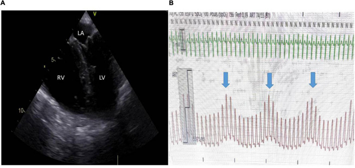 Figure 1: Acute cor pulmonale in a ventilated ARDS patient — severe right-ventricular dilatation with paradoxical septal motion, accompanied by a low arterial pressure with large pulse-pressure variation and an elevated central venous pressure. (Source: Petit M, Jullien E, Vieillard-Baron A. Frontiers in Physiology 2021;12:797252. DOI: 10.3389/fphys.2021.797252).
The Cardiopulmonary Interaction of Positive-Pressure Ventilation
Preload: intrathoracic pressure and venous return
Spontaneous breathing generates negative intrathoracic pressure that assists venous return; positive-pressure ventilation inverts this relationship. Each insufflation raises intrathoracic pressure, narrows the pressure gradient from the systemic veins to the right atrium, and lowers right-ventricular end-diastolic volume. With PEEP, the raised mean intrathoracic pressure continues to impede venous return throughout the cycle, so right-ventricular stroke volume falls cyclically. This is the physiological basis of respiratory variation in the arterial pulse and of the fall in cardiac output that follows an ill-judged fluid-and-pressure combination.
On the left side the effect is subtler: preload falls because pulmonary blood flow is transiently reduced, but the same rise in intrathoracic pressure lowers the transmural pressure the left ventricle must generate, reducing its effective afterload. Arterial pressure during insufflation therefore reflects both a fall in filling and a fall in load.
Afterload: lung distension and the pulmonary capillaries
The pulmonary microvasculature is a network of extra-alveolar vessels held open by radial traction and intra-alveolar vessels compressed by alveolar distension. Pulmonary vascular resistance is therefore minimal near functional residual capacity and rises at both extremes: at low lung volumes the extra-alveolar vessels collapse from loss of traction, and at high volumes the alveolar capillaries are compressed by overdistension. Its formal definition is purely haemodynamic:
$$PVR = \frac{mPAP - PAWP}{CO}$$where $PVR$ is pulmonary vascular resistance (Wood units, $\text{mmHg}\cdot\text{min}\cdot\text{L}^{-1}$, or $\times 80$ for $\text{dyn}\cdot\text{s}\cdot\text{cm}^{-5}$), $mPAP$ is mean pulmonary artery pressure ($\text{mmHg}$), $PAWP$ is pulmonary artery wedge pressure ($\text{mmHg}$), and $CO$ is cardiac output ($\text{L/min}$).
In the injured ARDS lung this relationship becomes treacherous because the aerated compartment is small. A tidal volume delivered to a "baby lung" overdistends the open units and leaves the collapsed units contributing nothing but hypoxic vasoconstriction. Positive end-expiratory pressure applied beyond the point of recruitment adds a fixed compressive load, so the sum of plateau pressure and regional inflation heterogeneity determines the pulmonary vascular impedance the right ventricle must face.
The interventricular septum and left-ventricular filling
Right-ventricular dilatation displaces the interventricular septum leftward and flattens it, so the left ventricle becomes a smaller, less compliant chamber that fills poorly and may be compressed within the pericardium — ventricular interdependence. Left-ventricular filling and stroke volume fall, aortic pressure drops, and right-ventricular wall tension rises, reducing right coronary perfusion at the very moment it is needed most. This positive-feedback loop terminates in cardiovascular collapse, and septal flattening on echo is its earliest warning.
Right-ventricular stroke volume is the pulmonary flow divided by heart rate, so the stroke-volume/pulmonary-vascular-resistance relation follows directly from the PVR definition:
$$SV_{RV} = \frac{CO}{HR} = \frac{mPAP - PAWP}{HR \times PVR}$$where $SV_{RV}$ is right-ventricular stroke volume (mL) and $HR$ is heart rate (beats/min). For a fixed pressure gradient, any rise in $PVR$ lowers stroke volume — the algebraic statement of afterload sensitivity.
The Right Ventricle: A Thin-Walled, Afterload-Sensitive Flow Generator
Ventriculo-arterial coupling
The right ventricle is a volume pump with a free wall only $3$–$5\text{ mm}$ thick, a crescentic cavity, and a low-pressure, high-compliance circulation. It tolerates wide swings in preload but tolerates acute afterload poorly. Ventriculo-arterial coupling is quantified by the ratio of end-systolic elastance $E_{es}$ (contractility) to effective arterial elastance $E_a$ (afterload); efficient transfer occurs when $E_{es}/E_a$ is approximately $1$–$2$. When pulmonary vascular resistance rises, $E_a$ rises; if the increase outruns contractile reserve, stroke volume falls and the ventricle dilates.
Acute cor pulmonale and its cost
Acute cor pulmonale is right-ventricular failure precipitated by an acute rise in pulmonary vascular resistance. In ARDS the accepted echocardiographic definition is a right-ventricular end-diastolic area to left-ventricular end-diastolic area ratio greater than $0.6$ with paradoxical septal motion; it occurs in roughly $20$–$25\%$ of moderate-to-severe ARDS and predicts worse outcome. Right-ventricular output falls, pulmonary blood flow falls, left-ventricular preload falls, and systemic cardiac output follows; a raised central venous pressure compounds the injury by reducing right coronary perfusion. The pulmonary artery pulsatility index (PAPi) captures this at the bedside:
$$PAPi = \frac{sPAP - dPAP}{CVP}$$where $PAPi$ is dimensionless, $sPAP$ and $dPAP$ are systolic and diastolic pulmonary artery pressures ($\text{mmHg}$), and $CVP$ is central venous pressure ($\text{mmHg}$). A value below approximately $1.85$ signals impaired right-ventricular function and rising right-sided filling pressures.
Sepsis and Septic Shock: Haemodynamics and Fluids
Definition and the Surviving Sepsis Campaign
Sepsis is life-threatening organ dysfunction from a dysregulated host response to infection, operationalised as an acute rise in the SOFA score of at least $2$ points. Septic shock is the subset with persistent hypotension requiring vasopressors to maintain a mean arterial pressure of $65\text{ mmHg}$ and a serum lactate above $2\text{ mmol/L}$ despite adequate fluid resuscitation, carrying a mortality approaching $40\%$. Surviving Sepsis Campaign bundles emphasise early lactate measurement, cultures before antibiotics, rapid $30\text{ mL/kg}$ crystalloid for hypotension or lactate at or above $4\text{ mmol/L}$, and vasopressors to that pressure target.
The septic haemodynamic profile and myocardial depression
Classic septic shock is a high-output, low-resistance state: cardiac output is often $6$–$10\text{ L/min}$, systemic vascular resistance is low, and mixed-venous oxygen saturation is high because oxygen extraction is impaired. This profile can hide a depressed myocardium. Sepsis-induced cardiomyopathy — biventricular dilatation, reduced ejection fraction, troponin release — is present in a substantial minority and is unmasked only when systemic vascular resistance is restored or the right ventricle is loaded. Those patients have no contractile reserve to meet a ventilatory afterload, which is why a normal-looking septic profile can deteriorate abruptly on the ventilator.
Fluid resuscitation: adequate but not excessive
Fluid is first-line therapy but not benign. In the ventilated patient a large positive balance raises intrathoracic pressure, fills a failing right ventricle, worsens septal shift, and causes pulmonary and peripheral oedema; it is independently associated with mortality. The modern approach assesses responsiveness dynamically, gives small aliquots with immediate reassessment, and moves early to vasopressors when the right ventricle is already loaded.
Ventilator Strategy in the Haemodynamically Unstable Patient
Low tidal volume, low plateau and driving pressures
Lung-protective ventilation is simultaneously right-ventricular protection. Tidal volume is set at $6\text{ mL/kg}$ predicted body weight, plateau pressure at or below $27$–$30\text{ cmH}_2\text{O}$, and driving pressure, $\Delta P = P_{plat} - PEEP$, at or below $15\text{ cmH}_2\text{O}$ (ideally $\leq 14$). Driving pressure is the ventilatory variable most closely linked both to ARDS mortality and to the cyclic mechanical load on the pulmonary circulation, so minimising it serves both the lung and the right ventricle.
PEEP titration, hyperinflation and auto-PEEP
PEEP has biphasic vascular effects: recruiting collapsed lung reduces shunt and may lower pulmonary vascular resistance, while overdistending open lung raises it. There is no single correct value; PEEP must be titrated against oxygenation, driving pressure, and haemodynamics. In a failing right ventricle the tell-tale is a fall in blood pressure with a simultaneous rise in central venous pressure at each increment — an overshoot to be reversed. In obstructive physiology, dynamic hyperinflation and auto-PEEP raise intrathoracic pressure throughout the cycle and can precipitate hypotension; reduce the respiratory rate and lengthen expiratory time until end-expiratory flow ceases.
Permissive hypercapnia and prone positioning
Hypercapnia and the accompanying acidosis are potent pulmonary vasoconstrictors, so permissive hypercapnia buys lung protection at the price of higher pulmonary vascular resistance and must be applied cautiously when the right ventricle is failing. Mild hypercapnia ($PaCO_2$ $45$–$60\text{ mmHg}$) is usually tolerated; severe hypercapnia with $pH < 7.20$ may tip the ventricle. Prone positioning recruits dorsal lung, homogenises inflation, and reduces shunt, thereby unloading the right ventricle; PROSEVA demonstrated a mortality benefit, and it also reduces the heart's compressive effect on dependent lung.
A driving pressure of $15\text{ cmH}_2\text{O}$ delivered to a heterogeneously inflated lung produces regional overdistension and regional hypoxic vasoconstriction simultaneously. Both raise pulmonary vascular resistance; limiting $\Delta P$ is therefore the most powerful ventilatory manoeuvre for the failing right ventricle.
Monitoring the Right Ventricle at the Bedside
Echocardiography
Critical-care echocardiography is the central tool. It reports right-ventricular size (the end-diastolic area ratio), septal kinetics (paradoxical motion, the "D-sign"), tricuspid regurgitation, tricuspid annular plane systolic excursion (TAPSE, abnormal below $17\text{ mm}$), and free-wall strain. Systolic pulmonary artery pressure is estimated from the tricuspid regurgitation velocity via the simplified Bernoulli relation:
$$sPAP \approx 4v^2 + RAP$$where $sPAP$ is systolic pulmonary artery pressure ($\text{mmHg}$), $v$ is the peak tricuspid regurgitation velocity ($\text{m/s}$), and $RAP$ is right atrial pressure ($\text{mmHg}$). The factor $4$ arises because the pressure drop across a valve is $4v^2$ in these units. Right-ventricular outflow-tract Doppler showing a cyclic reduction in flow with each insufflation, as in Figure 1, is a sensitive early marker of intolerance to the ventilator.
Catheters and dynamic indices
A central venous catheter provides central venous pressure and central venous oxygen saturation; a pulmonary artery catheter adds pulmonary pressures, cardiac output, and the pulsatility index. Dynamic indices — pulse-pressure variation above $13\%$ and stroke-volume variation — predict fluid responsiveness, but are unreliable in spontaneous breathing, arrhythmia, low tidal volume, right-ventricular failure, and raised intra-abdominal pressure.
Central venous pressure and pulmonary artery wedge pressure are poor surrogates for preload in the ventilated septic patient, being distorted by intrathoracic pressure and right-ventricular compliance. Treat the trend and dynamic response, never a single number.
Management of Acute Right-Ventricular Failure
Vasopressors and inotropes
Norepinephrine is first-line: it restores systemic vascular resistance and mean arterial pressure, with little effect on pulmonary vascular resistance at usual doses. Vasopressin, acting through V1 receptors, raises systemic resistance without pulmonary vasoconstriction and may even lower it, so it usefully spares norepinephrine dose. Dobutamine increases contractility and cardiac output but vasodilates and can worsen hypotension, so it is best combined with a vasopressor. Levosimendan, a calcium sensitiser, improves contractility without raising intracellular calcium and favourably alters ventriculo-arterial coupling; milrinone is a pulmonary vasodilator but causes systemic hypotension, limiting its use in shock. A mean arterial pressure of at least $65\text{ mmHg}$ is the minimum, and $70$–$80\text{ mmHg}$ better preserves right coronary perfusion. Pure $\alpha_1$ agonists such as phenylephrine raise pulmonary vascular resistance and should be avoided.
| Agent | Receptor / mechanism | Haemodynamic effect | Cautions |
|---|---|---|---|
| Norepinephrine | $\alpha_1 \gg \beta_1$ | Increases SVR and MAP; modest rise in PVR | First-line; very high doses raise pulmonary pressures |
| Vasopressin | V1 (vasopressin) | Increases SVR without pulmonary vasoconstriction | Adjunct to spare norepinephrine; digital and splanchnic ischaemia |
| Dobutamine | $\beta_1 > \beta_2$ | Increases contractility and cardiac output | Vasodilation can worsen hypotension; combine with pressor |
| Milrinone | PDE3 inhibition | Inotropy with pulmonary and systemic vasodilation | Systemic hypotension; renally cleared |
| Levosimendan | Calcium sensitiser, $\text{K}_{ATP}$ | Increases contractility, improves RV–PA coupling | Hypotension; long-lived active metabolite |
| Epinephrine | $\alpha_1$, $\beta_1$, $\beta_2$ | Increases cardiac output and MAP at higher dose | Arrhythmia, raised lactate, pulmonary vasoconstriction |
| Phenylephrine | $\alpha_1$ | Increases SVR | Pure vasoconstriction raises PVR; avoid in RV failure |
Pulmonary vasodilators and mechanical support
Inhaled nitric oxide and inhaled prostacyclin (epoprostenol or iloprost) act only on ventilated lung units, reducing pulmonary vascular resistance and improving right-ventricular–pulmonary-arterial coupling without systemic hypotension. In ARDS they produce transient improvements in oxygenation and pulmonary artery pressure but no consistent mortality benefit, so they are reserved for right-ventricular failure with high pulmonary vascular resistance. Phosphodiesterase-5 inhibitors and endothelin antagonists belong to chronic rather than acute pulmonary hypertension. In refractory right-ventricular failure, veno-venous extracorporeal membrane oxygenation or a right-ventricular assist device may be considered.
Rhythm and coronary perfusion
Sinus rhythm should be maintained. Atrial fibrillation and other tachyarrhythmias abolish the atrial contribution to right-ventricular filling and raise myocardial oxygen demand, while tachycardia shortens diastole and impairs right coronary perfusion. Rhythm control, electrolyte correction, and avoidance of negative inotropic agents are therefore priorities; right-ventricular ischaemia remains a common terminal event.
flowchart TD
    A["Septic patient on the ventilator with hypotension"] --> B["Assess volume status and right ventricle by critical-care echo"]
    B --> C{"Fluid responsive and no right-ventricular dilatation?"}
    C -->|"Yes"| D["Cautious fluid bolus of 250-500 mL, then reassess"]
    C -->|"No"| E["Start norepinephrine, target MAP >= 65 mmHg"]
    D --> E
    E --> F["Optimise ventilation: tidal volume 6 mL/kg, Pplat <= 27, driving pressure <= 14"]
    F --> G{"Echo shows acute cor pulmonale?"}
    G -->|"No"| H["Continue lung-protective ventilation and reassess"]
    G -->|"Yes"| I["Add vasopressin; consider dobutamine or levosimendan"]
    I --> J["Titrate PEEP; avoid overdistension and auto-PEEP"]
    J --> K["Add inhaled pulmonary vasodilator if PVR is high"]
    K --> L{"Persistent right-ventricular failure and shock?"}
    L -->|"No"| M["Wean vasoactive support as the right ventricle recovers"]
    L -->|"Yes"| N["Consider prone positioning; escalate to VV-ECMO or right-ventricular assist"]
Rising airway pressure lowers venous return and raises pulmonary vascular resistance, dilating the right ventricle, shifting the septum, and further reducing left-ventricular filling. If the patient deteriorates after a ventilator change that increased airway pressure, undo the change before adding another drug.
Key Takeaways
- Positive-pressure ventilation lowers right-ventricular preload, raises right-ventricular afterload, and shifts the interventricular septum, so it can precipitate right-ventricular failure even when the left ventricle is intrinsically normal.
- Pulmonary vascular resistance is minimal near functional residual capacity and rises with both atelectasis and overdistension; driving pressure is its bedside surrogate.
- The thin-walled right ventricle is afterload-sensitive; when effective arterial elastance outruns contractile reserve, acute cor pulmonale develops and systemic cardiac output falls.
- Septic shock is usually a high-output, low-resistance state, but coexisting myocardial depression and right-ventricular failure are common and are unmasked by vasopressors and by ventilation.
- Fluid resuscitation should be adequate but not excessive, and right-ventricular protection combines $6\text{ mL/kg}$ tidal volumes, low plateau and driving pressures, titrated PEEP, avoidance of auto-PEEP, prone positioning, and only limited permissive hypercapnia, since hypercapnic acidosis raises pulmonary vascular resistance.
- Norepinephrine and vasopressin are the vasopressors of choice, dobutamine and levosimendan support contractility, and inhaled pulmonary vasodilators reduce afterload selectively; echocardiography and the pulsatility index guide every step.