Veno-venous extracorporeal membrane oxygenation (VV-ECMO) is the most aggressive expression of the lung-protective principle. When an injured lung can no longer sustain gas exchange without being subjected to injurious pressure and volume, an extracorporeal circuit is interposed between the venous return and the pulmonary circulation to assume oxygenation and carbon dioxide elimination. The ventilator is then freed from its obligation to ventilate: it need only keep the lung gently inflated, distribute a small tidal volume, and preserve end-expiratory recruitment. This is ultra-protective ventilation, or lung rest.
The rationale is mechanical rather than merely gasometric. Ventilator-induced lung injury (VILI) arises from cyclic overdistension (volutrauma), repeated opening and closing of unstable units (atelectrauma), and the ensuing inflammatory biotrauma — all functions of the pressure and volume delivered to a lung whose aerated compartment has collapsed to a fraction of normal. Extracorporeal support does not repair the lung, but by taking over gas exchange it permits tidal volumes, driving pressures, and mechanical power far below what conventional protective ventilation can tolerate. The price is an invasive circuit with its own complications, a heavy anticoagulation and monitoring burden, and substantial resource cost.
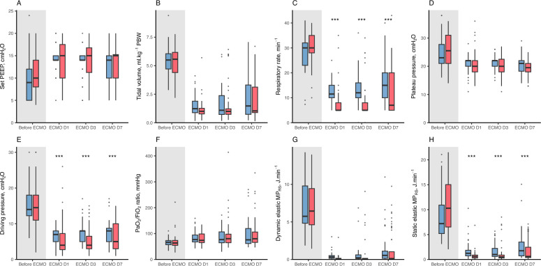 Figure 1: Once VV-ECMO assumes gas exchange, tidal volume, driving pressure and mechanical power fall sharply and are sustained at lung-rest levels, while PEEP and PaO2/FiO2 are preserved. (Source: Rodriguez Y, Thomachot A, Deniel G, et al. Annals of Intensive Care 2025;15:123. DOI: 10.1186/s13613-025-01525-0).
The Rationale for Extracorporeal Lung Rest
Mechanical stress, strain and the baby lung
In moderate-to-severe acute respiratory distress syndrome (ARDS) the aerated lung shrinks to the "baby lung" of Gattinoni while total lung volume is relatively preserved. A tidal volume of $6\text{ mL/kg}$ predicted body weight (PBW) that is innocuous in health therefore imposes far greater specific strain when distributed to a third of the normal aerated volume:
$$\varepsilon = \frac{V_T}{V_{aerated}}$$where $\varepsilon$ is dimensionless strain, $V_T$ is tidal volume (mL), and $V_{aerated}$ is end-expiratory aerated lung volume (mL). As $V_{aerated}$ falls, the same $V_T$ produces proportionally larger strain and a larger regional distending pressure.
Driving pressure and the accelerated-lung-injury model
Driving pressure links tidal volume to the mechanical properties of the respiratory system:
$$\Delta P = P_{plat} - PEEP = \frac{V_T}{C_{RS}}$$where $\Delta P$ is driving pressure ($\text{cmH}_2\text{O}$), $P_{plat}$ is end-inspiratory plateau pressure ($\text{cmH}_2\text{O}$), $PEEP$ is positive end-expiratory pressure ($\text{cmH}_2\text{O}$), $V_T$ is tidal volume (mL), and $C_{RS}$ is respiratory-system compliance ($\text{mL/cmH}_2\text{O}$). In the accelerated-lung-injury model the applied $\Delta P$ is amplified regionally by the ratio of total to aerated lung volume:
$$\Delta P_{reg} \approx \Delta P \times \frac{V_{total}}{V_{aerated}}$$where $\Delta P_{reg}$ is regional driving pressure ($\text{cmH}_2\text{O}$) and $V_{total}$ is total lung volume (mL). A $\Delta P$ of $15\text{ cmH}_2\text{O}$ may be tolerable in a nearly normal lung yet injurious in a lung with a small aerated fraction — which is why extracorporeal support, permitting a $V_T$ of only $1$–$3\text{ mL/kg PBW}$, is qualitatively different from lowering tidal volume within conventional protective ventilation.
The membrane oxygenator assumes gas exchange so that the native lung can rest. The ventilator's role shifts from ventilating to splinting: low-volume, low-pressure inflation with enough PEEP to maintain recruitment while cyclic strain and mechanical power approach zero.
Venous admixture and severity indices
Ultra-protective ventilation does not abolish hypoxaemia; it transfers its management to the circuit. Intrapulmonary shunting follows the shunt equation:
$$\frac{\dot{Q}_s}{\dot{Q}_t} = \frac{C_{c'O_2} - C_{aO_2}}{C_{c'O_2} - C_{v'O_2}}$$where $\dot{Q}_s/\dot{Q}_t$ is the shunt fraction (dimensionless), $C_{c'O_2}$ is end-capillary (ideal) oxygen content, $C_{aO_2}$ is arterial oxygen content, and $C_{v'O_2}$ is mixed-venous oxygen content (all in $\text{mL O}_2/\text{dL}$). The oxygenation index (OI) integrates oxygenation with the mean airway pressure that produces it:
$$OI = \frac{FiO_2 \times \overline{P}_{aw} \times 100}{PaO_2}$$where $FiO_2$ is fractional inspired oxygen (dimensionless), $\overline{P}_{aw}$ is mean airway pressure ($\text{cmH}_2\text{O}$), and $PaO_2$ is arterial oxygen partial pressure ($\text{mmHg}$). A rising OI reflects worsening oxygenation at escalating airway pressure, and a value above roughly $40$ identifies patients in whom extracorporeal referral should be considered.
Referral Criteria and the Evidence Base
Who to refer
Referral is considered when severe hypoxaemia or respiratory acidosis persists despite optimised conventional management — lung-protective ventilation, PEEP titration, neuromuscular blockade, prone positioning, and inhaled vasodilators. The widely used thresholds, from the enrolment criteria of the EOLIA trial, are any one of: $PaO_2/FiO_2 < 80\text{ mmHg}$ for more than $6\text{ h}$; $PaO_2/FiO_2 < 50\text{ mmHg}$ for more than $3\text{ h}$; or $pH < 7.25$ with $PaCO_2 \geq 60\text{ mmHg}$ for more than $6\text{ h}$ at a set respiratory rate below $35\text{ breaths/min}$.
Referral is not cannulation. Early contact lets the ECMO team advise on optimisation and transfer, and permits cannulation before irreversible multiorgan failure supervenes; outcome deteriorates markedly once ventilation has been prolonged beyond about $7$ days.
The EOLIA trial and its interpretation
EOLIA (Combes et al., 2018) randomised $249$ patients with very severe ARDS to immediate VV-ECMO or to continued conventional ventilation with ECMO as rescue. Sixty-day mortality was $35\%$ with ECMO versus $46\%$ with control (relative risk $0.76$, $95\%$ CI $0.55$–$1.04$, $P = 0.09$) — narrowly missing the conventional threshold, but best understood in light of two features: $28\%$ of control patients crossed over to ECMO, diluting any intention-to-treat effect, and the trial was underpowered for the observed effect size. As-treated, post-hoc, and Bayesian analyses have all favoured ECMO, the Bayesian reanalysis reporting a high posterior probability of mortality benefit.
The meta-analytic view and guidelines
A systematic review and meta-analysis of the randomised evidence (Munshi et al., Lancet Respiratory Medicine 2019) found VV-ECMO associated with reduced mortality (relative risk approximately $0.73$, $95\%$ CI $0.58$–$0.92$), the benefit concentrated in analyses accounting for crossover. Contemporary European and North American guidelines therefore recommend considering VV-ECMO in severe ARDS refractory to optimised conventional therapy — a rescue therapy rather than an experimental one. Modern ECMO, delivered with modern lung-protective background ventilation, saves lives in correctly selected patients.
Contraindications
Absolute contraindications identify patients with no realistic prospect of recovery or in whom the circuit cannot be safely managed; relative contraindications shift the risk–benefit balance without universally precluding support.
| Category | Absolute | Relative |
|---|---|---|
| Neurological | Devastating irreversible brain injury; brain death | Recent ischaemic stroke; uncontrolled seizures |
| Prognosis | Advanced malignancy or irreversible disease with no transplant option | Limited life expectancy from comorbidity |
| Bleeding | Active intracranial haemorrhage; uncontrollable bleeding; absolute contraindication to anticoagulation | High bleeding risk; recent major surgery or trauma |
| Respiratory | Non-recoverable lung disease with no transplant pathway | Ventilation beyond $7$–$10$ days without improvement |
| Organ failure | Severe irreversible multiorgan failure | Ongoing sepsis with vasopressor dependence |
| Vascular access | No feasible access | Extreme obesity ($BMI > 45$–$50$) |
Devastating neurological injury, uncontrolled haemorrhage or an inability to anticoagulate, and non-recoverable respiratory failure without a transplant destination are absolute barriers. Cannulating such patients prolongs dying and consumes a scarce resource.
Ultra-Protective Ventilation: The Lung-Rest Strategy
Once the circuit runs at full flow, tidal volume is reduced to $1$–$3\text{ mL/kg PBW}$ — a "quasi-apnoeic" strategy in which the ventilator's contribution to gas exchange becomes negligible. Plateau pressure is kept at or below $20$–$25\text{ cmH}_2\text{O}$ and driving pressure at or below $10\text{ cmH}_2\text{O}$ (many centres target $\leq 8$), well below the conventional protective thresholds of $P_{plat} \leq 30$ and $\Delta P \leq 15\text{ cmH}_2\text{O}$. A pressure-controlled mode with a long inspiratory time ($1.5$–$2.0\text{ s}$) delivers this gently; respiratory rate falls to $4$–$10\text{ breaths/min}$, and a truly apnoeic strategy (rate $0$–$4$) with the ventilator serving mainly to provide PEEP is a recognised variant. Ventilator $FiO_2$ is reduced to $0.21$–$0.40$, since systemic oxygenation depends on the circuit and a low inspired oxygen tension minimises absorption atelectasis. The deliberate acceptance of apnoea is the defining feature of lung rest.
| Parameter | Ultra-Protective Target | Conventional Protective Comparison |
|---|---|---|
| Mode | Pressure-controlled, long $T_i$ | Volume- or pressure-controlled |
| Tidal volume | $1$–$3\text{ mL/kg PBW}$ | $6\text{ mL/kg PBW}$ |
| Respiratory rate | $4$–$10\text{ /min}$ (apnoea accepted) | $12$–$20\text{ /min}$ |
| Plateau pressure | $\leq 20$–$25\text{ cmH}_2\text{O}$ | $\leq 30\text{ cmH}_2\text{O}$ |
| Driving pressure | $\leq 10\text{ cmH}_2\text{O}$ (target $\leq 8$) | $\leq 15\text{ cmH}_2\text{O}$ |
| PEEP | $10$–$15\text{ cmH}_2\text{O}$ | $5$–$24\text{ cmH}_2\text{O}$ titrated |
| Inspiratory time | $1.5$–$2.0\text{ s}$ | $0.8$–$1.2\text{ s}$ |
| Ventilator $FiO_2$ | $0.21$–$0.40$ | titrated to $SpO_2$ |
| Blood flow | $3$–$5\text{ L/min}$ (about $60\%$ of cardiac output) | — |
| Sweep gas flow | titrated to $PaCO_2$ | — |
When $V_T$ falls to $1$–$3\text{ mL/kg}$ and the rate to single digits, mechanical power — the integrated dose of injurious energy per minute — falls toward the apnoeic floor, the physiological essence of lung rest, as Figure 1 illustrates.
Trade-Offs: Atelectasis, Recruitment and Extracorporeal Gas Exchange
The risk of collapse at very low volumes
The principal hazard of extreme volume reduction is derecruitment. A lung ventilated with $1$–$2\text{ mL/kg}$ receives no meaningful cyclical inflation, and without adequate end-expiratory pressure the dependent alveoli collapse entirely; total collapse converts recoverable lung into consolidated, non-recruitable tissue and worsens shunt. Absorption atelectasis compounds the problem when a high ventilator $FiO_2$ is used. Lung rest is therefore not ventilatory abandonment: PEEP is deliberately maintained, and a cautious recruitment manoeuvre may occasionally be used.
The native–membrane interplay
PEEP is the anchor of the strategy, typically $10$–$15\text{ cmH}_2\text{O}$ individualised to oxygenation and compliance. Because the circuit, not the ventilator, sets gas exchange, the usual coupling between PEEP and $PaO_2$ loosens: oxygenation now depends chiefly on extracorporeal blood flow and the residual native shunt, while ventilator PEEP governs the amount of lung available to recover. Improving native compliance reduces the oxygenator's workload and permits weaning; progressive consolidation increases recirculation and reduces effective extracorporeal gas exchange.
Sweep, recirculation and flow limitation
Recirculation occurs when oxygenated blood returning from the return cannula is drawn back into a nearby drainage cannula before it can circulate; the recirculated fraction is processed twice by the membrane and is unavailable to the patient. It is minimised by separating the cannulae — drainage from the inferior vena cava (usually femoral) and return into the superior vena cava or right atrium (usually jugular) — and by avoiding excessive negative drainage pressure. A rising sweep requirement with poor systemic oxygenation despite adequate blood flow should prompt a search for recirculation.
Sweep gas flow, the volume of fresh gas passed across the membrane per minute, is the principal determinant of carbon dioxide clearance; the oxygen fraction in that sweep gas sets the oxygen tension of the returned blood. Oxygenation is far more dependent on blood flow, because oxygen transfer is limited by the rate at which desaturated haemoglobin is delivered to the membrane, whereas carbon dioxide is highly diffusible and largely swept away.
Carbon Dioxide and Oxygen Management on Support
Sweep gas, blood flow and the α and β phases
The membrane lung obeys the two-phase (α/β) logic of any diffusive exchanger. In the α phase, transfer is limited by the membrane and its sweep gas: raising sweep flow (for carbon dioxide) or the sweep oxygen fraction (for oxygen) increases transfer roughly linearly. In the β phase, the limiting step becomes delivery of substrate to the membrane by the blood: further increases in sweep flow yield little added carbon dioxide removal, and oxygen transfer plateaus regardless of sweep manipulation because it is constrained by blood flow. The corollaries are that carbon dioxide is controlled with sweep gas flow, oxygen content with blood flow (target $3$–$5\text{ L/min}$, roughly $60\%$ of cardiac output), and that the membrane's response lag means acute sweep changes produce abrupt, sometimes dangerous shifts in $PaCO_2$.
Targets and the danger of rapid change
The goal is a normal or near-normal $PaCO_2$ of $35$–$45\text{ mmHg}$ with a low or absent minute ventilation — gas exchange deliberately dissociated from ventilation. Arterial oxygen saturation is targeted at $88\%$–$92\%$; aiming higher demands disproportionate blood flow and $FiO_2$ and risks hyperoxia. Carbon dioxide should be lowered gradually, because a rapid fall in $PaCO_2$ after initiation alters cerebral blood flow and has been associated with intracranial haemorrhage; dual titration of minute ventilation and sweep flow smooths these excursions.
A large, sudden increase in sweep gas flow can crash $PaCO_2$ within minutes, causing cerebral vasoconstriction, alkalosis, and arrhythmia. Adjust sweep flow in small increments and verify with a blood gas, anticipating the membrane's response lag.
Complications and Monitoring
Bleeding, haemolysis and anticoagulation
Bleeding is the most frequent serious complication, driven by the continuous need for anticoagulation and by circuit-induced thrombocytopenia and platelet dysfunction. Unfractionated heparin is titrated to an activated partial thromboplastin time of roughly $40$–$60\text{ s}$ or an anti-factor-Xa level of $0.2$–$0.4\text{ IU/mL}$, with lower targets when bleeding risk is high; cannulation sites, the mediastinum, and the lungs are common sources. Haemolysis is detected by rising plasma free haemoglobin, lactate dehydrogenase, and bilirubin with falling haptoglobin and schistocytes on the film; it arises from pump trauma, high negative drainage pressures, and cannula malposition, and is managed by reducing shear and correcting position.
Cannula, vascular, neurological and infective complications
Cannula-related events include vessel perforation, limb ischaemia with femoral access, thrombosis, dislodgement, air embolism, and infection. Differential hypoxaemia belongs to veno-arterial (VA) rather than VV configurations: in peripheral VA-ECMO the poorly oxygenated blood ejected by the native heart competes with retrograde oxygenated flow, producing the "north–south" or Harlequin syndrome with relative upper-body, coronary, and cerebral hypoxaemia — a contrast worth remembering because it does not occur with VV support. Infection is common and often subtle and demands strict line care and surveillance. Neurological events — intracranial haemorrhage most feared for its relation to rapid $PaCO_2$ shifts and anticoagulation, but also ischaemic stroke and seizures — are among the strongest determinants of outcome.
Daily monitoring
Structured daily assessment includes arterial and pre- and post-membrane blood gases; haemoglobin, platelets, lactate, lactate dehydrogenase, plasma free haemoglobin, bilirubin, fibrinogen, and anticoagulation indices; examination of cannulation sites and distal perfusion; daily chest radiography or lung ultrasound for pneumothorax, effusion, consolidation, and cannula position; and echocardiography to confirm cannula placement, exclude intracardiac thrombus, and assess biventricular function.
Daily imaging detects cannula migration, recirculation from cannula proximity, pneumothorax, and progressive consolidation, each of which changes circuit management. Echocardiography additionally guides reduction of support and foresees right ventricular recovery.
Weaning from Extracorporeal Support
Assessing recovery, sweep reduction and decannulation
Weaning begins with the daily question of whether the native lung is recovering. Positive signals are improving compliance and tidal volume at a fixed driving pressure, a falling sweep requirement to maintain a normal $PaCO_2$, an improving $PaO_2/FiO_2$ on a fixed ventilator $FiO_2$, and increasing aeration on imaging; recovery is measured in days to weeks, and roughly two-thirds of patients assigned to ECMO in EOLIA were eventually decannulated.
The first step is to reduce and then stop the sweep gas flow while leaving the circuit in place, converting VV-ECMO into a passive conduit. An ECMO-free trial — sweep off, blood flow maintained or reduced, and the ventilator returned toward conventional settings — is observed for $1$–$2\text{ h}$ while gas exchange, respiratory effort, and haemodynamics are monitored. Weaning is appropriate when the $PaO_2/FiO_2$ exceeds about $200\text{ mmHg}$ on an $FiO_2$ of $0.5$ or less and $PEEP$ of $10\text{ cmH}_2\text{O}$ or less, respiratory drive is adequate, compliance is improving, and gas exchange remains stable with sweep off. Failure mandates resumption of full support and a renewed search for reversible pathology rather than premature decannulation.
flowchart TD
  A["Severe ARDS on optimised ventilation and prone positioning"] --> B["Referral criteria met: P/F below 80 for 6 h, or P/F below 50 for 3 h, or pH below 7.25 with PaCO2 60 mmHg or more"]
  B --> C{"Absolute contraindication?"}
  C -->|"Yes"| D["Do not cannulate: continue maximal conventional care"]
  C -->|"No"| E["VV-ECMO cannulation: drainage from IVC, return to SVC or right atrium"]
  E --> F["Set lung rest: tidal volume 1 to 3 mL/kg PBW, rate 4 to 10, PEEP 10 to 15, driving pressure 10 cmH2O or less, ventilator FiO2 0.21 to 0.40"]
  F --> G["Titrate blood flow 3 to 5 L/min and sweep gas to PaCO2 35 to 45 mmHg and SaO2 88 to 92 percent"]
  G --> H{"Daily assessment of native lung recovery"}
  H -->|"Not recovered"| G
  H -->|"Recovered"| I["Reduce sweep gas to zero: ECMO-free trial for 1 to 2 h"]
  I --> J{"Gas exchange stable on the trial?"}
  J -->|"No"| G
  J -->|"Yes"| K["Decannulate and return to conventional lung-protective ventilation"]
Key Takeaways
- VV-ECMO rests the lung by assuming gas exchange, enabling tidal volumes of $1$–$3\text{ mL/kg PBW}$, driving pressures $\leq 10\text{ cmH}_2\text{O}$, and mechanical power near the apnoeic floor.
- The rationale is mechanical: strain, cyclic atelectrauma, and biotrauma all scale with the pressure and volume delivered to the reduced aerated compartment of the baby lung.
- EOLIA narrowly missed significance ($P = 0.09$), but a $28\%$ crossover rate, as-treated and Bayesian analyses, and meta-analysis support a mortality benefit with modern management.
- Refer on the EOLIA thresholds ($P/F < 80$ for $6\text{ h}$, $P/F < 50$ for $3\text{ h}$, or $pH < 7.25$ with $PaCO_2 \geq 60$ for $6\text{ h}$) and before prolonged ventilation, recognising absolute and relative contraindications.
- PEEP ($10$–$15\text{ cmH}_2\text{O}$) anchors lung rest and must never be abandoned, because very low tidal volumes otherwise permit complete derecruitment and consolidation.
- Control carbon dioxide with sweep gas flow and oxygen content with blood flow, respecting the two-phase ($\alpha/\beta$) limits of the membrane and the danger of rapid $PaCO_2$ swings; target a normal $PaCO_2$ with minimal minute ventilation.
- Weaning is staged — reduce sweep, run an ECMO-free trial, assess native compliance and gas exchange, then decannulate — while monitoring for bleeding, haemolysis, cannula, infective, and neurological complications.