Liberation from mechanical ventilation is a graded process of shifting the work of breathing from the ventilator back to the patient. Weaning is that process — the systematic reduction of support as respiratory capacity recovers. Liberation is its successful endpoint, the patient sustaining spontaneous ventilation. Extubation is the separate mechanical act of removing the artificial airway. The distinction matters because each can fail independently: a patient may wean successfully yet fail extubation from laryngeal oedema or retained secretions, and a patient may be extubated only to need reintubation hours later.
The epidemiology is humbling. Around 50–60% of invasively ventilated adults are liberated at the first attempt (simple weaning); 20–30% need more than one spontaneous breathing trial (SBT) or up to seven days of reduction (difficult weaning); and 5–10% remain ventilator-dependent beyond seven days (prolonged weaning). Prolonged ventilation multiplies ventilator-associated pneumonia, critical illness myopathy, delirium, tracheostomy, and death. Weaning failure is therefore a distinct syndrome with its own pathophysiology, prediction, and treatment.
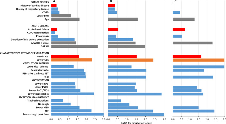 Figure 1: Pooled odds ratios across 26 variables show that extubation outcome is determined by the composite of respiratory, circulatory, and neurological function rather than by any single index. (Source: Torrini F, Gendreau S, Morel J, Carteaux G, Thille AW, Antonelli M, Mekontso Dessap A. Critical Care 2021. DOI: 10.1186/s13054-021-03802-3).
Definitions and the Epidemiology of Weaning Failure
Weaning, Liberation, and Extubation Distinguished
Weaning is a process, liberation is an outcome, and extubation is an airway decision. Liberation can be achieved with or without an artificial airway, since a tracheostomised patient may become ventilator-independent while remaining cannulated. Extubation adds airway-specific risks — oedema, aspiration, loss of airway protection — on top of the respiratory decision: wean the lungs, then decide whether the airway is safe to remove.
Simple, Difficult, and Prolonged Weaning
Three weaning categories are standardised by trajectory, not duration alone. Simple weaning is extubation at the first SBT. Difficult weaning is failure of the first SBT, requiring up to three SBTs or as long as seven days from the first attempt. Prolonged weaning is failure of at least three SBTs, or ventilation beyond seven days after the first attempt. The categories are prognostic: mortality rises from roughly 5% with simple weaning to 10–15% and up to 25–30% with difficult and prolonged weaning respectively.
Why Weaning Fails
Weaning failure is an imbalance between respiratory load and neuromuscular capacity, modulated by the cardiovascular and neurological systems. The load may be excessive — tube resistance, reduced compliance from oedema or atelectasis, intrinsic positive end-expiratory pressure in obstructive disease — while capacity may fail from diaphragmatic weakness caused by sepsis, steroids, and disuse, phrenic injury, or critical illness polyneuromyopathy. Cardiovascular limitation is the most under-recognised mechanism: spontaneous ventilation raises venous return and left-ventricular afterload, precipitating pulmonary oedema in patients with limited cardiac reserve. Because the causes are multiple, no single measurement predicts outcome reliably and a composite assessment is superior.
Readiness Assessment and Daily Screening
Resolution of the Acute Illness, Oxygenation, and Haemodynamics
Weaning should not begin until the process that precipitated intubation is resolving, with no escalating vasopressor requirement, uncontrolled sepsis, or anticipated procedure requiring re-intubation within 24 hours. Objective criteria include a ratio of arterial oxygen tension to inspired oxygen fraction ($P_{a\text{O}_2}/\text{FiO}_2$) of at least 150–200 $\text{mmHg}$ on a fraction of inspired oxygen ($\text{FiO}_2$) no greater than 0.4–0.5, a PEEP of 5–8 $\text{cmH}_2\text{O}$ or less, haemodynamic stability, and pH above 7.30. A rise in natriuretic peptide or an echocardiographic abnormality during a trial identifies the cardiac-limitation phenotype and should prompt optimisation of volume status and afterload before further attempts.
Respiratory Drive, Cough, and Secretions
The patient must initiate breaths, requiring intact respiratory drive and neuromuscular transmission. Cough strength is assessed clinically and, where available, by peak cough expiratory flow of at least 60 $\text{L/min}$; a weak cough cannot clear secretions and predicts extubation failure. Secretions should be manageable with suctioning no more often than every one to two hours.
Sedation, Neurological Status, and Delirium
An awake, cooperative patient who follows commands and tolerates a spontaneous awakening trial is far more likely to be extubated successfully. Deep sedation is a common and modifiable cause of delayed weaning, so sedation should be interrupted daily, delirium screened with the Confusion Assessment Method for the ICU (CAM-ICU), and the Richmond Agitation–Sedation Scale documented. Neurological injury, raised intracranial pressure requiring controlled hyperventilation, or inability to protect the airway contraindicates extubation, though not necessarily weaning.
Daily Screening Checklist
| Domain | Bedside criterion | Met |
|---|---|---|
| Precipitant | Cause resolving; no procedure planned in 24 h | $\square$ |
| Oxygenation | $P_{a\text{O}_2}/\text{FiO}_2 \geq 150\text{–}200\ \text{mmHg}$ on $\text{FiO}_2 \leq 0.4\text{–}0.5$ | $\square$ |
| PEEP | $\leq 5\text{–}8\ \text{cmH}_2\text{O}$ | $\square$ |
| Haemodynamics | No escalating vasopressors; no ischaemia or arrhythmia | $\square$ |
| Drive | Initiates breaths; rate 8–35 breaths/min | $\square$ |
| Cough | Effective; peak cough flow $\geq 60\ \text{L/min}$ | $\square$ |
| Secretions | Suctioning no more often than every 1–2 h | $\square$ |
| Neurological | Awake, follows commands, RASS 0 to $-$1, CAM-ICU negative | $\square$ |
| Biochemistry | pH $\geq 7.30$; adequate phosphate, magnesium, potassium | $\square$ |
Readiness is dynamic, so the checklist should be completed on every morning round. A daily paired spontaneous awakening trial and spontaneous breathing trial is the most reliable way to avoid leaving an eligible patient ventilated by omission rather than by decision.
The Spontaneous Breathing Trial
Techniques
The SBT places the patient on minimal or no support for a defined interval. The T-piece imposes the full work of breathing, including tube resistance. Low-level pressure support (5–8 $\text{cmH}_2\text{O}$) offsets part of the tube resistance and is the most widely used. Low-level pressure support with PEEP (typically 5 $\text{cmH}_2\text{O}$) counterbalances intrinsic PEEP and improves triggering in obstructive patients. Automatic tube compensation applies a flow-proportional pressure to cancel the tube's resistive load. No technique is clearly superior; the choice should follow local protocol.
Duration and Physiological Rationale
A 30-minute trial is standard: a landmark randomised comparison found it non-inferior to a 120-minute trial for successful extubation, with less fatigue. The rationale is to reproduce the post-extubation load while the airway is still secured, so that failure occurs under supervision.
Prediction: RSBI, P0.1, and Maximal Inspiratory Pressure
The rapid shallow breathing index (RSBI) integrates respiratory rate and tidal volume into a single measure of the rapid, shallow pattern that characterises impending failure:
$$RSBI = \frac{f}{V_T}$$where $f$ is the respiratory rate in breaths per minute and $V_T$ is the tidal volume in litres, so the index carries units of $\text{breaths}\cdot\text{min}^{-1}\cdot\text{L}^{-1}$. Measured after one minute of unassisted breathing, the threshold of less than 105 $\text{breaths}\cdot\text{min}^{-1}\cdot\text{L}^{-1}$ retains good negative predictive value but only moderate positive predictive value.
The airway occlusion pressure ($P_{0.1}$) measures the pressure generated in the first 100 milliseconds of an occluded inspiration and reflects respiratory drive independent of airway resistance:
$$P_{0.1} = P_{ao}(t = 0.1\ \text{s})$$where $P_{ao}$ is the airway pressure in $\text{cmH}_2\text{O}$ during a brief end-expiratory occlusion. A normal $P_{0.1}$ is below 2 $\text{cmH}_2\text{O}$; a value above 4–6 $\text{cmH}_2\text{O}$ indicates excessive drive and predicts weaning failure, while an abnormally low value suggests inadequate central drive. The maximal inspiratory pressure (MIP, or negative inspiratory force) quantifies muscle strength; a value more negative than $-20$ to $-30\ \text{cmH}_2\text{O}$ is generally required, though it is effort-dependent.
| Predictor | Threshold suggesting success | Comment |
|---|---|---|
| RSBI ($f/V_T$) | $< 105\ \text{breaths}\cdot\text{min}^{-1}\cdot\text{L}^{-1}$ | Best negative predictive value; measure at 1 min unassisted |
| $P_{0.1}$ | $< 2\text{–}4\ \text{cmH}_2\text{O}$ | Reflects drive; $> 4\text{–}6\ \text{cmH}_2\text{O}$ predicts failure |
| MIP / NIF | More negative than $-20$ to $-30\ \text{cmH}_2\text{O}$ | Effort-dependent supportive criterion |
| Composite | Tolerance of a 30-min SBT | Most robust single predictor of success |
The RSBI and $P_{0.1}$ are screening tools. A meta-analysis of predictors confirms that extubation outcome reflects the composite of respiratory, circulatory, and neurological function (Figure 1). Never extubate, or withhold extubation, on the basis of one number.
Weaning Strategy: Protocolised, Bundle-Based Weaning
How weaning is conducted matters as much as whether it is attempted. Physician-directed weaning relies on bedside judgement, tends to be conservative, and leaves patients ventilated longer than necessary. A protocolised approach — algorithm-driven daily screening, a standardised SBT, and explicit extubation criteria — shortens ventilation by roughly one to two days. Automated weaning, in which closed-loop systems reduce pressure support or conduct automated SBTs, produces a further modest reduction in weaning time in randomised trials and Cochrane reviews, though not a consistent mortality benefit.
Weaning is inseparable from sedation and rehabilitation. Daily sedation interruption paired with the SBT — the paired "wake up and breathe" strategy — reduces ventilator days, intensive-care stay, and mortality, and anchors the ABCDEF bundle: Assess and manage pain; Both spontaneous awakening and breathing trials; Choice of analgesia and sedation; Delirium assessment; Early mobility; and Family engagement. Early mobilisation attenuates critical illness myopathy, and routine delirium screening identifies the confusional state that both delays weaning and predicts reintubation.
Predictors and Prevention of Extubation Failure
Extubation failure — reintubation or death within 48–72 hours of planned extubation — occurs in 10–15% of unselected patients and carries a mortality of 25–50%, rising the longer reintubation is delayed. Risk factors include age above 65 years, chronic cardiac or respiratory disease, a weak cough, abundant or difficult-to-clear secretions, impaired neurological status or delirium, prolonged ventilation, positive fluid balance, a failed prior extubation, and hypercapnia or a high RSBI at the end of an SBT.
The cuff-leak test addresses the airway rather than the lungs. With the cuff deflated, the difference between inspired and expired tidal volume estimates the leak around the tube; a leak below about 110 $\text{mL}$ or below 10–15% of delivered tidal volume suggests laryngeal oedema and predicts post-extubation stridor. Accuracy is modest, so the test is best used where risk factors for laryngeal injury are present — prolonged or traumatic intubation, a large tube, or female sex — because an absent leak should change management. Where concern exists, corticosteroids (for example hydrocortisone 100 mg every eight hours or methylprednisolone 20–40 mg every four to six hours) begun at least four hours before extubation reduce stridor and reintubation.
For airway-independent risk, prophylactic post-extubation support changes outcome. In high-risk patients, non-invasive ventilation (NIV) started immediately after extubation reduces respiratory failure and intensive-care mortality; in lower-risk patients, high-flow nasal oxygen (HFNO) halves reintubation compared with conventional oxygen; and combining intermittent NIV with HFNO during breaks is superior to HFNO alone in the highest-risk and hypercapnic patients. The interface should therefore follow the pre-extubation risk profile and arterial carbon dioxide tension rather than unit habit.
Post-Extubation Care
The hours after extubation are the highest-risk period of the ventilatory course, since most reintubations occur within 24–48 hours. Patients require continuous pulse oximetry and regular assessment of respiratory rate, work of breathing, and conscious level. Post-extubation stridor is treated with systemic corticosteroids, nebulised adrenaline as a temporising bridge acting within minutes, and helium–oxygen mixtures, with a low threshold for reintubation using a smaller tube. Video laryngoscopy, a range of tube sizes, and a skilled operator should be immediately available where difficulty is anticipated.
A failed extubation is an airway emergency: reintubate early, while the patient is stable enough for a controlled procedure, rather than late, when tiring or hypoxaemic. The threshold for reintubation is lower than for initial intubation, because outcome worsens the longer it is deferred.
Liberation from the ventilator is not the end of recovery. Post-intensive-care syndrome (PICS) is the constellation of physical, cognitive, and psychological impairments that persist after critical illness: critical illness polyneuromyopathy with weakness and reduced exercise capacity; cognitive deficits in attention, memory, and executive function; and anxiety, depression, and post-traumatic stress disorder. Early mobilisation and delirium prevention during ventilation, minimising sedation and antipsychotic exposure, and structured follow-up after discharge are the principal mitigations, and screening should begin before discharge. Attention to swallowing, nutrition, sleep, and mental health completes the liberation pathway.
The Evidence Base and a Bedside Algorithm
The modern practice of liberation rests on a few landmark trials. The finding that a once-daily SBT is superior to multiple daily trials, and that a 30-minute trial is non-inferior to a 120-minute trial, shortened exposure to fatigue. The "wake up and breathe" protocol showed that paired daily sedation interruption and SBT reduces ventilator days, intensive-care stay, and mortality. Systematic reviews of protocolised versus physician-directed weaning confirm shorter ventilation, Cochrane reviews of automated weaning show a further modest reduction, and the post-extubation trials — NIV in high-risk patients, HFNO in lower-risk patients, and the combination in the highest-risk and hypercapnic groups — define the risk-stratified support that follows a successful trial.
flowchart TD
    A["Daily screen: precipitant resolving, FiO2 <= 0.4, PEEP <= 8, stable haemodynamics, awake, effective cough"] --> B{"Sedation interrupted and delirium assessed?"}
    B -- "No" --> C["Optimise sedation, mobility and secretions; reassess next day"]
    B -- "Yes" --> D["Spontaneous breathing trial: 30 min T-piece or pressure support 5-8 cmH2O"]
    D --> E{"Tolerated? rate < 35, SpO2 >= 90%, no distress, stable haemodynamics"}
    E -- "No" --> F["Return to rest ventilation, treat cause, reassess; enter difficult-weaning pathway if recurrent"]
    E -- "Yes" --> G{"High-risk features? age > 65, cardiac or respiratory disease, weak cough, abundant secretions, prolonged ventilation"}
    G -- "Yes" --> H["Cuff-leak assessment; plan prophylactic HFNO or NIV with high-flow breaks"]
    G -- "No" --> I["Extubate to conventional oxygen"]
    H --> J["Extubate; monitor closely for stridor and failure in the first 48 h"]
    I --> J
    J --> K{"Signs of failure or stridor?"}
    K -- "Yes" --> L["Treat stridor; reintubate early and escalate to invasive ventilation"]
    K -- "No" --> M["Continue post-extubation care; screen for post-intensive-care syndrome"]
Key Takeaways
- Weaning is a process, liberation an outcome, and extubation an airway decision; each can fail independently and must be judged separately.
- Categorise every patient as simple, difficult, or prolonged weaning, because the category predicts mortality and sets the intensity of the pathway.
- Screen for readiness each morning with a composite checklist covering the precipitant, oxygenation, PEEP, haemodynamics, drive, cough, secretions, sedation, and biochemistry.
- Perform a 30-minute SBT by T-piece or low-level pressure support, and document the physiological response.
- Use the RSBI (threshold below 105 $\text{breaths}\cdot\text{min}^{-1}\cdot\text{L}^{-1}$), $P_{0.1}$, and MIP as adjuncts; the meta-analysis in Figure 1 confirms that no single index determines outcome.
- Pair spontaneous awakening and breathing trials, provide early mobility, and screen for delirium within the ABCDEF bundle to shorten ventilation.
- Identify high-risk patients before extubation — age, cardiac and respiratory comorbidity, weak cough, abundant secretions, neurological impairment, previous failure — and plan prophylactic HFNO or NIV accordingly.
- Treat post-extubation stridor promptly with corticosteroids, adrenaline, and a low threshold for reintubation with a smaller tube, and screen survivors for post-intensive-care syndrome.