The immunocompromised patient who develops acute hypoxaemic respiratory failure presents the broadest differential diagnosis and narrowest margin for error in critical care. Cytotoxic chemotherapy, prolonged neutropenia, transplantation, and checkpoint inhibition each dismantle specific immunological barriers, leaving the lung vulnerable to opportunistic pathogens, to the treatments themselves, and to a diffuse inflammatory response easily mistaken for infection.
The population is heterogeneous and the therapeutic window short. Precision means matching exposure history and its timing to the likely mechanism of injury, pursuing a definitive diagnosis in parallel with oxygen escalation, and ventilating a fragile alveolar–capillary barrier with the least injurious pressures possible. Every PEEP change here is also a haemodynamic decision.
The Spectrum of Pulmonary Complications
Infectious Pneumonia
Infection remains the commonest and most treatable cause, but the differential shifts with the nature and duration of the immunological defect. Bacterial pathogens, including Pseudomonas aeruginosa, Stenotrophomonas maltophilia, and enteric Gram-negatives, dominate during profound neutropenia and the first weeks after stem-cell transplantation. Opportunistic moulds, chiefly Aspergillus species and the Mucorales, become prominent with prolonged corticosteroid exposure and graft-versus-host disease (GVHD). Viral pathogens — cytomegalovirus (CMV), herpes simplex virus, influenza, respiratory syncytial virus, and SARS-CoV-2 — follow a similar pattern, with CMV typically reactivating one to four months after allogeneic transplantation.
Pneumocystis jirovecii pneumonia (PJP) is common, treatable, and frequently missed. It presents with subacute dyspnoea, a dry cough, and hypoxaemia disproportionately severe for the diffuse ground-glass opacification seen on imaging. Risk is highest with prolonged corticosteroids, HIV infection with a CD4 count below 200 cells/$\mu$L, transplantation, and rituximab or temozolomide exposure. Because it is a fungus, serum $\beta$-D-glucan is characteristically elevated while galactomannan is not, separating PJP from invasive aspergillosis at the bedside.
Non-Infectious and Treatment-Related Entities
Non-infectious entities mimic infection closely. Radiation pneumonitis appears two to six months after thoracic irradiation and classically respects the radiation field with a geometric border. Engraftment syndrome occurs in the peri-engraftment period after transplantation and combines fever, rash, capillary leak, and non-cardiogenic pulmonary oedema that often responds to corticosteroids. Diffuse alveolar haemorrhage (DAH) carries a mortality above 50 percent in transplant recipients and is suggested by haemoptysis, a falling haemoglobin, and progressively bloodier bronchoalveolar lavage (BAL) returns.
Misclassification is penalised both ways: escalating antimicrobials for immune-mediated pneumonitis adds toxicity without benefit, while high-dose corticosteroids for infection can worsen fungal and viral disease. Exposure timeline, CT pattern, and BAL cell count resolve most cases.
Drug-Induced and Treatment-Related Lung Toxicity
Classic Cytotoxic and Immunosuppressive Agents
Bleomycin causes dose-related pneumonitis progressing to fibrosis, with risk rising beyond about 400 cumulative units and amplified by concurrent oxygen, radiation, and granulocyte colony-stimulating factor. Methotrexate produces an acute hypersensitivity pneumonitis with fever, eosinophilia, and migratory infiltrates that often resolves on withdrawal alone. Busulfan and carmustine cause late fibrosis, gemcitabine and the mammalian target of rapamycin inhibitors cause an organising pneumonia pattern, and amiodarone uniquely produces several phenotypes — chronic interstitial pneumonitis, organising pneumonia, eosinophilic pneumonia, and acute respiratory distress syndrome (ARDS); its very long half-life means injury can progress for weeks after withdrawal.
Immune Checkpoint Inhibitor Pneumonitis
Checkpoint inhibitors unleash T-cell activity against tumour and against normal tissue. Immune checkpoint inhibitor-related pneumonitis (ICI-P) presents with cough, dyspnoea, and ground-glass or organising pneumonia. Real-world lung-cancer cohorts report an incidence approaching 10 percent, a median time to diagnosis of about 50 days, and mortality in roughly a quarter of patients; baseline fibrosis and pre-existing obstructive disease are independent risk factors. Management follows CTCAE grade: hold the agent, and for grade 2 or higher start prednisone-equivalent 1–2 mg/kg/day, escalating to infliximab or mycophenolate if steroid-refractory.
Cardiotoxic Agents and the Cardio-Oncology Interface
Anthracyclines such as doxorubicin cause dose-dependent, largely irreversible type I cardiomyopathy through topoisomerase-II$\beta$-mediated cardiomyocyte injury, with risk rising appreciably above a cumulative doxorubicin-equivalent dose of 400 mg/m$^2$. Trastuzumab causes typically reversible type II dysfunction, and combining it with an anthracycline multiplies risk. These agents spare the lung directly, but their cardiac legacy shapes ventilation: an occult cardiomyopathy may tolerate positive pressure and high PEEP very poorly.
| Agent or class | Predominant pulmonary toxicity | Typical timing | Management |
|---|---|---|---|
| Bleomycin | Pneumonitis progressing to fibrosis | Weeks to months; dose-related | Stop drug; corticosteroids; limit FiO$_2$ |
| Methotrexate | Acute hypersensitivity pneumonitis, eosinophilia | Days to weeks | Withdraw; corticosteroids if severe |
| Checkpoint inhibitors | Organising pneumonia, hypersensitivity, ARDS | Median ~50 days | Hold agent; steroids 1–2 mg/kg/day; infliximab if refractory |
| Amiodarone | Interstitial pneumonitis, organising pneumonia, ARDS | Months; cumulative | Withdraw; corticosteroids; avoid hyperoxia |
| Trastuzumab deruxtecan | Interstitial lung disease or pneumonitis | Early to delayed | Hold; corticosteroids; CTCAE-based |
| Anthracyclines | Cardiomyopathy with cardiogenic pulmonary oedema | Weeks to years | Dexrazoxane prophylaxis; heart-failure therapy |
Diagnostic Strategy
Non-Invasive Testing First
The initial workup should be fast, broad, and largely non-invasive. High-resolution CT narrows the differential more than any single test: ground-glass opacity suggests PJP, a nodular or halo pattern suggests angioinvasive mould, and a field-restricted opacity suggests radiation pneumonitis. Blood and sputum cultures, respiratory viral polymerase chain reaction (PCR), serum galactomannan, $\beta$-D-glucan, and procalcitonin each contribute; none is definitive alone. Galactomannan has moderate sensitivity for invasive aspergillosis, better on BAL than in serum; $\beta$-D-glucan is a pan-fungal marker whose negative predictive value is its main strength, with false positives from certain antibiotics, albumin, and haemodialysis. Empirical broad-spectrum therapy alone is inadequate: it neither secures a diagnosis nor prevents the accumulation of drug toxicity.
Early Bronchoscopy and BAL
When the non-invasive panel is non-diagnostic — as it is in perhaps half of cases — bronchoscopy with BAL is the highest-yield procedure: immunofluorescence and PCR for PJP, galactomannan for Aspergillus, viral PCR, culture, cytology for malignant infiltration, and cell count with Prussian blue staining for DAH. It is a physiological stressor that can precipitate intubation, and thrombocytopenia below about 20–50$\times$10$^9$/L should be corrected first. Expert consensus favours early rather than rescue bronchoscopy, because a definitive diagnosis reduces unnecessary antimicrobial exposure and directs immunosuppression.
Within the first hour: arterial blood gas and lactate, blood cultures, respiratory viral PCR, serum galactomannan and $\beta$-D-glucan, and high-resolution CT. Within 24 hours if undiagnosed: bronchoscopy with BAL for cytology, cell count, stains, culture, PCR, and fungal markers.
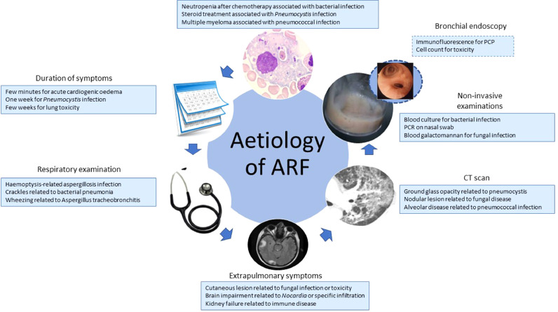 Figure 1: Six clinical axes — symptom duration, examination, CT, biomarkers, and BAL — identify the cause of acute respiratory failure in the immunocompromised host. (Source: Lemiale V, et al. Journal of Intensive Medicine 2021. DOI: 10.1016/j.jointm.2021.09.003).
Integrated Diagnostic and Ventilatory Algorithm
The algorithm links diagnosis to oxygen escalation and mechanical support.
flowchart TD
    A["Immunocompromised host with acute hypoxaemic respiratory failure"] --> B["Bundle: ABG, lactate, cultures, viral PCR, galactomannan, beta-D-glucan, HRCT"]
    B --> C{"Diagnosis established or highly probable?"}
    C -->|"Yes"| D["Targeted therapy: antibacterials, TMP-SMX plus steroids for PJP, antifungals, antivirals, withdraw culprit drug, steroids"]
    C -->|"No"| E["Early bronchoscopy with BAL: cytology, cell count, Prussian blue, culture, PCR, fungal markers"]
    E --> F{"Diagnosis now established?"}
    F -->|"Yes"| D
    F -->|"No"| G["Reassess exposures, repeat HRCT, consider empirical therapy and multidisciplinary review"]
    D --> H["Oxygen escalation: HFNO 50-60 L/min, FiO2 to SpO2 92-96 percent"]
    G --> H
    H --> I{"Improving within 1 hour with no distress?"}
    I -->|"Yes"| J["Continue non-invasive support with hourly reassessment"]
    I -->|"No"| K["Prompt intubation; avoid delayed intubation"]
    K --> L["Lung-protective ventilation: VT 6 mL/kg PBW, Pplat <= 30 cmH2O, driving pressure <= 15 cmH2O, titrated PEEP"]
    L --> M{"Refractory hypoxaemia despite optimised ventilation?"}
    M -->|"No"| N["Continue protective ventilation, prone if P/F < 150, daily liberation assessment"]
    M -->|"Yes"| O["Assess ECMO candidacy against reversibility and goals of care; time-limited trial"]
    O --> P["Concurrent goals-of-care discussion with oncology, patient, and family"]
Ventilation Strategy in the Immunocompromised Host
Non-Invasive Support and the Intubation Question
Non-invasive ventilation (NIV) was long favoured to avoid intubating neutropenic or thrombocytopenic patients, but two randomised trials tempered that enthusiasm. In the HIGH trial, 776 immunocompromised patients with acute hypoxaemic failure received high-flow nasal oxygen (HFNO) or standard oxygen; day-28 mortality was 35.6 versus 36.1 percent, with no difference in intubation rate, although HFNO improved PaO$_2$/FiO$_2$ and reduced respiratory rate. In the GRRR-OH trial, 374 patients given early NIV versus oxygen alone showed no mortality benefit (24.1 versus 27.3 percent), with oxygenation failure in 38.2 versus 44.8 percent.
HFNO is therefore a reasonable first-line support at 50–60 L/min with a precise FiO$_2$, but only under frequent reassessment. NIV has a limited role in de novo hypoxaemic failure, where it risks generating injurious tidal volumes and, critically, delaying a necessary intubation — a delay independently associated with worse outcome. Patients not improving within one hour of optimised non-invasive support should be intubated rather than subjected to prolonged trial-and-failure.
Invasive Ventilation and Lung Protection
Once intubated, lung protection applies with particular force, because the injured immunocompromised lung is exquisitely vulnerable to ventilator-induced lung injury (VILI). Tidal volume is set at 6 mL/kg predicted body weight, plateau pressure at or below 30 cmH$_2$O, and driving pressure minimised. Shunt fraction and oxygenation index quantify gas-exchange failure:
$$\dot{Q}_s/\dot{Q}_t = \frac{C_cO_2 - C_aO_2}{C_cO_2 - C_{\bar{v}}O_2}$$where $\dot{Q}_s/\dot{Q}_t$ is the shunt fraction (dimensionless), $C_cO_2$ is end-capillary, $C_aO_2$ arterial, and $C_{\bar{v}}O_2$ mixed-venous oxygen content, each in mL O$_2$/dL. Arterial content is $C_aO_2 = 1.34 \times Hb \times SaO_2 + 0.003 \times PaO_2$, with haemoglobin $Hb$ in g/dL, saturation $SaO_2$ as a fraction, and $PaO_2$ in mmHg. The oxygenation index is a practical surrogate:
$$OI = \frac{FiO_2 \times \overline{P}_{aw} \times 100}{PaO_2}$$where $FiO_2$ is the inspired oxygen fraction (dimensionless), $\overline{P}_{aw}$ is mean airway pressure in cmH$_2$O, and $PaO_2$ is in mmHg. Driving pressure is the ventilatory variable most strongly associated with survival:
$$\Delta P = P_{plat} - PEEP_{tot} = \frac{V_T}{C_{RS}}$$where $\Delta P$ is driving pressure in cmH$_2$O, $P_{plat}$ is plateau pressure, $PEEP_{tot}$ is total end-expiratory pressure including intrinsic PEEP, $V_T$ is tidal volume in mL, and $C_{RS}$ is respiratory-system compliance in mL/cmH$_2$O. In a pooled analysis of 3,562 patients with ARDS, each 1-SD increment in $\Delta P$ — about 7 cmH$_2$O — carried a relative risk of death of 1.41. Reducing $\Delta P$, rather than simply lowering tidal volume or raising PEEP, is the mechanistically meaningful target.
Every increment in tidal volume, plateau pressure, and driving pressure amplifies mechanical power delivered to an already leaky membrane. Target $SpO_2$ 92–96 percent, accept permissive hypercapnia, and titrate PEEP to oxygenation and haemodynamics rather than a fixed number.
Specific Hazards of Positive-Pressure Ventilation
Barotrauma and Dissemination of Infection
Positive pressure is not neutral in this population. In PJP, thin-walled cysts and necrotic alveoli predispose to pneumothorax and pneumomediastinum, which may be bilateral and sudden, so the threshold for chest imaging should be low. In DAH, raised intrathoracic pressure can worsen alveolar bleeding; the strategy is the lowest mean airway pressure compatible with oxygenation, permissive hypercapnia, and aggressive correction of coagulopathy. Barotrauma also risks bronchopleural fistula. Concern that positive pressure or aerosol-generating procedures disseminate infection is largely theoretical; the dominant hazard is the direct mechanical injury of VILI on a diffuse inflammatory process.
Procedures in the Neutropenic and Thrombocytopenic Patient
Procedural risk is amplified by haematological failure. BAL, central venous access, and chest-drain insertion each carry bleeding risk when platelets are low; a count above 50$\times$10$^9$/L is desirable before bronchoscopy or invasive line placement. Neutropenia also blunts sputum culture yield and delays radiographic evolution, so imaging frequently lags deterioration.
Never attribute new hypoxaemia in a neutropenic patient to the underlying disease without completing the diagnostic bundle. Never persist with non-invasive support once it is failing, because delayed intubation is independently lethal.
Extracorporeal Membrane Oxygenation, Futility, and Goals of Care
Veno-venous extracorporeal membrane oxygenation (VV-ECMO) can rescue refractory hypoxaemia when ventilation cannot maintain gas exchange without injurious pressures. Reported mortality in immunocompromised patients is high — historically 60–80 percent — but it is not uniformly futile. Survival concentrates among those with a reversible or bridgeable cause, such as PJP with preserved organ function, and among those in whom ECMO permits genuine lung rest while definitive therapy works. Immunosuppression and even neutropenia are not absolute contraindications in contemporary extracorporeal guidance, but candidacy must be judged against the trajectory of the malignancy and the reversibility of the respiratory failure.
Futility is a judgement about trajectory, not a single number. The defensible approach is an explicit, time-limited trial of advanced support with pre-agreed physiological benchmarks and an early goals-of-care conversation with the patient, family, and oncology team. ECMO should not become a bridge to nowhere; it belongs where a plausible path to recovery or transplantation exists, and should be withdrawn when that path closes.
Cardio-Oncology: Cardiomyopathy, PEEP, and Pulmonary Hypertension
Chemotherapy-Induced Cardiomyopathy
Anthracycline cardiomyopathy is cumulative, dose-dependent, and often irreversible, whereas trastuzumab-related dysfunction is typically reversible on withdrawal. Surveillance with ejection fraction and global longitudinal strain detects subclinical injury. When such a patient requires ventilation, anaemia, sepsis, and positive intrathoracic pressure can unmask a compensated cardiomyopathy and precipitate cardiogenic pulmonary oedema that mimics ARDS — a distinction with major therapeutic consequences, since one demands decongestion and the other lung protection with often generous fluid tolerance.
Heart–Lung Interaction under Positive Pressure
PEEP increases right-ventricular afterload by compressing the pulmonary vasculature and reduces left-ventricular preload by impeding venous return. In a patient with chemotherapy-induced right-ventricular dysfunction or pulmonary hypertension these effects compound: a modest PEEP increase may reduce cardiac output and worsen oxygenation by lowering mixed-venous oxygen content even as it recruits alveoli. Echocardiography, central venous oxygen saturation, and lactate trend outperform any static pressure.
Pulmonary Hypertension in the Cancer Patient
Pulmonary hypertension here is usually group 2 (left-heart disease) or group 3 (hypoxic lung disease), though dasatinib causes group 1 pulmonary arterial hypertension. Non-invasive ventilation or CPAP helps when obstructive sleep apnoea contributes. Systemic vasodilators risk worsening ventilation–perfusion matching and hypotension and should be used only with invasive or echocardiographic monitoring, never as a substitute for treating the underlying process.
Treat lungs and heart as one system. Before every PEEP change ask what it does to the right ventricle; before every fluid bolus in a hypoxaemic cancer patient ask whether the left ventricle can accept it.
Key Takeaways
- Infection, drug-induced injury, radiation pneumonitis, engraftment syndrome, and DAH all present as acute hypoxaemic respiratory failure; exposure history and timing are the fastest route to diagnosis.
- Galactomannan supports invasive aspergillosis and $\beta$-D-glucan supports PJP or other fungal disease; both are adjuncts to imaging, culture, and BAL.
- Early bronchoscopy with BAL is the highest-yield test when the non-invasive workup is non-diagnostic, but requires corrected thrombocytopenia and a secured airway.
- HFNO is reasonable first-line support; NIV does not reduce mortality in de novo hypoxaemic failure and must never delay intubation.
- Tidal volume 6 mL/kg predicted body weight, plateau pressure at or below 30 cmH$_2$O, and minimised driving pressure are the core defence against ventilator-induced lung injury.
- Positive pressure carries specific hazards: pneumothorax in PJP, worsened alveolar bleeding in DAH, and procedural bleeding with neutropenia or thrombocytopenia.
- VV-ECMO can rescue selected patients with reversible causes, but candidacy must be judged by trajectory and paired with a time-limited goals-of-care plan.
- Anthracycline and HER2-targeted cardiotoxicity interacts with ventilation because PEEP alters right- and left-ventricular loading.