Inhalation injury complicates roughly one in five major burns and is, independently, the strongest single predictor of death in the burned patient. It is not one disease. A single exposure delivers three insults, each with its own anatomy, tempo and treatment: a thermal burn of the supraglottic airway, a chemical burn of the tracheobronchial tree, and a systemic poisoning of oxygen transport and cellular respiration by carbon monoxide and cyanide. The clinician who collapses these into one diagnosis will intubate too late, ventilate too roughly and overlook the toxin that is actually killing the patient.
The three injuries run on different clocks. The upper airway is burned instantly but closes over hours, so its crisis is an airway decision made in anticipation. The lower airway is injured over the first day and fails over the first week, so its crisis is ventilation and secretion clearance. The systemic poisoning is present at the door and is a metabolic emergency. Recognition is therefore not one judgement but three parallel ones, and the classical signs — facial burns, singed nasal hair, oropharyngeal soot, hoarseness — support a decision without ever excluding injury.
The Triad of Inhalation Injury
Thermal Injury Is an Upper-Airway Disease
The upper airway is an efficient heat exchanger, so inspired gas at several hundred degrees cools below $50\ ^\circ\text{C}$ by the carina; thermal necrosis is therefore largely confined to the nose, pharynx and larynx, except with steam inhalation, which carries latent heat of vaporisation. Laryngospasm and reflex mucosal oedema follow, amplified by the systemic capillary leak and by the crystalloid given to resuscitate it. The result is a rapidly narrowing, collapsible supraglottic airway whose rate of closure governs the first six hours.
Chemical Injury Reaches the Lower Airway on Soot
Thermal energy is spent above the cords; chemical energy is not. Burning modern furnishings and synthetics generates an aerosol in which $0.1$–$1\ \mu\text{m}$ soot particles carry aldehydes such as acrolein, hydrogen chloride, sulfur dioxide, ammonia and free radicals into the distal airway. Solubility determines depth: highly soluble agents injure the proximal airway, whereas less soluble gases such as phosgene and nitrogen dioxide reach the alveoli. The result is epithelial destruction with ciliary loss, sloughing and surfactant inactivation, compounded by bronchospasm and a delayed capillary leak indistinguishable from ARDS, as neutrophils, cytokines and reactive oxygen species convert the chemical burn into diffuse lung injury over $24$–$72$ hours.
Systemic Toxicity: The Alveolus as a Portal
The third injury is not structural. Carbon monoxide and hydrogen cyanide, from incomplete combustion and pyrolysis of nitrogen-containing polymers, cross the alveolar membrane and poison both ends of the oxygen cascade: carriage in blood and use in the mitochondrion. This injury is silent on the chest radiograph and normal on the arterial oxygen tension, which is precisely why it is missed.
Recognition and Diagnosis
History and the Bedside Signs
The key historical feature is a fire in an enclosed space, because confined combustion consumes oxygen and concentrates the products of pyrolysis for rebreathing. Clinicians look for burns of the face and neck, singed nasal hairs, oropharyngeal soot, carbonaceous sputum, hoarseness, dysphagia, odynophagia and stridor. The extent of cutaneous burns does not predict the airway, and no sign is individually sensitive or specific: soot in the mouth may accompany a trivial airway, and a normal examination after a closed-space fire does not exclude lethal injury.
Stridor means the airway has already narrowed critically; waiting for it converts a controlled intubation into an emergency and lets oedema destroy the landmarks that make laryngoscopy straightforward. Hoarseness or odynophagia after a closed-space fire is enough to secure the airway electively, with an experienced operator.
Bronchoscopy: Diagnosis, Grading and Prognosis
Flexible bronchoscopy, ideally within the first $24$ hours, is the reference standard. Findings evolve from erythema and oedema through soot deposition and ulceration to necrosis and obliteration of normal anatomy, and grading systems — the endoscopic Abbreviated Injury Score for the respiratory tract — correlate with ventilation duration, pneumonia incidence and mortality. The examination is also therapeutic, allowing clearance of secretions and early casts. Because findings progress, a normal early study does not exclude injury and mandates reassessment; the admission radiograph is frequently normal even in severe injury.
Airway Management: The Primacy of Early Intubation
The Natural History of Progressive Oedema
Airway resistance rises with the fourth power of a falling radius, so modest circumferential swelling produces a disproportionate fall in flow. Oedema from thermal injury, systemic inflammation and fluid resuscitation accumulates in the loose pharyngeal and laryngeal tissues, and the airway can progress from patent to impassable within hours. Early intubation is therefore not escalation but a pre-emptive act that preserves control of a deteriorating airway.
Technique and the Pitfalls of Delay
Intubate with a large-bore cuffed tube — $8.0$–$9.0\ \text{mm}$ internal diameter in an adult — because the patient will need bronchoscopy, effective suction and possibly high-frequency ventilation; a small tube trades an easier intubation for a week of inadequate pulmonary toilet. Treat the airway as difficult: senior operator, videolaryngoscopy, suction and a prepared surgical airway, with cervical spine precautions if warranted. Delayed intubation is the dangerous path: the cords become invisible, the neck swollen and the surgical airway hazardous, while hypoxic brain injury accrues in the interval.
Intubate now for stridor, hoarseness or odynophagia after a closed-space fire; deep facial, neck or perioral burns; soot below the cords or carbonaceous sputum; reduced consciousness; or transfer to a burn centre. Anticipate difficult laryngoscopy in every case.
Carbon Monoxide and Cyanide Poisoning
Carbon Monoxide: A Defect of Content, Not of Tension
Carbon monoxide binds haemoglobin with roughly $210$–$250$ times the affinity of oxygen, reduces the haemoglobin available to carry oxygen, shifts the dissociation curve to the left and inhibits cytochrome c oxidase. The resulting tissue hypoxia is invisible to the arterial oxygen tension, because $PaO_2$ measures dissolved oxygen and not content, and invisible to pulse oximetry, which cannot distinguish oxyhaemoglobin from carboxyhaemoglobin at its two wavelengths. Only multi-wavelength co-oximetry measures carboxyhaemoglobin and methaemoglobin and reports the true saturation. The oxygen content relationship makes the defect explicit:
$$CaO_2 = 1.34 \times [Hb] \times (1 - F_{COHb}) \times S_pO_2 + 0.003 \times PaO_2$$where $CaO_2$ is arterial oxygen content ($\text{mL}\ \text{O}_2\cdot\text{dL}^{-1}$), $[Hb]$ haemoglobin ($\text{g}\cdot\text{dL}^{-1}$), $1.34$ the Hüfner coefficient ($\text{mL}\ \text{O}_2\cdot\text{g}^{-1}$ Hb), $F_{COHb}$ the carboxyhaemoglobin fraction, $S_pO_2$ the pulse-oximetric saturation, $0.003$ the plasma solubility of oxygen ($\text{mL}\ \text{O}_2\cdot\text{dL}^{-1}\cdot\text{mmHg}^{-1}$) and $PaO_2$ the arterial oxygen tension ($\text{mmHg}$). In severe poisoning a normal $PaO_2$ and a normal oximeter trace coexist with a materially reduced oxygen content.
Treating Carbon Monoxide Poisoning
Treatment begins at once with high-concentration oxygen by tight-fitting reservoir mask, or through the endotracheal tube once the airway is secure. Oxygen accelerates dissociation: the carboxyhaemoglobin half-life falls from about $300$ minutes in room air to roughly $90$ minutes on $100\%$ oxygen at one atmosphere, and to approximately $30$ minutes at $3\ \text{atm}$ in a hyperbaric chamber. Hyperbaric oxygen is considered for loss of consciousness, severe neurological signs, myocardial ischaemia, acidosis or carboxyhaemoglobin above roughly $25\%$ (about $15$–$20\%$ in pregnancy), though trial evidence for a mortality benefit is inconsistent and the level correlates poorly with severity.
Cyanide: Mitochondrial Asphyxia
Hydrogen cyanide is released by combustion of nitrogen-containing polymers — wool, silk, polyacrylonitrile and polyurethane foam. It inhibits cytochrome c oxidase, arresting oxidative phosphorylation so that cells cannot use the oxygen delivered to them; anaerobic metabolism follows and lactate rises steeply. Hydroxocobalamin, $5\ \text{g}$ intravenously over $15$ minutes and repeated if needed, binds cyanide to form cyanocobalamin, which is excreted in urine. It is preferred over nitrite antidotes in the fire victim, because nitrites induce methaemoglobinaemia and further reduce oxygen content in a patient who may also be poisoned by carbon monoxide.
A smoke-inhalation patient with a normal $PaO_2$, a normal-looking saturation, severe lactic acidosis and vasopressor-dependent shock has cyanide poisoning until proven otherwise. Treat empirically with hydroxocobalamin rather than awaiting a confirmatory assay.
Mechanical Ventilation Strategy
Lung-Protective Ventilation, PEEP and Permissive Hypercapnia
The injured lung is ventilated as any ARDS lung: tidal volume $6\ \text{mL}\cdot\text{kg}^{-1}$ predicted body weight, plateau pressure below $30\ \text{cmH}_2\text{O}$ and driving pressure below about $15\ \text{cmH}_2\text{O}$. PEEP is titrated to oxygenation and compliance; higher values are often needed because surfactant inactivation, casts and atelectasis create a large shunt, but titration must respect a patient undergoing major fluid resuscitation.
The shunt responsible for the hypoxaemia is quantified as:
$$\frac{Q_s}{Q_t} = \frac{CcO_2 - CaO_2}{CcO_2 - CvO_2}$$where $Q_s/Q_t$ is the shunt fraction and $CcO_2$, $CaO_2$ and $CvO_2$ are the end-capillary, arterial and mixed-venous oxygen contents ($\text{mL}\ \text{O}_2\cdot\text{dL}^{-1}$). Permissive hypercapnia is acceptable in most patients, with the caveat that the burn patient is hypermetabolic and may generate carbon dioxide at $1.5$–$2$ times basal rates. Active heated humidification is mandatory, because dry gas inspissates carbonaceous secretions and promotes cast formation.
High-Frequency Ventilation and the Bronchoscopic Signature
When conventional ventilation fails, high-frequency percussive and oscillatory ventilation improve oxygenation and mobilise secretions; data suggest percussive ventilation improves the $P_aO_2/F_iO_2$ ratio compared with low-tidal-volume ventilation early after injury, though a mortality benefit is unproven. The characteristic bronchoscopic findings carry prognostic weight: the higher the endoscopic grade, the longer the ventilation, the more pneumonia and the greater the mortality.
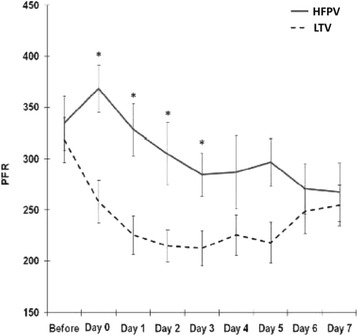 Figure 1: High-frequency percussive ventilation improved the PaO2/FiO2 ratio compared with low-tidal-volume ventilation during the first days after smoke inhalation injury, supporting its use as a rescue mode. (Source: Walker PF, Buehner MF, Wood LA, et al. Critical Care 2015. DOI: 10.1186/s13054-015-1077-4).
Assessment and Airway Algorithm
The algorithm integrates recognition, airway control, toxicology and ventilation; the airway decision deliberately precedes all others.
flowchart TD
    A["Closed-space fire, soot, hoarseness, stridor or reduced consciousness"] --> B{"Hard airway trigger present? Stridor, hoarseness, deep facial burns, soot below the cords or need for transfer"}
    B -->|"No"| C["Humidified oxygen, upright posture, serial airway examination, low threshold to intubate"]
    B -->|"Yes"| D["Intubate early with a large-bore cuffed tube, videolaryngoscopy and a prepared surgical airway"]
    D --> E["Bronchoscopy: grade injury and clear casts and secretions"]
    E --> F["Co-oximetry for carboxyhaemoglobin; arterial blood gas and lactate"]
    F --> G{"Carboxyhaemoglobin elevated?"}
    G -->|"Yes"| H["100% oxygen; consider hyperbaric oxygen if severe"]
    G -->|"No"| I["Proceed to ventilation"]
    H --> J{"Refractory lactic acidosis or unexplained shock?"}
    I --> J
    J -->|"Yes"| K["Hydroxocobalamin 5 g IV for suspected cyanide poisoning"]
    J -->|"No"| L["Ventilate 6 mL/kg predicted body weight, titrated PEEP, permissive hypercapnia, heated humidification"]
    K --> L
    L --> M{"Persistent hypoxaemia or bronchial casts?"}
    M -->|"Yes"| N["Consider percussive or oscillatory ventilation, serial bronchoscopy and extracorporeal rescue"]
    M -->|"No"| O["Continue lung-protective ventilation; reassess liberation daily"]
Special Considerations in the Burned Patient
Fluid Resuscitation and the Lung
Resuscitation is titrated to urine output around $0.5\ \text{mL}\cdot\text{kg}^{-1}\cdot\text{h}^{-1}$, with crystalloid formulae as a starting point rather than a target. Over-resuscitation — "fluid creep" — is common, and the same capillary leak that mandates volume for skin and gut also floods the airway and pulmonary interstitium; the airway can swell hours after admission purely from administered fluid.
Ventilator-Associated Pneumonia, Tracheostomy and Casts
Inhalation injury independently predicts ventilator-associated pneumonia, and diagnosis is confounded by the persistent systemic inflammatory response and by tracheobronchitis rather than new infection. Prevention follows the usual bundle — semi-recumbent positioning, oral care, subglottic secretion drainage and daily sedation interruption — with vigilance for the immunosuppression of a lost skin barrier and altered cell-mediated immunity. Tracheostomy is often considered when liberation stalls or secretions demand frequent suction, though a mortality benefit is unproven and the swollen neck makes it technically demanding. Bronchial casts of sloughed epithelium, fibrin, mucus and carbon are a feared cause of major airway obstruction and death; management combines humidification, nebulised mucolytics such as acetylcysteine, nebulised heparin, chest physiotherapy and therapeutic bronchoscopy, with coagulopathy considered before heparin is used.
Toxic Inhalations Beyond the Fireground
Exposures away from fires follow the same principles of decontamination, oxygenation and support, but each agent has a characteristic mechanism and tempo that set the observation period and the specific therapy.
| Agent | Typical source | Mechanism | Treatment |
|---|---|---|---|
| Chlorine | Bleach and acid mixing, pools, industry | Hydrolysis to hydrochloric and hypochlorous acid; oxidant epithelial injury and delayed oedema | Remove from source, humidified oxygen, inhaled bronchodilator, supportive care |
| Ammonia | Fertiliser, refrigeration, cleaning products | Alkaline injury; saponifies lipids and causes liquefactive necrosis of upper airway and proximal bronchi | Copious water irrigation of eyes and skin, early airway control, humidified oxygen, bronchodilator |
| Phosgene | Welding, decomposition of chlorinated solvents | Acylation of alveolar proteins; capillary leak after a $6$–$24$ hour latent period | Strict rest, observe for delayed pulmonary oedema, oxygen, PEEP, early intubation if oedema develops |
| Hydrogen sulfide | Manure, sewers, oil and gas, pulp mills | Inhibition of cytochrome c oxidase, identical to cyanide; olfactory paralysis at high concentration | Rescuer protection, $100\%$ oxygen, supportive care, consider hydroxocobalamin |
| Cyanide | Combustion of wool, silk, polyacrylonitrile and polyurethane | Inhibition of cytochrome c oxidase; anaerobic metabolism and severe lactic acidosis | Hydroxocobalamin $5\ \text{g}$ IV, high-flow oxygen, supportive care |
Decontamination and Supportive Care
Remove the patient from the source, with rescuers protected, particularly against hydrogen sulfide and cyanide, where secondary exposure has killed rescuers. Remove contaminated clothing and irrigate skin and eyes copiously. Emesis and activated charcoal have no role in inhalational exposure. Titrate oxygen, give bronchodilators for bronchospasm, and let the same principles of anticipated oedema govern intubation. For phosgene the cardinal principle is rest, because exertion provokes the delayed capillary leak; observe for at least $24$ hours. Mechanical ventilation is supportive, using lung-protective settings and PEEP, with extracorporeal support for refractory hypoxaemia.
Key Takeaways
- Inhalation injury is three overlapping diseases: thermal injury confined to the supraglottic airway, chemical injury of the tracheobronchial tree delivered on soot, and systemic poisoning by carbon monoxide and cyanide.
- The classical signs — closed-space fire, facial burns, singed nasal hair, oropharyngeal soot, hoarseness and stridor — support but never exclude the diagnosis; cutaneous burn extent does not predict airway injury.
- Bronchoscopy within $24$ hours is the reference standard, and the endoscopic grade correlates with ventilation duration, pneumonia and mortality.
- Oedema progresses rapidly and destroys airway landmarks; intubate early and electively with a large-bore tube ($8.0$–$9.0\ \text{mm}$ internal diameter in adults), anticipating a difficult airway.
- In carbon monoxide poisoning the $PaO_2$ and pulse oximetry are falsely reassuring; co-oximetry is required, and treatment is high-flow oxygen with hyperbaric oxygen reserved for severe poisoning.
- Suspect cyanide poisoning in smoke inhalation with unexplained severe lactic acidosis or shock and a normal oxygen tension; treat empirically with hydroxocobalamin $5\ \text{g}$ intravenously.
- Ventilate protectively at $6\ \text{mL}\cdot\text{kg}^{-1}$ predicted body weight with titrated PEEP and permissive hypercapnia, using heated humidification, cast clearance and high-frequency or extracorporeal rescue when refractory.
- In the burned patient, over-resuscitation worsens airway and pulmonary oedema, and ventilator-associated pneumonia, bronchial casts and sepsis drive late mortality.