Thoracic surgery and major chest trauma both require the intensivist to ventilate a lung that was never designed to receive the whole cardiac output, while the other lung is collapsed, injured or leaking air. One-lung ventilation (OLV) is the deliberate isolation of one lung so that the operative side can be deflated, protected from soiling or rested. It is not two-lung ventilation halved: the non-ventilated lung remains perfused but cannot take up oxygen, creating a shunt that only hypoxic pulmonary vasoconstriction partly corrects. The same physiology reappears in the trauma patient with a contused lung, a pleural space full of blood or air, and a chest wall that no longer forms a stable bellows.
The unifying principle is that the single ventilated lung must be protected from receiving twice its usual blood flow, while oxygenation is restored by physiological manoeuvres rather than by reflexive increases in tidal volume or inspired oxygen. Emergency presentations — tension pneumothorax, massive haemothorax and open pneumothorax — form a time-critical pathway that overrides ventilator optimisation.
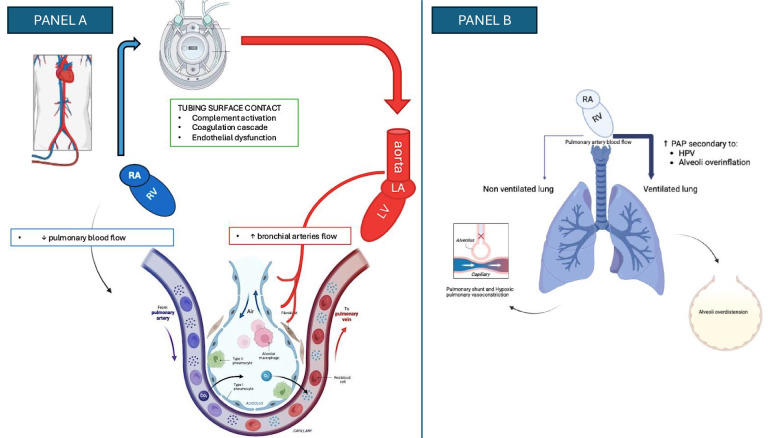 Figure 1: During one-lung ventilation the non-ventilated lung remains perfused, producing an intrapulmonary shunt; hypoxic pulmonary vasoconstriction diverts some of that flow to the ventilated lung but simultaneously raises right-ventricular afterload, so the whole cardiac output is delivered to a single lung. (Source: Montisci A, Maj G, La Via L, et al. Journal of Anesthesia, Analgesia and Critical Care 2026. DOI: 10.1186/s44158-026-00387-7).
One-Lung Ventilation: Indications and Devices
Indications
Lung isolation is indicated whenever ventilation must be directed to one lung or withheld from the other. In thoracic surgery — lobectomy, pneumonectomy, oesophagectomy, thoracoscopic and anterior spinal procedures — deflation of the operative lung creates the surgical working space and prevents retraction injury to ventilated parenchyma. Isolation also prevents cross-contamination of a healthy lung by pus, blood or secretions, and is essential in massive haemoptysis, where flooding of the dependent lung is rapidly fatal. A fourth indication is severe unilateral lung disease, in which the diseased lung consumes ventilation while contributing little gas exchange.
Lung isolation is used for (1) surgical exposure of the lung, mediastinum, oesophagus or spine; (2) prevention of cross-contamination of a healthy lung; (3) massive haemoptysis or unilateral pulmonary haemorrhage; and (4) severe unilateral lung disease that contributes little gas exchange.
Double-Lumen Tubes
The double-lumen tube (DLT) is the default device for adult thoracic surgery. Two lumens and two cuffs divide the airway: a tracheal lumen ending above the carina and a bronchial lumen that passes into a main-stem bronchus. Inflating or deflating the bronchial cuff and clamping the appropriate lumen converts two-lung ventilation to OLV within seconds. Left-sided tubes are preferred because the left main bronchus is long enough to seat the bronchial cuff without occluding the right upper-lobe orifice, which lies close to the carina. A right-sided DLT is reserved for lesions that prevent left main-stem intubation, such as a left tracheobronchial tumour, and its bronchial cuff carries a port to ventilate the right upper lobe.
Position is never assumed from insertion depth. Auscultation is unreliable once the chest is open and the patient lateral, so fibreoptic bronchoscopy should confirm the bronchial cuff just below the carina, the tracheal lumen above it, and a patent right upper-lobe orifice.
Bronchial Blockers
An endobronchial blocker is a balloon-tipped catheter advanced through or beside a single-lumen tube and inflated in the target bronchus. Blockers suit a small airway, an anticipated difficult intubation, a patient already intubated in intensive care, or a child. Their limitations are slower lung collapse, because the blocked lung deflates only through collateral channels; restricted suction and isolation; and a tendency to migrate when the patient is repositioned or positive pressure is lost.
Choosing the Device
Selection reflects the airway, the procedure and the urgency. A DLT offers the fastest and most reliable collapse, separate suction and separate lung ventilation, whereas a blocker is gentler on the airway and works through a conventional tube. Difficult airways, brief procedures and incomplete collapse favour a blocker; pneumonectomy or absolute isolation favours a DLT.
The Pathophysiology of the Open Chest and the Non-Ventilated Lung
The Open Chest
Opening the pleura abolishes the negative intrapleural pressure that holds the lung against the chest wall, and the operative lung collapses to its hilum by elastic recoil. If the chest wall is destabilised, a flail segment may move paradoxically. Positive pressure restores tidal volume, but delivers it through a chest of reduced mechanical integrity: dependent regions are compressed by the mediastinum and abdominal contents, while non-dependent regions receive most of the tidal volume and are exposed to over-distension.
Shunt Through the Non-Ventilated Lung
The defining problem of OLV is shunt, not hypoventilation. The non-ventilated lung keeps receiving blood flow, but its alveoli contain no fresh gas, so that blood is not oxygenated; mixing it with blood from the ventilated lung lowers arterial oxygen content. The shunt fraction is:
$$\frac{\dot{Q}_s}{\dot{Q}_t} = \frac{C_{cO_2} - C_{aO_2}}{C_{cO_2} - C_{vO_2}}$$where $\dot{Q}_s/\dot{Q}_t$ is the shunt fraction (dimensionless, expressed as a percentage), $\dot{Q}_t$ is total cardiac output ($\text{L}\cdot\text{min}^{-1}$), $C_{cO_2}$ is end-capillary oxygen content ($\text{mL O}_2\cdot\text{dL}^{-1}$), $C_{aO_2}$ arterial oxygen content and $C_{vO_2}$ mixed-venous oxygen content. Because the shunt is a true right-to-left pathway, raising $F_iO_2$ corrects it poorly: blood that bypasses ventilation never sees the extra oxygen. Shunt fractions of $20$–$30\%$ are common during OLV, and frank hypoxaemia appears when they are higher.
Hypoxic Pulmonary Vasoconstriction
The compensation is hypoxic pulmonary vasoconstriction (HPV): small pulmonary arteries constrict in response to low alveolar oxygen tension, diverting blood away from poorly ventilated regions. During OLV this reduces perfusion of the non-ventilated lung and improves matching in the ventilated lung. Its efficiency is expressed as the fraction of baseline perfusion diverted:
$$\dot{Q}_{NVL}^{HPV} = \dot{Q}_{NVL}\,(1 - E_{HPV})$$where $\dot{Q}_{NVL}$ is baseline perfusion of the non-ventilated lung ($\text{L}\cdot\text{min}^{-1}$), $\dot{Q}_{NVL}^{HPV}$ its perfusion after vasoconstriction, and $E_{HPV}$ the efficiency of HPV (dimensionless, typically $0.4$–$0.6$). HPV is powerful but finite — even at full efficiency a substantial fraction of cardiac output still crosses the non-ventilated lung — and the rise in pulmonary vascular resistance it creates loads the right ventricle.
What Blunts Hypoxic Pulmonary Vasoconstriction
HPV acts within minutes, peaks by about $15$–$30\ \text{min}$, and is easily weakened. Volatile anaesthetics depress it dose-dependently, whereas propofol-based total intravenous anaesthesia preserves it better. Hypoxia, hypercapnia and the resulting acidosis blunt it, as do vasodilators such as nitrates, calcium-channel blockers and dobutamine. High mixed-venous oxygen tension reduces the stimulus, and a high left-atrial pressure can mechanically divert blood back to the non-ventilated lung.
Two common reactions to desaturation worsen it. Raising $F_iO_2$ to $1.0$ blunts HPV through high mixed-venous oxygen tension while doing little for the shunted blood; indiscriminate PEEP can over-distend the ventilated lung, compress its capillaries and divert blood back to the non-ventilated side.
Ventilation Strategy During One-Lung Ventilation
Tidal Volume and Driving Pressure
The ventilated lung receives nearly the whole cardiac output and is at risk of volutrauma and hyperperfusion injury. Tidal volume is therefore $4$–$6\ \text{mL}/\text{kg}$ predicted body weight (PBW), lower than the $6$–$8\ \text{mL}/\text{kg}$ of two-lung ventilation. The pressure that actually distends alveoli is the driving pressure:
$$\Delta P = P_{plat} - P_{EEP_{tot}} = \frac{V_T}{C_{rs}}$$where $\Delta P$ is driving pressure ($\text{cmH}_2\text{O}$), $P_{plat}$ end-inspiratory plateau pressure ($\text{cmH}_2\text{O}$), $P_{EEP_{tot}}$ total end-expiratory pressure ($\text{cmH}_2\text{O}$), $V_T$ tidal volume ($\text{mL}$) and $C_{rs}$ respiratory-system compliance ($\text{mL}\cdot\text{cmH}_2\text{O}^{-1}$). Keeping $\Delta P$ below about $15\ \text{cmH}_2\text{O}$ is associated with fewer postoperative pulmonary complications and is more informative than plateau pressure alone, because it removes the PEEP contribution. A rising driving pressure should prompt a search for malposition, secretions, pneumothorax or atelectasis.
PEEP to the Dependent Lung
PEEP applied to the dependent lung counteracts compression atelectasis and reduces shunt through that lung. The response is biphasic: too little leaves dependent alveoli collapsed, whereas too much over-distends non-dependent alveoli, compresses their capillaries and diverts blood to the non-ventilated lung. PEEP is therefore titrated from about $5\ \text{cmH}_2\text{O}$ by oxygenation, compliance and haemodynamics.
Permissive Hypercapnia
Because a low tidal volume must clear the whole carbon-dioxide production, some hypercapnia is expected. Permissive hypercapnia accepts a pH of about $7.25$–$7.35$ in exchange for lower airway pressures. It is usually well tolerated, but it vasodilates the pulmonary circulation and can blunt HPV, so it is avoided with raised intracranial pressure, severe pulmonary hypertension or cardiac instability.
Improving Oxygenation
When saturation falls, correct the cause first: verify tube position and haemodynamics. Then apply continuous positive airway pressure (CPAP) of $1$–$5\ \text{cmH}_2\text{O}$ with oxygen to the non-ventilated lung, which oxygenates the shunted blood while leaving the lung mostly collapsed for the surgeon — often dramatically effective. Intermittent reinflation, coordinated with the surgeon, restores ventilation temporarily, and gentle recruitment of the dependent lung opens atelectatic units. Inhaled vasodilators and reduction of the volatile agent are later steps; extracorporeal support is reserved for refractory hypoxaemia.
Before increasing $F_iO_2$ or PEEP, ask the surgeon's permission and apply $1$–$5\ \text{cmH}_2\text{O}$ CPAP with oxygen to the non-ventilated lung. It treats the shunt at its source, without over-distending the ventilated lung.
Recommended Initial Settings
| Parameter | Initial setting during OLV | Physiological target or safety threshold |
|---|---|---|
| Tidal volume | $4$–$6\ \text{mL}/\text{kg}$ PBW | Scaled to height, never to actual weight |
| Respiratory rate | $12$–$16\ \text{min}^{-1}$ | Titrated to $pH$ and $PaCO_2$; accept permissive hypercapnia |
| PEEP (dependent lung) | $5\ \text{cmH}_2\text{O}$, titrated | Increase for atelectasis; avoid over-distension |
| Plateau pressure | $< 25$–$30\ \text{cmH}_2\text{O}$ | Interpret with driving pressure and compliance |
| Driving pressure | $< 15\ \text{cmH}_2\text{O}$ | The pressure that distends the single ventilated lung |
| $F_iO_2$ | Lowest maintaining $SpO_2$ $92$–$96\%$ | Avoid prolonged $1.0$; it blunts HPV |
| CPAP to non-ventilated lung | $1$–$5\ \text{cmH}_2\text{O}$ with oxygen | First-line rescue, coordinated with the surgeon |
| Recovery of two-lung ventilation | Re-expand and confirm with bronchoscopy | Reassess pressures after the lung is reopened |
Complications of One-Lung Ventilation
Hypoxaemia
Hypoxaemia is the commonest complication, arising from shunt through the non-ventilated lung compounded by atelectasis in the ventilated lung. It is most likely before HPV fully develops, in patients with poor baseline lung function, and when the ventilated lung is dependent in the lateral position.
Barotrauma and Ventilator-Induced Lung Injury
The single ventilated lung is vulnerable because the tidal volume distributes into half the usual volume. Over-distension presents as rising plateau and driving pressures, rising $PaCO_2$ from increased dead space, and haemodynamic compromise from high intrathoracic pressure.
Double-Lumen Tube Malposition
Malposition is a common and under-recognised cause of sudden deterioration. A cuff that migrates outwards ventilates both lungs and breaches isolation; one that advances too far occludes a lobar orifice and produces atelectasis; one that herniates over the carina obstructs both lumens. An abrupt rise in peak airway pressure with desaturation after repositioning or a change in retraction is the cardinal sign. Bronchoscopy is both diagnostic and therapeutic, and the tube is re-verified after every patient movement.
Post-Thoracotomy Respiratory Failure
Residual atelectasis, pain-related splinting, weak cough, fluid overload and loss of resected lung volume can precipitate respiratory failure in the first $48$–$72\ \text{h}$. Management prioritises analgesia, secretion clearance, physiotherapy, careful fluid balance and non-invasive support, with reintubation if gas exchange deteriorates.
Chest Trauma
Rib Fractures and Flail Chest
Rib fractures impair ventilation through pain: each breath is splinted, cough is suppressed, and atelectasis and pneumonia follow. A flail chest — fractures of several adjacent ribs in two places — creates a segment that moves paradoxically. The paradoxical motion matters less than the pulmonary contusion that almost always accompanies it; flail chest is a marker of a severely injured lung, and treatment is analgesia and ventilatory support rather than fixation of the segment.
Three or more rib fractures, or any fracture in an elderly patient, mandates aggressive analgesia and monitoring. The danger is not the bone but the retained secretions and pneumonia that follow splinting, so the plan is judged by cough and spirometry, not by the pain score alone.
Pulmonary Contusion
Pulmonary contusion is haemorrhage and oedema in the alveolar space, producing consolidation, reduced compliance and a shunt that worsens over $24$–$48\ \text{h}$. It is often underestimated initially, so a normal admission film after a high-energy mechanism does not exclude profound later hypoxaemia. Treatment is supportive: protective ventilation, secretion clearance and avoidance of fluid overload, because the injured capillary bed is volume-sensitive.
Pneumothorax and Haemothorax
Traumatic pneumothorax may enlarge rapidly under positive pressure if undrained. Haemothorax is dangerous for its volume and its mechanical effect, and is monitored by the initial drainage and the hourly rate. Both require thoracostomy; their tension and massive variants are emergencies.
Analgesia and Secretion Clearance
Effective analgesia is the single most important intervention after rib fracture, because it converts a splinted chest into one the patient can clear. A multimodal approach is used, with thoracic epidural or paravertebral blockade for multiple or bilateral fractures, paracetamol and non-steroidal agents where safe, and opioids sparingly to preserve respiratory drive.
Lung-Protective Ventilation of the Contused Lung
The contused lung is focally injured and non-homogeneous. It is ventilated with $6\ \text{mL}/\text{kg}$ PBW, a driving pressure below $15\ \text{cmH}_2\text{O}$ and titrated PEEP that recruits without over-distending healthy lung. Severe unilateral contusion occasionally warrants independent lung ventilation, a specialist decision.
Non-Invasive Support
Non-invasive ventilation and high-flow nasal oxygen help the cooperative trauma patient with hypoxaemia but no failing airway or haemodynamic instability, splinting the chest wall and recruiting alveoli. They do not replace analgesia and are unsafe in a tiring or obtunded patient, or with an untreated pneumothorax.
Emergency Presentations Requiring Immediate Action
Tension Pneumothorax
Tension pneumothorax is a clinical diagnosis that must not wait for imaging. Positive pressure accelerates it; severe distress, unilateral absent breath sounds, distended neck veins, hypotension and an enlarging hemithorax are the signs. Management is immediate needle decompression or finger thoracostomy in the fifth intercostal space, followed by a chest drain. In the ventilated patient, decompression must precede any further increase in airway pressure.
A chest radiograph must never delay decompression. After decompression, confirm placement, insert a definitive chest drain, and reassess the ventilator.
Massive Haemothorax
Massive haemothorax — more than about $1{,}500\ \text{mL}$ drained immediately, or more than $200\ \text{mL}\cdot\text{h}^{-1}$ for several hours — causes respiratory and haemorrhagic shock. Resuscitation with blood products and decompression with a large-bore drain proceed simultaneously, with surgical evaluation if bleeding continues. Clamping the drain is avoided because it may convert the injury into a tension haemothorax.
Open Pneumothorax
An open chest wound creates a pathway between the pleural space and the atmosphere; when the defect exceeds about two-thirds of the tracheal diameter, air moves preferentially through it. A three-sided occlusive dressing acts as a flutter valve, followed by a chest drain at a separate site and definitive closure. Complete occlusion of the wound can convert it into a tension pneumothorax.
Chest Trauma Priorities
| Priority | Intervention | Key threshold or caveat |
|---|---|---|
| 1. Airway | Secure and re-secure; assess for expanding neck haematoma | Recheck after every move |
| 2. Breathing | Decompress tension pneumothorax; drain haemothorax | Clinical diagnosis; do not delay for imaging |
| 3. Circulation | Blood products for massive haemothorax | Avoid clamping the drain |
| 4. Chest wall | Multimodal analgesia; regional block for multiple fractures | Judge by cough and spirometry |
| 5. Ventilation | $6\ \text{mL}/\text{kg}$ PBW, $\Delta P < 15\ \text{cmH}_2\text{O}$, titrated PEEP | Lung-protective in contusion |
| 6. Secretions | Physiotherapy, cough assistance, early mobilisation | Commonest cause of late deterioration |
Clinical Pathways
Desaturation During One-Lung Ventilation
flowchart TD
    A["Desaturation during one-lung ventilation"] --> B["Increase FiO2 to 1.0 and check the circuit for obstruction or disconnection"]
    B --> C["Emergency check: haemodynamics, pneumothorax, endobronchial cuff position"]
    C --> D{"Tube or drain problem?"}
    D -->|"Yes"| E["Re-verify the double-lumen tube with bronchoscopy; decompress a pneumothorax"]
    D -->|"No"| F["Optimise cardiac output and haemoglobin; reduce volatile agent or switch to TIVA"]
    F --> G{"SpO2 still below 90 percent?"}
    G -->|"No"| H["Continue protective one-lung ventilation; avoid prolonged FiO2 of 1.0"]
    G -->|"Yes"| I["CPAP 1-5 cmH2O with oxygen to the non-ventilated lung, coordinated with the surgeon"]
    I --> J{"Improved?"}
    J -->|"No"| K["Intermittent reinflation and gentle recruitment of the dependent lung"]
    K --> L{"Improved?"}
    L -->|"No"| M["Inhaled pulmonary vasodilator; consider extracorporeal support"]
    L -->|"Yes"| H
    J -->|"Yes"| H
    E --> H
Chest Trauma Management Pathway
flowchart TD
    A["Chest trauma with respiratory compromise"] --> B["Primary survey: airway, breathing, circulation"]
    B --> C{"Tension pneumothorax, massive haemothorax or open pneumothorax?"}
    C -->|"Yes"| D["Immediate decompression or three-sided dressing; chest drain; blood products"]
    C -->|"No"| E["Analgesia: thoracic epidural or paravertebral block for multiple rib fractures"]
    D --> E
    E --> F["Secretion clearance, physiotherapy and early mobilisation"]
    F --> G{"Hypoxaemia or respiratory failure?"}
    G -->|"Mild"| H["High-flow nasal oxygen or non-invasive ventilation if cooperative and stable"]
    G -->|"Severe or failing"| I["Intubate; lung-protective ventilation of the contused lung"]
    H --> J["Reassess gas exchange, cough and chest radiograph"]
    I --> J
    J --> K{"Deteriorating?"}
    K -->|"Yes"| L["Reassess for retained haemothorax, pneumonia, contusion progression or ARDS"]
    K -->|"No"| M["Wean support as analgesia and secretion clearance improve"]
Key Takeaways
- One-lung ventilation isolates one lung for surgical exposure, cross-contamination, massive haemoptysis or unilateral disease; left-sided double-lumen tubes are the default, with bronchial blockers for difficult airways and small tubes.
- The central problem is shunt through the still-perfused, non-ventilated lung, described by $\dot{Q}_s/\dot{Q}_t = (C_{cO_2} - C_{aO_2})/(C_{cO_2} - C_{vO_2})$, and poorly corrected by oxygen alone.
- Hypoxic pulmonary vasoconstriction diverts $40$–$60\%$ of non-ventilated lung perfusion but is blunted by volatile anaesthetics, hypoxia, hypercapnia and vasodilators, raising shunt and right-ventricular afterload.
- Ventilate the dependent lung with $4$–$6\ \text{mL}/\text{kg}$ PBW, titrated PEEP of about $5\ \text{cmH}_2\text{O}$, and a driving pressure $\Delta P = P_{plat} - P_{EEP_{tot}}$ below $15\ \text{cmH}_2\text{O}$, accepting some permissive hypercapnia.
- For desaturation, correct tube position and haemodynamics first, then apply $1$–$5\ \text{cmH}_2\text{O}$ CPAP to the non-ventilated lung and recruit the dependent lung before escalating $F_iO_2$ or PEEP.
- Chest trauma is dominated by pain, contusion and pleural air or blood; analgesia and secretion clearance matter as much as the ventilator, and the contused lung is ventilated protectively.
- Tension pneumothorax, massive haemothorax and open pneumothorax are immediate-action diagnoses: decompress on clinical grounds, drain with a large-bore tube without clamping, and never let imaging delay treatment.