Severe obesity — a body-mass index of $40\ \text{kg}\cdot\text{m}^{-2}$ or more — is not merely a comorbidity accompanying respiratory failure, but a primary mechanical disease of the respiratory system. Adipose tissue in the chest wall, mediastinum and abdomen loads the diaphragm, displaces it cephalad and compresses the lung from below, while the rib cage stiffens. The resulting physiology is restrictive: functional residual capacity contracts, peripheral airways close within the tidal range, and the pressure required to inflate the respiratory system climbs even when the lung parenchyma is intrinsically healthy. The same physiology follows kyphoscoliosis, neuromuscular weakness and fibrothorax.
The distinction between a stiff respiratory system and a stiff lung is the centre of this chapter. When a severely obese patient's airway pressures appear alarming, the reflex is to reduce tidal volume or withdraw support; often the opposite is correct, because much of the applied pressure is consumed by the chest wall and abdomen rather than by distending alveoli. The clinician's task is to partition the pressure and decide whether the lung is genuinely over-distended. Ventilation here is an exercise in pressure accounting, not pressure avoidance.
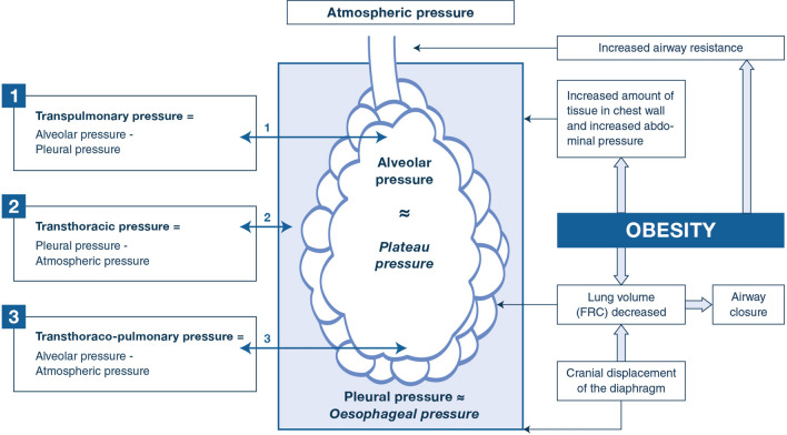 Figure 1: In obesity the applied airway pressure is shared between the lung (transpulmonary) and the chest wall and abdomen (transthoracic); a high plateau pressure commonly reflects an elevated transthoracic component rather than alveolar over-distension, which is why oesophageal manometry is needed to interpret it. (Source: De Jong A, Wrigge H, Hedenstierna G, et al. Intensive Care Med 2020. DOI: 10.1007/s00134-020-06286-x).
The Respiratory Physiology of Severe Obesity
Mass Loading of the Chest Wall and Abdomen
Adipose tissue is mechanically active, and the abdomen is its most consequential reservoir. Fat pushes the thin diaphragmatic dome upward, flattening it so that its fibres shorten onto an unfavourable part of the length–tension curve and its force falls; the rib cage is loaded from outside, so both the inspiratory pump and the container it acts upon are compromised. The elastic load is expressed as compliance:
$$C = \frac{\Delta V}{\Delta P}$$where $C$ is compliance ($\text{L}\cdot\text{cmH}_2\text{O}^{-1}$), $\Delta V$ the change in volume ($\text{L}$) and $\Delta P$ the change in distending pressure ($\text{cmH}_2\text{O}$). Lung and chest wall behave as two elastic structures in series:
$$\frac{1}{C_{rs}} = \frac{1}{C_L} + \frac{1}{C_{cw}}$$where $C_{rs}$, $C_L$ and $C_{cw}$ are respiratory-system, lung and chest-wall compliance respectively. In severe obesity $C_{cw}$ may halve while $C_L$ is comparatively preserved, so $C_{rs}$ is dominated by the abnormal chest wall.
Reduced Functional Residual Capacity and Airway Closure
Functional residual capacity (FRC) is the volume at which the outward recoil of the chest wall balances the inward recoil of the lung; both determinants are deranged in obesity, and FRC falls further when the patient lies supine. Expiratory reserve volume may approach zero. Closing capacity consequently exceeds FRC, so dependent airways collapse during quiet breathing and reopen only with a deep inspiration. The resulting atelectasis produces shunt, a widened alveolar–arterial gradient and hypoxaemia poorly responsive to inspired oxygen alone. It also explains the small oxygen reservoir available for preoxygenation, which is why the obese patient desaturates within a minute or two of apnoea.
Partitioned Compliance: a Stiff Chest Wall with a Relatively Preserved Lung
The essential point is that a high airway pressure in obesity is usually not a sign of lung injury. At end-inspiration the plateau pressure represents the pressure needed to distend the chest wall plus the lung. Because the chest wall and abdomen dominate the load, plateau pressure rises while transpulmonary pressure — the true distending stress on the alveoli — may remain modest. Pleural pressure, by contrast, is abnormally high, with haemodynamic as well as respiratory consequences.
Two patients may both have a plateau pressure of $32\ \text{cmH}_2\text{O}$. In ARDS this reflects a stiff, small lung at genuine risk of volutrauma; in severe obesity the same number may reflect an almost normal lung inside an extremely stiff container. The number alone cannot distinguish them; the partition of pressure can.
Intra-Abdominal Pressure and the Supine Position
Intra-abdominal pressure is frequently elevated to $12$–$20\ \text{mmHg}$ and rises further when the patient is flat. Transmitted through the diaphragm into the pleural space, it makes the supine position the single most adverse posture for the obese respiratory system: FRC is lowest, atelectasis maximal and pleural pressure highest. Any measure that unloads the abdomen — sitting the patient up or applying positive airway pressure — improves mechanics immediately.
Obesity Hypoventilation Syndrome and Obstructive Sleep Apnoea
Obesity hypoventilation syndrome (OHS) is defined by obesity, daytime hypercapnia with $PaCO_2 > 45\ \text{mmHg}$ and sleep-disordered breathing, after other causes of hypoventilation are excluded. It represents decompensation of a respiratory system that has exhausted its reserve. The mechanical load is compounded by central factors: leptin resistance blunts the ventilatory response to carbon dioxide, and chronic nocturnal hypoxaemia further depresses chemosensitivity. Obstructive sleep apnoea is present in most such patients, because pharyngeal fat narrows the upper airway and sleep-related loss of dilator tone allows it to collapse.
The overlap makes ventilation position-dependent. During rapid-eye-movement sleep, atonia combines with the supine posture — when FRC is lowest and the airway most collapsible — to produce the deepest desaturations and greatest carbon dioxide retention. Sleep is effectively a nightly mechanical stress test that the obese respiratory system repeatedly fails. Daytime ventilation may be adequate while awake and upright, yet reserve is minimal, so sedation, pneumonia, surgery or a supine transfer precipitates overt hypercapnic failure.
Sleeping posture is as physiologically significant as minute ventilation in this population. Avoiding the supine posture during sleep, and nursing the ventilated patient semi-recumbent or in reverse Trendelenburg, reduces the mechanical load measurably. In OHS the treatment target is correction of daytime hypercapnia, not merely nocturnal oxygenation.
Tidal Volume, Predicted Body Weight, and Airway Pressure
The Predicted Body Weight Formulas
Scaling tidal volume to actual body weight is the most dangerous error in this population. Adipose tissue consumes almost no oxygen and produces almost no carbon dioxide, so it has no legitimate claim on alveolar ventilation; the metabolically active lean mass is best estimated from height. Tidal volume is therefore referenced to predicted body weight (PBW):
$$PBW_{male} = 50 + 0.91 \times [\text{height (cm)} - 152.4]$$$$PBW_{female} = 45.5 + 0.91 \times [\text{height (cm)} - 152.4]$$where PBW is in kilograms and height in centimetres. A $170\ \text{cm}$ man has a PBW of about $66\ \text{kg}$; at $6\ \text{mL}/\text{kg}$ his tidal volume is about $400\ \text{mL}$, whereas indexing the same tidal volume to an actual weight of $150\ \text{kg}$ would give $900\ \text{mL}$.
There is no situation in which tidal volume should be indexed to actual body weight. In a $180\ \text{kg}$ patient the error generates breaths over a litre, causing volutrauma, dynamic hyperinflation and haemodynamic collapse. If tidal volumes "look small" for the size of the patient, that impression is the point: the lungs are the size their height dictates, not the size the scale suggests.
Interpreting High Airway Pressures
A raised peak pressure with a normal plateau pressure suggests a resistive or tube-related problem rather than alveolar distension. A raised plateau pressure requires the partition described above. Only when transpulmonary pressure is genuinely high should tidal volume fall below $6\ \text{mL}/\text{kg}$ PBW, and even then modestly and by measurement rather than reflex. Under-ventilating a patient whose lung is not injured merely exports the harm to carbon dioxide retention and atelectasis.
Counteracting the Chest-Wall Load: PEEP, Recruitment, and Oesophageal Manometry
The Transpulmonary Pressure Principle
PEEP plays a different role in obesity than in classical ARDS. It is not primarily a recruitment device for a diffusely injured lung but a counterweight to the external and abdominal load collapsing dependent airways: it holds the airway open through expiration, restores FRC and reduces atelectatic shunt. The quantity that matters is the transpulmonary pressure:
$$P_{tp} = P_{aw} - P_{pl}$$where $P_{tp}$ is transpulmonary pressure ($\text{cmH}_2\text{O}$), $P_{aw}$ airway opening pressure ($\text{cmH}_2\text{O}$) and $P_{pl}$ pleural pressure ($\text{cmH}_2\text{O}$). The related transthoracic pressure, $P_{cw} = P_{pl} - P_{atm}$, describes the chest-wall and abdominal load. Because pleural pressure is high, a given airway pressure yields a lower transpulmonary pressure than in a lean patient.
Oesophageal Manometry at the Bedside
When airway pressures appear alarming, oesophageal manometry resolves the ambiguity. A balloon in the lower oesophagus provides a surrogate for pleural pressure, allowing end-inspiratory and end-expiratory transpulmonary pressures to be estimated. The usual targets are an end-expiratory $P_{tp}$ that is non-negative, so that dependent airways do not collapse, and an end-inspiratory $P_{tp}$ below about $15$–$20\ \text{cmH}_2\text{O}$, above which alveolar over-distension becomes a concern. An obese patient with a plateau pressure of $35\ \text{cmH}_2\text{O}$ but an end-inspiratory transpulmonary pressure of $8\ \text{cmH}_2\text{O}$ is not sustaining lung injury; a patient with identical airway pressures and a transpulmonary pressure of $25\ \text{cmH}_2\text{O}$ is.
Recruitment manoeuvres can transiently open collapsed lung, but higher and repeated pressures carry haemodynamic cost and barotrauma risk in a chest already loaded by raised intra-abdominal pressure. Reserve them for selected, haemodynamically stable patients, deliver them briefly, and confirm benefit by compliance or gas exchange rather than pressure alone.
Practical Measures: Positioning, Interface, and Non-Invasive Ventilation
Positioning
The ramped or reverse-Trendelenburg position is the single most useful bedside intervention. Elevating the trunk $30$–$45^\circ$ lets the abdominal contents fall away from the diaphragm, increasing FRC, reducing atelectasis and lowering pleural pressure. The same posture should be used for preoxygenation and intubation: ear-to-sternal-notch alignment improves the laryngoscopic view and prolongs safe apnoea time. The flat supine position should be regarded as actively harmful and avoided unless a procedure demands it.
Interface and Mode
Non-invasive ventilation is the preferred support for OHS and for many obese patients with acute hypercapnic failure, and its success depends on the interface. An oronasal mask is usually required because obese patients often breathe through the mouth; a helmet may be considered when mask intolerance or high pressure requirements limit conventional NIV. A pressure-support mode is used with an adequate expiratory positive airway pressure, often $8$–$12\ \text{cmH}_2\text{O}$, to stent the upper airway and counteract the abdominal load. In OHS a back-up rate is essential, because the defect is a loss of central drive; without a guaranteed minimum rate the patient may simply hypoventilate when asleep or fatigued.
Ventilator Settings and Physiological Targets
| Parameter | Initial setting | Physiological target or safety threshold |
|---|---|---|
| Tidal volume | $6\ \text{mL}/\text{kg}$ PBW | Never indexed to actual weight; accept $6$–$8$ if $P_{tp}$ is low |
| Respiratory rate | $14$–$20\ \text{min}^{-1}$ | Titrated to pH and $PaCO_2$; avoid hypoventilation in OHS |
| PEEP / EPAP | $8$–$12\ \text{cmH}_2\text{O}$ (up to $15$–$20$ if tolerated) | Keep end-expiratory $P_{tp} \geq 0$; monitor haemodynamics |
| Peak / plateau pressure | Interpret, do not target blindly | Plateau $> 30\ \text{cmH}_2\text{O}$ prompts oesophageal manometry |
| End-inspiratory $P_{tp}$ | $< 15$–$20\ \text{cmH}_2\text{O}$ | The true index of alveolar over-distension |
| Driving pressure | $< 15\ \text{cmH}_2\text{O}$ where measurable | Read with the transpulmonary caveat above |
| $SpO_2$ | $92$–$96\%$ | Higher targets increase absorption atelectasis |
| Position | Reverse Trendelenburg $30$–$45^\circ$ | Never nurse flat supine |
| Recruitment | Selected, stable patients only | Confirm benefit by compliance or gas exchange |
Before escalating PEEP or accepting a high plateau pressure, sit the patient up. A $30$–$45^\circ$ reverse-Trendelenburg tilt frequently improves compliance and oxygenation within minutes and lowers the pressures required, at no cost and no risk.
Chest-Wall Restriction from Other Causes
Kyphoscoliosis
Severe kyphoscoliosis distorts the rib cage so that its compliance is markedly reduced and its geometry inefficient. The physiology mirrors obesity: high pleural pressure, a stiff respiratory system with a relatively preserved lung, and higher inflation pressures to achieve the same transpulmonary pressure. Diurnal hypercapnia supervenes as the load outlasts the inspiratory muscles, and nocturnal hypoventilation is the earliest manifestation. Nocturnal non-invasive ventilation is the mainstay and improves both symptoms and survival.
Neuromuscular Disease and Cough Assistance
Neuromuscular disease restricts the chest wall indirectly by weakening the respiratory pump. Vital capacity and cough flows fall, atelectasis accumulates and secretions cannot be cleared. Ventilatory support is usually non-invasive and often needed at night before it is needed by day. Cough assistance is a parallel priority: mechanical insufflation–exsufflation augments peak cough flow, and assisted coughing should be taught before an exacerbation makes secretion clearance impossible. Invasive ventilation is reserved for acute failure or an unprotected airway.
Fibrothorax and Pleural Restriction
Fibrothorax, whether post-infective, post-traumatic or post-surgical, encases the lung in a rigid pleural peel. Lung volumes fall and positive-pressure ventilation must overcome both the peel and any residual parenchymal disease. As in the other restrictive conditions, higher airway pressures are often required and must be interpreted against transpulmonary rather than airway pressure alone. Definitive treatment is surgical decortication where feasible; ventilation is supportive in the interim.
- Do not extubate a patient with a weak cough and vital capacity below about $15\ \text{mL}/\text{kg}$ without a plan for non-invasive support and secretion clearance.
- Start nocturnal NIV early in chronic restrictive hypercapnia, before daytime failure declares itself.
- Teach and apply mechanical insufflation–exsufflation before secretions become unmanageable.
- Treat reversible components — pleural collection, bronchospasm, infection — promptly.
- Reassess with oesophageal manometry when airway pressures and clinical response diverge.
Sedation, Positioning Hazards, and the Timing of Tracheostomy
Sedation and Induction
The obese patient has a small oxygen reservoir and a high closing capacity, so the interval between apnoea and critical desaturation is short. Preoxygenation should be performed in the semi-recumbent or ramped position with non-invasive ventilation, which recruits collapsed lung far better than a face mask, and apnoeic oxygenation may extend the safe period. Induction doses should be based on lean or predicted body weight, since drug distribution in adipose tissue differs greatly; opioid and sedative sensitivity is increased in OSA, and residual sedation after extubation is a common cause of reintubation.
Positioning Hazards
Positioning is a therapy but also a hazard. The ramped and reverse-Trendelenburg positions require vigilance for skin shear and for the patient sliding down the bed. When prone positioning is used for severe ARDS in obesity, chest and pelvic supports must let the abdomen hang free rather than compressing it, which would raise intra-abdominal and pleural pressures further. Lines, tubes and the airway must be secured before any turn, with enough staff to move the patient safely.
Early Tracheostomy
Prolonged ventilation is common in severe obesity and restrictive chest-wall disease because weaning is slow: the load is fixed, the muscles are deconditioned and the work of breathing stays high. When liberation has not occurred by roughly one to two weeks, tracheostomy should be considered rather than deferred indefinitely. It reduces dead space and airway resistance, facilitates secretion clearance, and allows non-invasive support to be resumed during the day once invasive ventilation is needed only at night.
A Ventilation Strategy for Obesity and Chest-Wall Restriction
The algorithm integrates positioning, predicted-body-weight tidal volume, PEEP titration, oesophageal manometry and non-invasive support; each step follows optimisation of the last.
flowchart TD
    A["Severe obesity (BMI >= 40 kg/m2) or chest-wall restriction"] --> B["Calculate predicted body weight from height; set tidal volume 6-8 mL/kg PBW"]
    B --> C["Position: reverse Trendelenburg 30-45 degrees; never flat supine"]
    C --> D{"Awake, cooperative, no NIV contraindication?"}
    D -->|"Yes"| E["Non-invasive ventilation: pressure support with EPAP 8-12 cmH2O; add back-up rate in obesity hypoventilation syndrome"]
    D -->|"No"| F["Intubate in the ramped position; preoxygenate with NIV; consider apnoeic oxygenation"]
    F --> G["PEEP 8-15 cmH2O titrated to best compliance and haemodynamic tolerance"]
    G --> H{"Plateau pressure > 30 cmH2O?"}
    H -->|"No"| K["Maintain lung-protective settings; reassess daily"]
    H -->|"Yes"| I["Oesophageal manometry: estimate pleural pressure and compute Ptp = Paw - Ppl"]
    I --> J{"End-inspiratory transpulmonary pressure high?"}
    J -->|"No"| K
    J -->|"Yes"| L["Reduce tidal volume toward 6 mL/kg PBW; treat atelectasis; consider selected recruitment"]
    K --> M{"Persistent hypercapnia or failure to wean beyond 1-2 weeks?"}
    M -->|"Yes"| N["Optimise position and interface; consider early tracheostomy; resume NIV when possible"]
    M -->|"No"| O["Continue support; assess liberation and extubation readiness daily"]
Key Takeaways
- Severe obesity is a primary mechanical disease of the respiratory system: adipose mass loads the chest wall and abdomen, reduces functional residual capacity and expiratory reserve volume, promotes dependent atelectasis, and raises intra-abdominal pressure, especially supine.
- Respiratory-system compliance falls while lung compliance is relatively preserved, so airway and plateau pressures are dominated by the transthoracic (chest-wall and abdominal) load rather than by alveolar distension.
- Tidal volume must always be scaled to predicted body weight, derived from height, and never to actual body weight; a $170\ \text{cm}$ man receives about $400\ \text{mL}$ at $6\ \text{mL}/\text{kg}$ PBW regardless of his weight on the scale.
- PEEP counteracts the abdominal and chest-wall load, restores end-expiratory lung volume and reduces atelectatic shunt; end-expiratory transpulmonary pressure should be kept non-negative, typically with $8$–$12\ \text{cmH}_2\text{O}$ and sometimes more.
- Oesophageal manometry converts an alarming airway pressure into an interpretable one: $P_{tp} = P_{aw} - P_{pl}$, with end-inspiratory $P_{tp}$ held below about $15$–$20\ \text{cmH}_2\text{O}$ to avoid over-distension.
- Positioning is therapy: the ramped or reverse-Trendelenburg posture unloads the diaphragm, and the supine position should be avoided; pressure-support non-invasive ventilation with adequate EPAP and, in obesity hypoventilation syndrome, a back-up rate is the preferred initial support.
- Kyphoscoliosis, neuromuscular disease and fibrothorax share the same restrictive physiology of high pleural pressure and high inflation pressure; non-invasive ventilation, cough assistance and early consideration of tracheostomy are the pillars of management.