Prone positioning — rotating a mechanically ventilated patient to the ventral decubitus position — is the only adjunct to lung-protective ventilation that has reproducibly reduced mortality in moderate-to-severe acute respiratory distress syndrome (ARDS). Computed tomography long ago showed that ARDS injury concentrates in the dependent dorsal lung, yet for two decades proning improved oxygenation without a survival signal. The PROSEVA trial (2013) resolved that ambiguity: early, prolonged, protocolised proning in severe ARDS cut 28-day mortality from 32.8% to 16.0%, by combining rigorous selection, early initiation, long daily duration, and disciplined team execution.
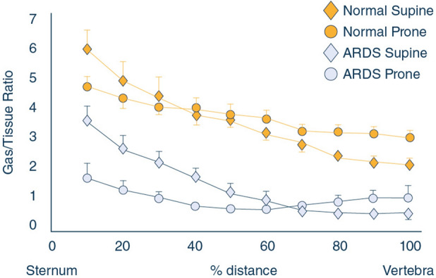 Figure 1: The gas/tissue ratio falls steeply from sternum to vertebrae when supine but becomes nearly uniform when prone, so the same airway pressure produces far more even regional distension. (Source: Guérin C, Albert RK, Beitler J, et al. Intensive Care Medicine 2020;46(12):2385–2396. DOI: 10.1007/s00134-020-06306-w).
The Transpulmonary Pressure Gradient and Its Reversal
The supine dorsal–ventral gradient
In the supine patient, gravity, the abdominal contents, and the mass of the heart impose a vertical gradient of pleural pressure ($P_{pl}$). Pleural pressure is least negative — most positive — in the dependent dorsal regions near the diaphragm and mediastinum, and most negative beneath the sternum. Because the pleural space transmits the weight of the thoracic contents hydrostatically, the gradient approximates $\partial P_{pl}/\partial h \approx \rho g$, where $\rho$ is tissue density ($\approx 1\text{ g/mL}$) and $g$ gravitational acceleration; clinically this is roughly $1\text{ cmH}_2\text{O}$ per centimetre of height, and steeper beneath the heart, whose weight compresses the adjacent dorsal lung.
The pressure distending any alveolar unit is the transpulmonary pressure:
$$P_{tp} = P_{aw} - P_{pl}$$where $P_{tp}$, $P_{aw}$, and $P_{pl}$ are the transpulmonary, airway, and pleural pressures in $\text{cmH}_2\text{O}$ ($P_{pl}$ estimated by oesophageal balloon manometry). At end-inspiration $P_{aw}$ is essentially uniform throughout the airway tree, so regional differences in $P_{pl}$ pass directly into $P_{tp}$: dorsal units are under-distended and collapse, while ventral units are over-distended.
Redistribution of transpulmonary pressure and regional compliance
The mechanical consequence follows from regional compliance:
$$C_{reg} = \frac{\Delta V_{reg}}{\Delta P_{tp,reg}}$$where $C_{reg}$ is regional compliance ($\text{mL/cmH}_2\text{O}$), $\Delta V_{reg}$ the regional tidal volume change (mL), and $\Delta P_{tp,reg}$ the regional transpulmonary pressure change ($\text{cmH}_2\text{O}$). Dorsal units with a high $P_{pl}$ sit on the flat portion of their pressure–volume curve, while ventral units approach the upper inflection where compliance falls. Ventilation therefore concentrates in a small non-dependent fraction — the "baby lung" — while the dorsal majority is repeatedly recruited and derecruited, generating atelectrauma and biotrauma.
Proning reverses this geometry. The dorsal lung becomes non-dependent, its $P_{pl}$ falls and its $P_{tp}$ rises; the ventral lung becomes dependent but is protected by the rigid sternum and the small anterior mediastinal mass. The heart rests against the sternum and the abdominal contents fall ventrally, relieving the dorsal compression that dominated supine. The distribution of $P_{tp}$ becomes far more homogeneous, as Figure 1 shows: the steep supine gas/tissue gradient flattens when prone.
The entire mechanical effect is captured by $P_{tp} = P_{aw} - P_{pl}$: the ventilator sets $P_{aw}$, but position-dependent $P_{pl}$ determines which alveoli are actually distended. Proning does not raise $P_{aw}$; it redistributes $P_{pl}$ so that the same $P_{aw}$ becomes a more uniform distending force.
Ventilation–perfusion matching, shunt and right ventricular unloading
Improved aeration brings improved perfusion matching. Hypoxic pulmonary vasoconstriction diverts blood from non-aerated dorsal units, but incompletely, so a substantial shunt persists; when those units open in prone they become available for ventilation and perfusion, and shunt falls. Oxygenation often rises within the first hour, although the mortality benefit does not depend on it — non-responders still benefit. Proning also unloads the right ventricle: recruitment lowers pulmonary vascular resistance and afterload, and relieving the heart of the dorsal lung's weight improves filling. Echocardiography during prolonged proning shows regression of right ventricular dilatation and septal normalisation.
The survival advantage is not contingent on the oxygenation response: recruitment, reduced shunt, and right ventricular unloading occur independently of the $PaO_2$ trajectory.
Beyond Oxygenation: Non-Respiratory Mechanisms
Secretion drainage and airway clearance
Secretion pooling is a constant feature of the supine ventilated patient. Rotation to prone converts the dorsal airways from the most dependent to the least dependent segment, so retained secretions drain centrally where suction clears them. This postural drainage effect is most striking when secretions are abundant, making rigorous pre- and post-turn airway care integral to the protocol.
Cardiac and abdominal decompression
Two masses compress the supine dorsal lung: the heart, lying over the left lower lobe, and the abdominal contents, which displace the diaphragm cranially, particularly in obesity or distension. Proning relocates the heart against the sternum and lets the abdomen fall away from the diaphragm. This explains why proning helps even when global compliance is unchanged, and why patients with raised intra-abdominal pressure gain disproportionately.
Computed tomographic evidence of aeration homogeneity
Quantitative CT in supine ARDS shows well-aerated non-dependent tissue, a transitional zone, and a large non-aerated dependent fraction. After a prone session, repeat CT shows a substantial reduction in non-aerated tissue and more even aeration along the ventral–dorsal axis. The reduction in non-aerated lung exceeds any increase in overdistension, so proning recruits lung rather than merely redistributing tidal volume.
The Evidence Base
PROSEVA: design and mortality results
PROSEVA was a multicentre randomised trial of 466 patients with severe ARDS. Eligibility required a $P/F$ below $150\text{ mmHg}$, $FiO_2 \ge 0.6$, $PEEP \ge 5\text{ cmH}_2\text{O}$, and tidal volume near $6\text{ mL/kg}$ predicted body weight; patients were assigned to prone sessions of at least 16 hours or to remain supine. Mortality at 28 days was $16.0\%$ versus $32.8\%$ ($P<0.001$; hazard ratio $0.39$, $95\%$ CI $0.25$–$0.63$), and 90-day mortality was $23.6\%$ versus $41.0\%$ ($P<0.001$; hazard ratio $0.44$, $95\%$ CI $0.29$–$0.67$). Complications were similar between groups except that cardiac arrest was more frequent supine — consistent with the haemodynamic and right ventricular benefits above.
Why earlier trials were negative
Four differences distinguish PROSEVA from the negative trials that preceded it:
- Population. Earlier trials enrolled heterogeneous hypoxaemic patients, many without moderate-to-severe ARDS; PROSEVA restricted enrolment to a $P/F$ below $150\text{ mmHg}$, where the recruitable dorsal compartment is large and the potential benefit greatest.
- Duration. Sessions typically lasted 6–8 hours, insufficient to sustain recruitment; PROSEVA mandated at least 16 consecutive hours daily.
- Timing. Proning began within roughly the first 36 hours of ARDS and mechanical ventilation, before fibroproliferation rendered the dorsal lung irreversibly consolidated.
- Background ventilation. Every PROSEVA patient received tidal volumes near $6\text{ mL/kg}$ predicted body weight, with deep sedation or neuromuscular blockade to suppress dyssynchrony during turns; earlier trials using 10–12 mL/kg may have masked any benefit.
The trial did not discover a new mechanism — it validated a protocol. Selection ($P/F<150$), duration ($\ge 16\text{ h}$), timing (early), and background lung-protective ventilation were each necessary, and no earlier trial combined all four.
Meta-analyses, guidelines and the COVID-19 experience
Meta-analyses pooling the later, longer-duration trials confirmed a mortality benefit concentrated in patients proned beyond 12 hours daily, with no consistent benefit from shorter sessions. Guidelines from the American Thoracic Society, the European Society of Intensive Care Medicine, and the Society of Critical Care Medicine therefore recommend prone positioning in moderate-to-severe ARDS, strongly for sessions exceeding 12 hours daily.
The COVID-19 pandemic provided the largest real-world test: observational cohorts of invasively ventilated patients reported improved oxygenation and respiratory mechanics with survival associations consistent with PROSEVA. Practice extended in two directions — many centres prolonged sessions beyond 24 hours when oxygenation failed to improve on return to supine, although randomised evidence remains limited, and awake proning of non-intubated patients reduced intubation in meta-trials without a clear mortality benefit. The pandemic also exposed implementation failure, with proning markedly underused despite unambiguous evidence, prompting quality-improvement programmes focused on team training and protocol standardisation.
The Prone Positioning Protocol
Initiation, duration and monitoring
Proning should be considered as soon as the patient meets oxygenation criteria on lung-protective settings, and started early rather than reserved as a last resort. The target is at least 16 consecutive hours daily — in practice one prolonged overnight session, not several short turns. Duration is cumulative, and a session should not be stopped merely because oxygenation improves transiently, since much of the benefit accrues from sustained recruitment and rest of the dorsal lung. Eligibility is reconfirmed before every session because ARDS severity evolves.
| Criterion | Threshold / Requirement | Notes |
|---|---|---|
| Oxygenation | $P/F$ ratio $< 150\text{ mmHg}$ | Assess on $PEEP \ge 5\text{ cmH}_2\text{O}$ and $FiO_2 \ge 0.6$ |
| Ventilator setting | $FiO_2 \ge 0.6$, $PEEP \ge 5\text{ cmH}_2\text{O}$ | Lung-protective ventilation, $V_T \approx 6\text{ mL/kg}$ PBW |
| Timing | Within roughly the first 36 hours | Before fibroproliferation |
| Daily duration | $\ge 16$ consecutive hours | One prolonged session preferred |
| Sedation | Deep sedation or neuromuscular blockade | Suppresses dyssynchrony during turns |
| Haemodynamics | Stable on modest vasopressor support | Reassess after every turn |
| Airway | Secure tube, documented depth | Reconfirm depth and cuff pressure each turn |
Continuation and stopping rules
Sessions repeat daily until sustained improvement. The conventional stopping rule is a $P/F$ of at least $150\text{ mmHg}$ sustained for four hours supine, on $PEEP \le 10\text{ cmH}_2\text{O}$ and $FiO_2 \le 0.6$. Improvement is judged over a full supine interval, not a single blood gas, and a patient who deteriorates rapidly on return to supine should be re-proned without delay. Failure to respond to a correctly executed 16-hour session should prompt reassessment of the diagnosis and of rescue options such as extracorporeal membrane oxygenation.
flowchart TD
    A["Meet eligibility criteria (P/F < 150, FiO2 >= 0.6, PEEP >= 5 cmH2O)"] --> B["Confirm no contraindication"]
    B --> C["Assemble team of 4-5 trained staff"]
    C --> D["Pre-turn checklist: secure ETT and lines, empty stomach, preoxygenate"]
    D --> E["Log-roll turn to prone position"]
    E --> F["Confirm ETT depth, cuff pressure, line patency, haemodynamics"]
    F --> G["Position head, arms and pressure-relieving padding"]
    G --> H["Maintain prone for >= 16 consecutive hours"]
    H --> I{"Sustained improvement supine (P/F >= 150 on PEEP <= 10, FiO2 <= 0.6)?"}
    I -->|"No"| H
    I -->|"Yes"| J["Stop daily proning and continue lung-protective ventilation"]
Practical Execution and Team Safety
Team composition and roles
Prone positioning is a coordinated procedure whose safety depends on staffing and clearly assigned roles. A minimum of four to five trained staff is required. The airway physician leads, stands at the head, controls the head and endotracheal tube throughout, and gives the verbal commands; this role is never delegated to an inexperienced operator. Two to three nurses, one on each side, control the trunk, limbs, drains, and intravascular catheters. A respiratory therapist manages the ventilator circuit, preventing traction and disconnection. A runner retrieves padding, dressings, and suction so no team member leaves the bedside mid-procedure.
The log-roll turn
The patient is positioned supine with arms at the sides and all leads, catheters, and drains reorganised so nothing crosses beneath the body; circuit tubing is routed over the head of the bed with slack. The team then log-rolls the patient: to the edge of the bed away from the ventilator, onto one side while the airway physician stabilises the head and tube, then fully onto the front. Once prone, the patient is centred and the arms placed in the "swimming" or "superman" position — one arm abducted overhead, the other alongside — with a pillow or gel roll supporting chest and pelvis to reduce abdominal compression. The head rests turned to one side on a soft cradle. Arms and head alternate every one to two hours to prevent brachial plexus stretch and unilateral facial pressure injury.
Assign one person solely to the endotracheal tube and one solely to the ventilator circuit. The commonest catastrophic complications — tube displacement and circuit disconnection — arise when a single operator tries to manage both.
Securing the airway, lines and tubes
Before the turn, preoxygenate with $FiO_2$ of $1.0$ for five minutes, confirm and record tube depth and cuff pressure, suction the airway, empty the gastric tube, verify that every infusion line is labelled, has adequate slack, and will remain on top of the patient, and cap all stopcocks. During the turn, keep the head neutral, avoid tube traction, and support the circuit; if disconnection or displacement occurs, revert to supine and re-secure the airway before proceeding. After the turn, reconfirm depth and cuff pressure, recheck line patency, obtain a blood gas, and suction again, because prone posture mobilises previously pooled dorsal secretions.
Head, arm and pressure-point care
Facial pressure ulcers and oedema are the archetypal complications of prolonged proning. The face, anterior shoulders, chest, iliac crests, knees, and toes bear the body's weight and need pressure-redistributing foam or gel supports. Eyes are protected with lubricant and closure to prevent corneal abrasion and chemosis, and ears must not be folded beneath the head. Pressure points are inspected and relieved at least every two hours, in synchrony with arm and head rotation. Facial oedema usually resolves within hours of returning to supine but can obstruct the airway after extubation, so it is anticipated at liberation.
Enteral nutrition
Enteral nutrition is continued during prone sessions and need not be routinely withheld: feeding prone is well tolerated and gastric residual volumes are not systematically increased, so continuing preserves caloric delivery over a prolonged course. Practical measures include prokinetics, head-of-bed elevation or reverse-Trendelenburg tilt where tolerated, and pausing the infusion around the rotation itself to reduce regurgitation and aspiration. Feeding resumes once the patient is stable and tube position is confirmed.
Contraindications, Cautions and Complications
Absolute and relative contraindications
Contraindications fall into two groups: conditions in which rotation or ventral pressure would be immediately dangerous, and conditions in which the risk outweighs the expected benefit.
Spinal instability, unstable pelvic or long-bone fractures, open abdomen or recent major abdominal or sternal surgery, raised intracranial pressure without adequate monitoring, life-threatening arrhythmia, and haemodynamic instability requiring escalating vasopressor support are contraindications to elective prone positioning. A relative contraindication requires an individualised decision by the senior intensivist.
| Category | Contraindication or Caution | Rationale |
|---|---|---|
| Spine / skeleton | Unstable spinal injury; unstable pelvic, rib, or long-bone fractures | Cord injury or fracture displacement during the log-roll |
| Central nervous system | Raised intracranial pressure without invasive monitoring | Dorsal venous congestion raises intracranial pressure further |
| Abdomen / thorax | Open abdomen; recent abdominal surgery; recent sternotomy | Wound dehiscence, evisceration, or sternal instability |
| Cardiovascular | Life-threatening arrhythmia; haemodynamic instability | Rotation provokes transient hypotension and arrhythmia |
| Airway | Difficult airway; facial trauma; recent tracheal surgery | Tube displacement and inability to rescue the airway |
| Other | Massive haemoptysis; pregnancy (relative); body size beyond bed capacity | Aspiration; compression; inability to position safely |
Complications and their prevention
Most complications are preventable, and their incidence falls with experience and protocol adherence.
- Pressure ulcers. The commonest complication, affecting face, chest, iliac crests, knees, and toes; prevented by pressure-redistributing surfaces, systematic padding, and two-hourly relief of pressure points.
- Facial and periorbital oedema. From dependent venous congestion; self-limiting, with eye care preventing corneal injury and anticipation of airway swelling guiding extubation timing.
- Endotracheal tube displacement or obstruction. Prevented by a dedicated airway operator, secure fixation, pre- and post-turn depth verification, and closed suction.
- Accidental removal of catheters and drains. Prevented by securing and labelling every line with adequate slack on the patient's upper surface.
- Aspiration. Reduced by emptying the stomach, pausing feed during rotation, prokinetics, and prompt oropharyngeal suctioning.
- Turn-related haemodynamic instability. Anticipated by optimising volume status and vasopressors and avoiding immediate re-proning of a patient still unstable after the turn.
- Rare complications. Brachial plexus injury, arm venous congestion, retinal ischaemia, and temporomandibular joint injury — minimised by alternating arm and head position every one to two hours.
flowchart TD
    subgraph Pre["PRE-TURN SAFETY CHECKLIST"]
        P1["Confirm eligibility and absence of contraindications"]
        P2["Preoxygenate FiO2 1.0 for 5 minutes and suction airway"]
        P3["Verify ETT depth and cuff pressure"]
        P4["Label and secure all lines, catheters and drains"]
        P5["Empty stomach and pause enteral feed"]
        P6["Assemble team and assign roles verbally"]
    end
    subgraph Turn["TURN EXECUTION"]
        T1["Airway operator controls head and ETT"]
        T2["Coordinated log-roll onto the front"]
        T3["Centre patient and support chest and pelvis"]
        T4["Position head and arms in swimming position"]
    end
    subgraph Post["POST-TURN VERIFICATION"]
        V1["Recheck ETT depth, cuff pressure and ventilation"]
        V2["Recheck line patency and haemodynamics"]
        V3["Blood gas and secretion clearance"]
        V4["Inspect and pad all pressure points"]
    end
    Pre --> Turn --> Post
Abort the session and return the patient to supine for endotracheal tube displacement, ventilator circuit disconnection that cannot be rapidly corrected, refractory haemodynamic collapse, or a new life-threatening arrhythmia. Re-secure the airway before considering re-proning.
Key Takeaways
- Prone positioning reduces mortality in moderate-to-severe ARDS by redistributing pleural pressure and homogenising regional transpulmonary pressure ($P_{tp} = P_{aw} - P_{pl}$), recruiting dorsal lung, reducing shunt, and unloading the right ventricle.
- The PROSEVA protocol — enrolment at a $P/F$ below $150\text{ mmHg}$ with $FiO_2 \ge 0.6$ and $PEEP \ge 5\text{ cmH}_2\text{O}$, initiation within roughly 36 hours, and sessions of at least 16 consecutive hours daily — reduced 28-day mortality from $32.8\%$ to $16.0\%$.
- Earlier negative trials used shorter sessions, later initiation, broader populations, and less rigorous lung-protective ventilation; PROSEVA's success reflects combining all four.
- Safe execution requires four to five trained staff with a dedicated airway operator, a pre-turn checklist, secure tube and line management, and two-hourly rotation of head, arms, and pressure points.
- Enteral nutrition should be continued during prone sessions, with feed paused only around the rotation.
- Spinal instability, unstable fractures, raised intracranial pressure, open abdomen or recent abdominal or sternal surgery, life-threatening arrhythmia, and haemodynamic instability contraindicate elective proning.
- Sessions continue daily until sustained supine improvement ($P/F \ge 150\text{ mmHg}$ on $PEEP \le 10\text{ cmH}_2\text{O}$ and $FiO_2 \le 0.6$).